
11-05-2026 12:32
 Bernard CLESSE
Pourriez-vous m'aider à identifier cette héloti
13-05-2026 15:26
 François Freléchoux
Bonjour,Voici une récolte faite il y a quelques j
12-05-2026 15:41
 Nicolas VAN VOOREN
Dear Ascolovers, especially interested in Pezizale
13-05-2026 12:05
 Thierry Blondelle
Bonjour à tous,J'aimerais avoir confirmation de c
10-05-2026 23:17
 Andreas Gminder
Hello,today we found in a moist steep decidous for
28-04-2026 20:07
 Lothar Krieglsteiner
... on twig in the air at standing Ceratonia siliq
27-04-2026 20:52
Lothar Krieglsteiner
Found on hanging tiwg of Olea europaea in dried-ou
11-05-2026 20:22
Lothar Krieglsteiner
on attached twig of standing Ficus caricaquite uns
29-04-2026 10:44
Lothar Krieglsteiner
growing at moist, drying-out soil at the side of a
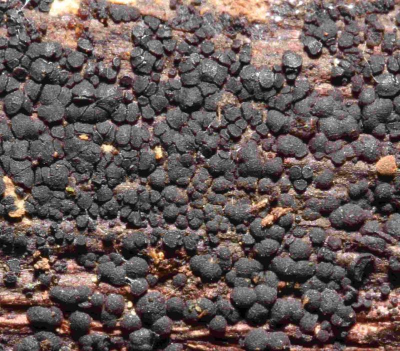
Here's already my next problem:
Substrate: (Yet) Unidentified deciduous wood
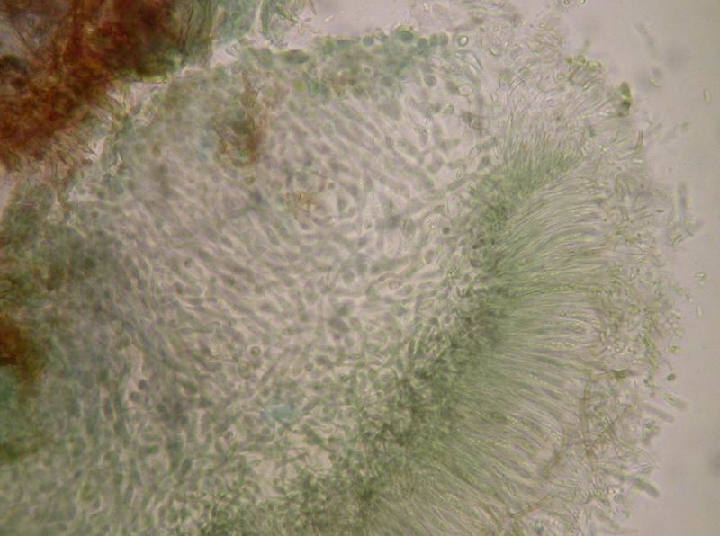
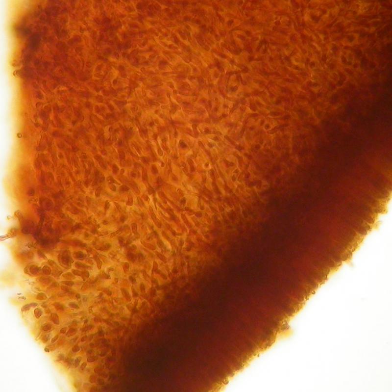
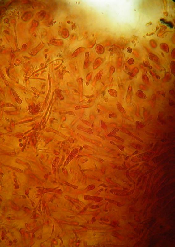
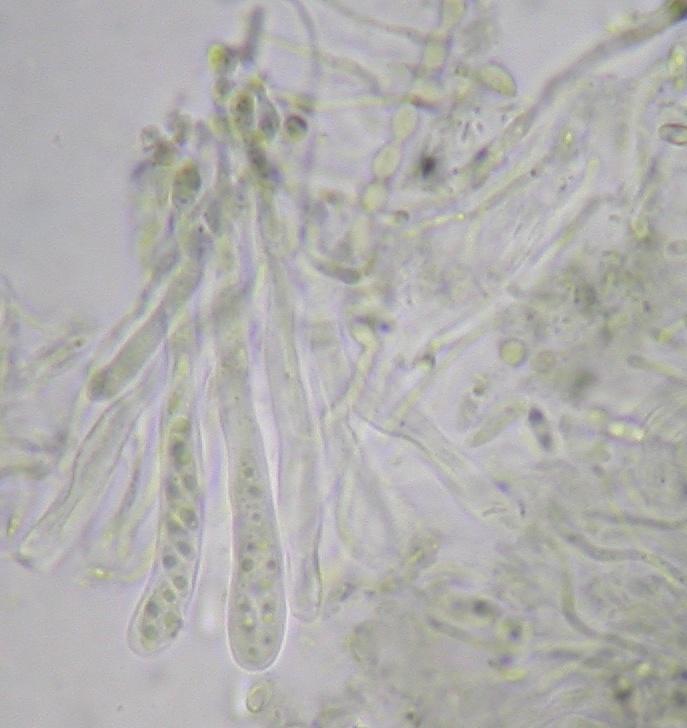
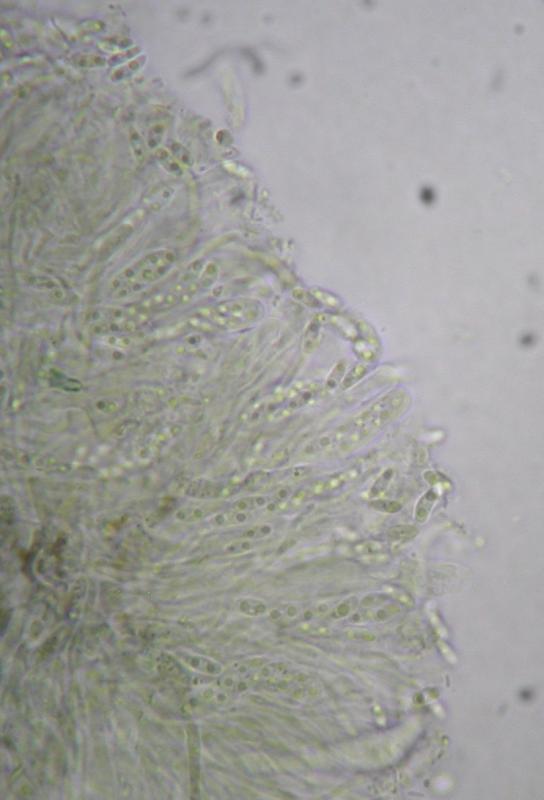
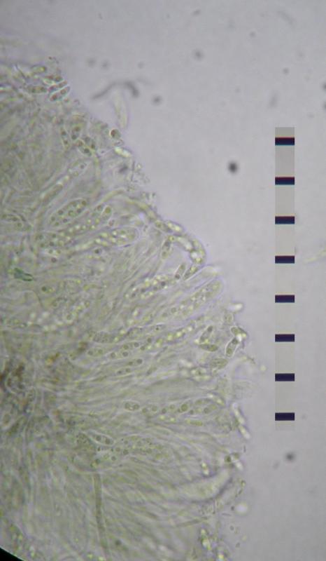
Macro: Apothecia, up to 0.5 mm diam. (as single apothecia), exceeding 1 mm in confluent parts. Densely aggregated and often more or less coalescing to confluent parts. Black, with a distinct margin when young, then flat without margin and later convex. Exuding as a red-brown colour in KOH.
Micro: The whole disc is incrusted with a brown substance and structures are difficult to see when prepared in water. This Substance dissolves in KOH as a reddish exsudate and then the structures are hyaline to distinctly green. Asci 35-40 x 5 µm, IKI-, Melzer-, Spores hyaline, 4.5-5.5 x 1.2-1.6 µm, with two oildrops, aseptate. Paraphyses about 1 µm diameter. Excipulum not clearly differentiated from medulla, the medulla consisting of gelatinous tissue with loosely intertwined, irregularly expanded hyphae (Photos).
Thank you for any help
Stefan

Ist was Durelloides.
Hier noch zwei Fotos von Sporen/Asci
LG
Stefan
danke, das hilft! Vergleiche mal meine Zeichnungen im Verzeichnis Phaeangella = Durella redbrown, und da "bigutt spores narrow ionom". Da sind zwei Funde, einer aus USA, der andere aus Luxemburg. Die wuchsen aber an ansitzenden Ästen.
Hast du keine Skala in deinen Fotos? Dann könnte ich etwas nachmessen.
Das Auflösen und Austreten des rotbraunen Pigments ist typisch für diese Art (ionomidotisch). Irgendwie erinnert er auch an Ionomitodis fulvotingens, aber nur mikroskopisch inklusive Excipulum.
Grüße
Zotto
Habe hier mal noch einen 10 mü Massstab reingeflickt. Was besseres habe ich im Moment nicht. Hilft wohl nicht viel. Kann aber gerne nochmals gewisse Sachen nachmessen...
LG
Stefan
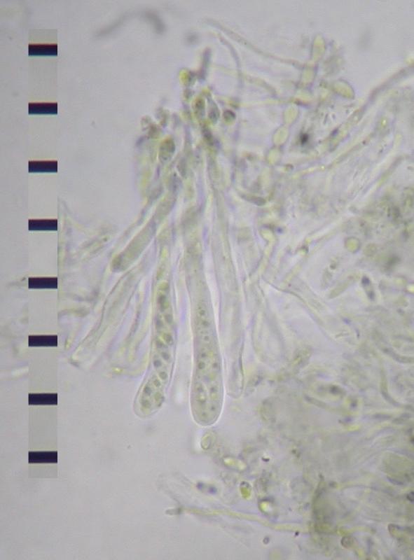
Asci tot 45 x 4.5-5.5 µm,
Sporen etwa 4,5-5,5 x 1,8-2 µm