

22-04-2026 20:17
 Marian Jagers
Is anyone familiar with the Hyphomycetes genus Pse
22-04-2026 20:54
 Enrique Rubio
Hi to everybody.This Pyrenopeziza grew in moist le
22-04-2026 01:06
 Richard VALERI
Bonjour à tous.Je vous présente cette Nectria s.
21-04-2026 13:36
Gernot FriebesHi,I am out of ideas for this one. I collected Sal
21-04-2026 13:19
Gernot FriebesHi,this Lophodermium on Typha has ascospores measu
21-04-2026 13:05
Gernot FriebesHi,this hyphomycete feels familiar but I was not a
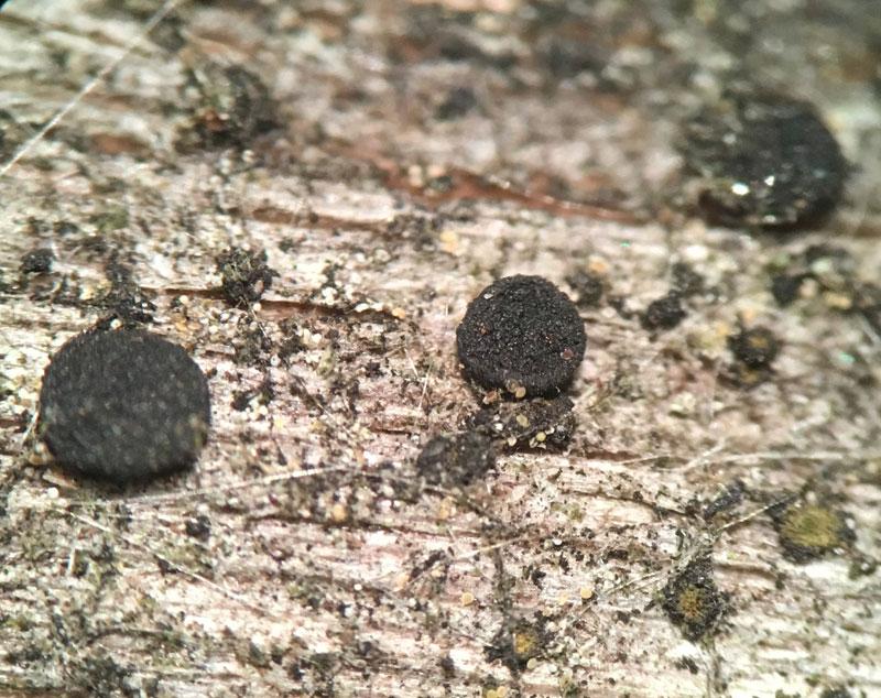
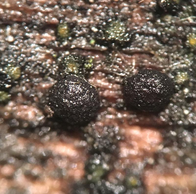
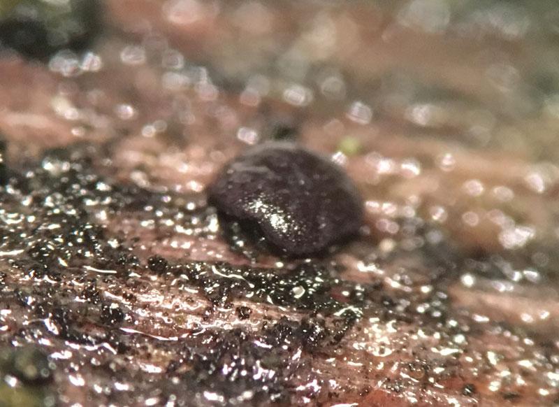
Patellariaceae? Tryblidaria? Murangium?
Ethan Crenson,
28-09-2017 04:26
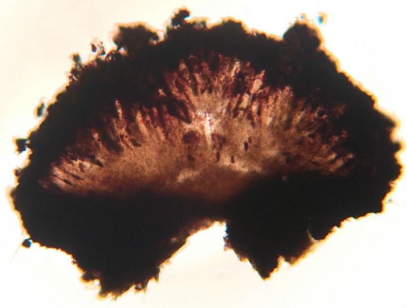
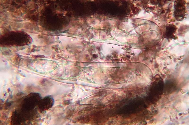
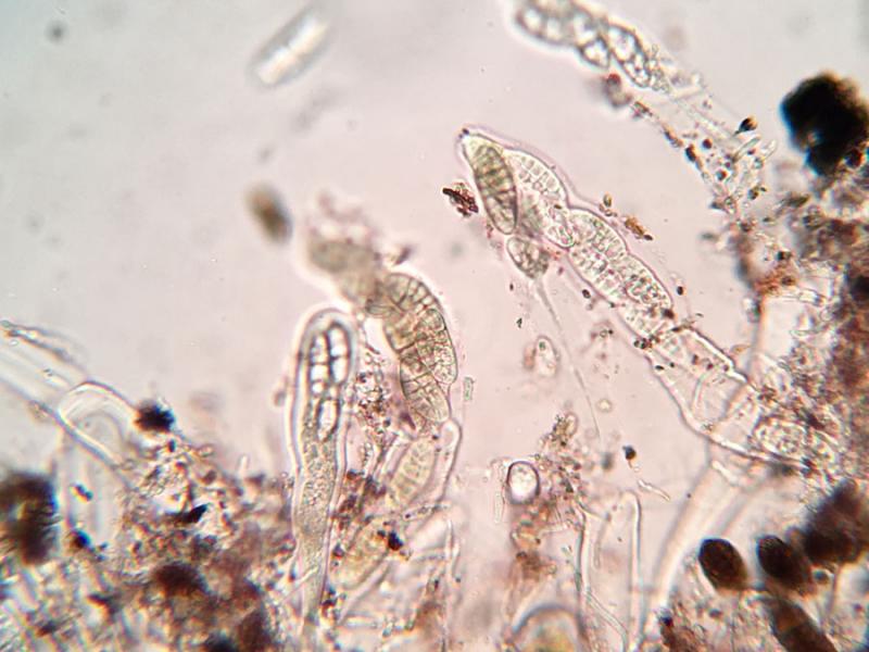
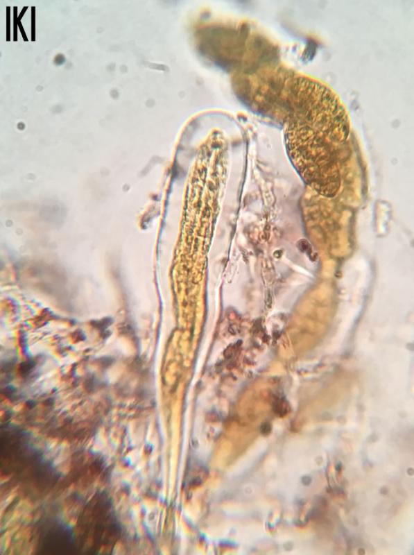
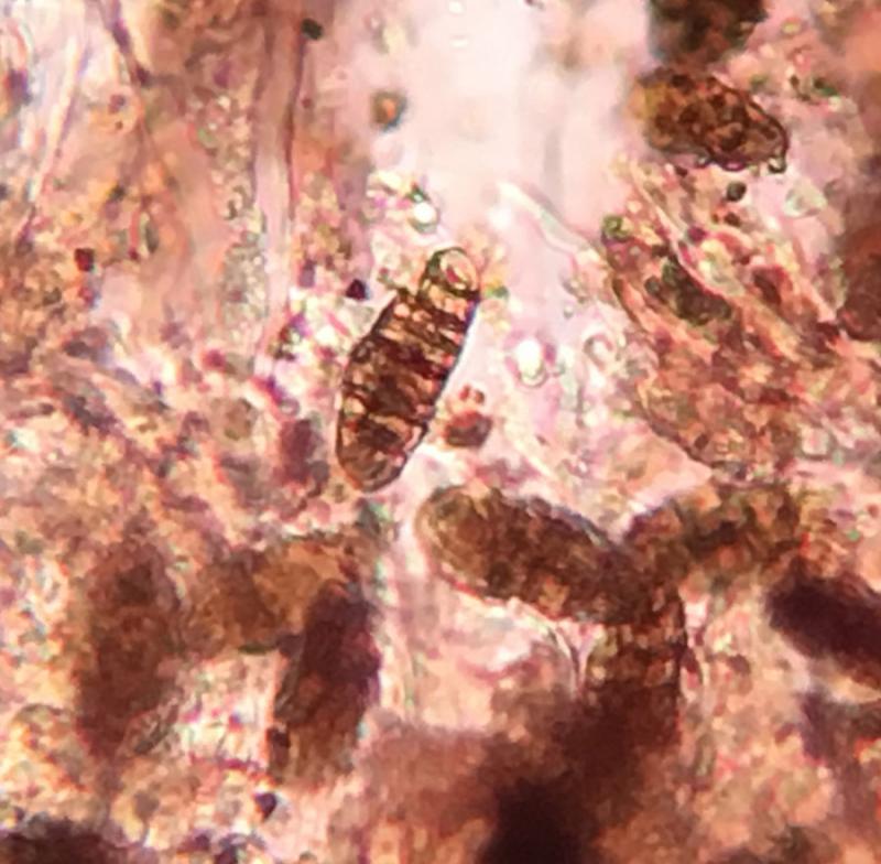
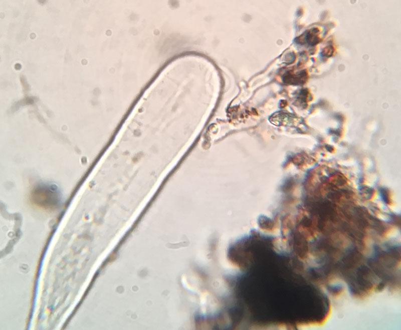
Asci with 8 spores, bitunicate, about 138-148 x 24-25µm. IKI-
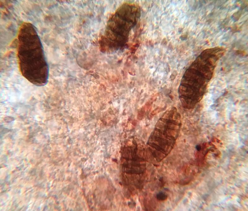
Spores muriform, at first hyaline, finally dark brown, 4-7 septate with longitudinal septa. 25-33 x 10-13µm. It appears that the spores inside the asci are at the larger end of the scale 31-33µm long while the darker spores loose in the centrum are somewhat shorter 23-27µm.
The outer layer appears to be a dark, agglutinated (?) layer appearing red in projected light. It was very difficult to discern any paraphyses or other features.
Because of the resemblance to Patellarioid ascos, I had thought it might be Tryblidaria, but perhaps that's not a very good fit. Can anyone provide me with a shove in the right direction? Thanks!

Hans-Otto Baral,
28-09-2017 07:49

Re : Patellariaceae? Tryblidaria? Murangium?
Hi Ethan
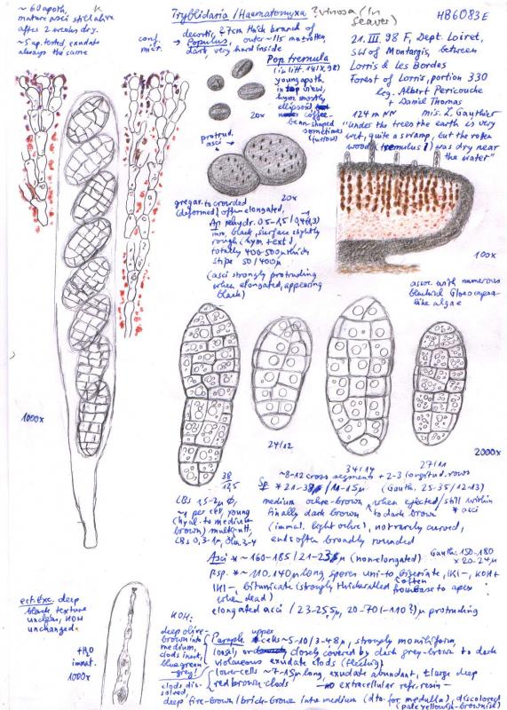
your sample is not in good shape but I think it is a Haematomyxa. I once studied a collection that I identified as Haematomyxa cf. vinosa. It is an American species, originally from New Jersey on Quercus, but I had it on Populus from France (near Montargis, leg. A. Pericouche).
This collection yielded a strong ionomidotic reaction: when adding KOH a deep olive-brown sap extruded and the exudate became blue-green-gray.
Zotto
Ethan Crenson,
28-09-2017 17:34
Re : Patellariaceae? Tryblidaria? Murangium?
Zotto,
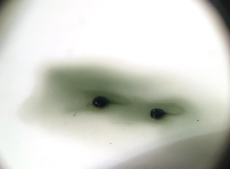
Thank you for your help! I crushed two fruiting bodies and got an immediate olive green reaction when I added KOH. In your notes you mention Tryblidaria/Haematomyxa ? vinosa (in Seaver). I have looked at Seaver's volume on Inoperculates, but I only see "Tryblidium" but no vinosa. Where I can find out more about Haematomyxa?
Regards,
Ethan
Ethan Crenson,
28-09-2017 19:04
Re : Patellariaceae? Tryblidaria? Murangium?
Aha! Disregard my comment about Seaver's Inoperculates. I have found "Haematomyxa vinosa" on page 372.
Hans-Otto Baral,
28-09-2017 20:26
Re : Patellariaceae? Tryblidaria? Murangium?
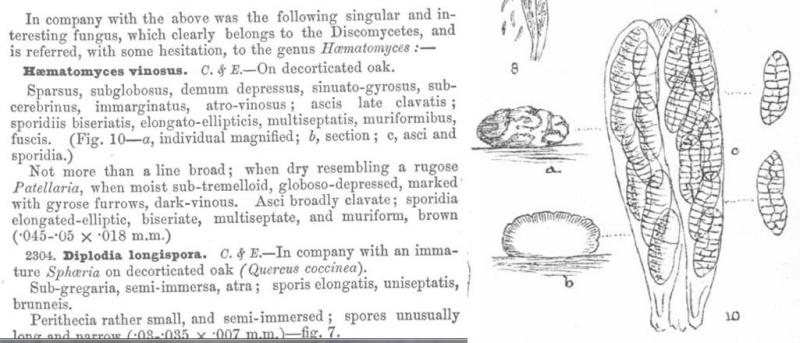
Here is the Cooke description. The spores are longer while the width in Cooke & Ellis seems a bit too high compared to the length (45-50 x 18).
Jason Karakehian,
30-09-2017 00:41

Re : Patellariaceae? Tryblidaria? Murangium?
Hi, I was just working on this recently from simillar collections from West Virginia and Kentucky. We thought about Haematomyxa but ultimately came to Tryblidaria fenestrata.
Hans-Otto Baral,
30-09-2017 08:40
Re : Patellariaceae? Tryblidaria? Murangium?
Hi Jason
do your samples show the wine-red-brown pigment (in water) and olive-brown ionomidotic reaction? My studies on T. fenestrata and the close T. azarae show olive-black exudate (in water) that does not change or dissolve in KOH (as in Patellaria) as far as I remember. T. fenestrata seems to differ from T. azarae in hyaline spores with rounded ends vs. yellow-ochre spores (always inside the living asci) with pointed ends.
The colour of the particles among the paraphyses appear to be diagnostic between the two genera: refractive and hyaline in Tryblidaria, red-brown in Haematomyxa. The swollen paraphysis cells with constrictions at the septa might be a further difference between the genera. See my drawings in the Patellaria-Tryblidaria folder:
https://drive.google.com/drive/folders/0B5SeyOEkxxZhejBuVzBCX2tqQTg
Zotto
Jason Karakehian,
30-09-2017 16:09
Re : Patellariaceae? Tryblidaria? Murangium?
Thank you! I will try to get to my notes and specimens, which are not here right now and get back here with some answers! Best - Jason