27-04-2026 20:52
 Lothar Krieglsteiner
Found on hanging tiwg of Olea europaea in dried-ou
28-04-2026 22:51
 Bernard CLESSE
Bonsoir à toutes et tous,Pourriez-vous m'aider à
29-04-2026 08:01
Lothar Krieglsteiner
... on twig attached to small tree of Citrus auran
29-04-2026 10:44
Lothar Krieglsteiner
growing at moist, drying-out soil at the side of a
28-04-2026 20:33
Vitus SchäfftleinHello, I found Trochila ilicina on Ilex aquifoliu

28-04-2026 21:50
 Pablo Sandoval
Hola a todos,Espero se encuentren bien. Hace mucho
27-04-2026 18:05
Lothar Krieglsteiner
... still attached at standing tree. The green con
28-04-2026 20:07
Lothar Krieglsteiner
... on twig in the air at standing Ceratonia siliq
27-04-2026 18:48
Tony MoverleyCollected 23rd April 2026, Norfolk, EnglandSwarms
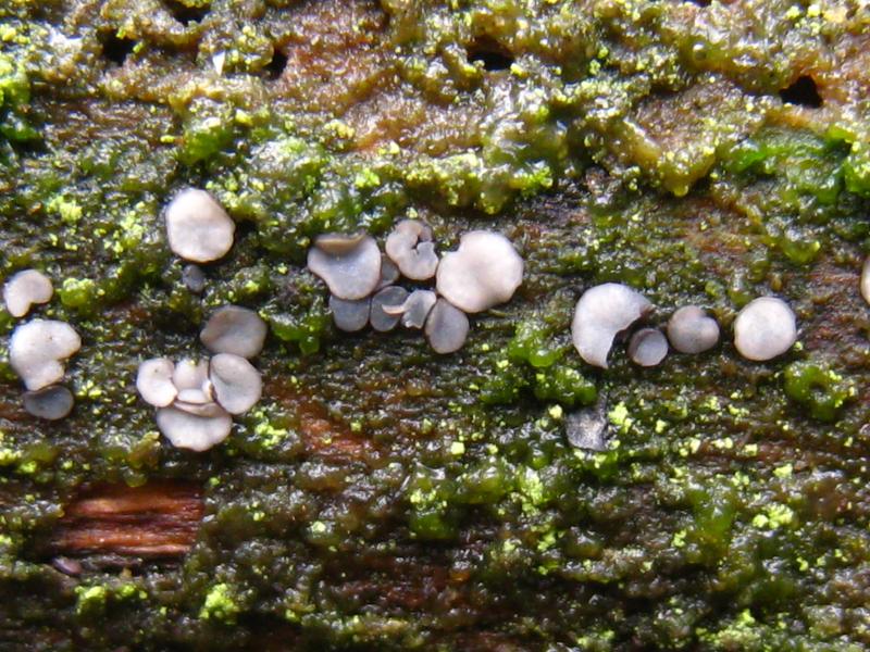
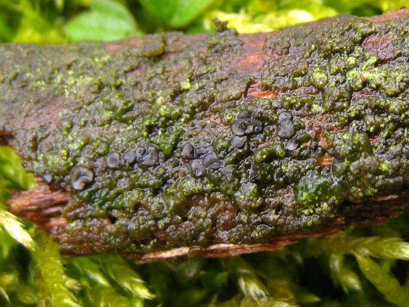
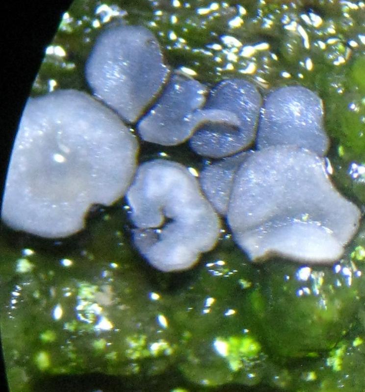
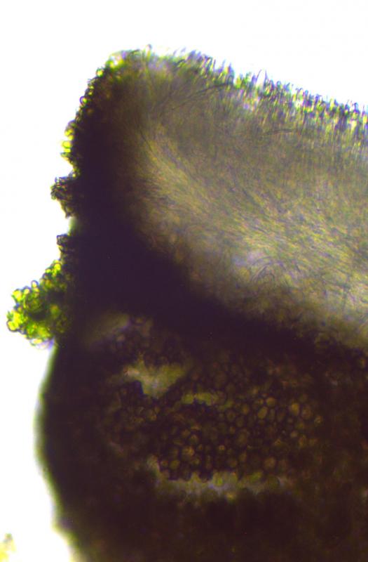
Mollisia found on decorticated branch covered with algae.
FRB: diameter 1.5 mm; gregarious in small clusters, hymenium light grey/blue, edge and bottom dark black brown,
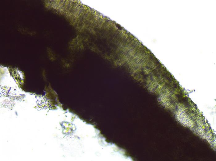
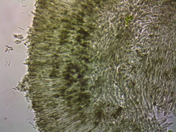
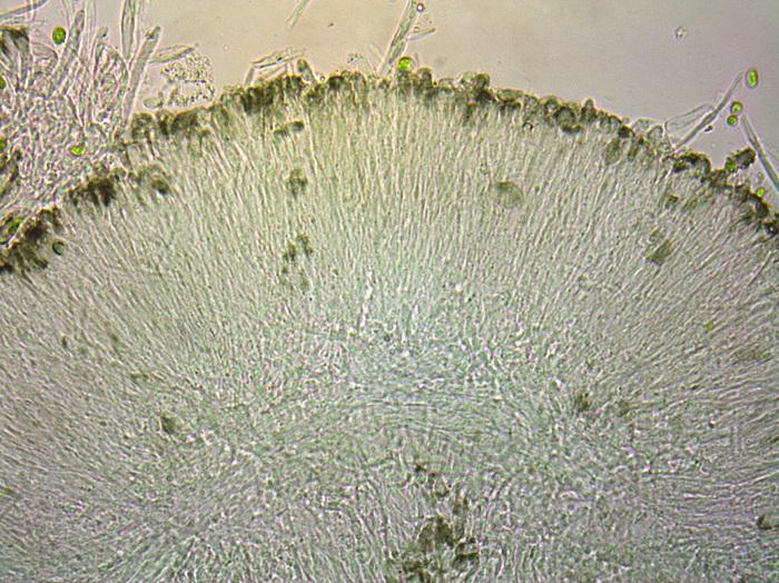
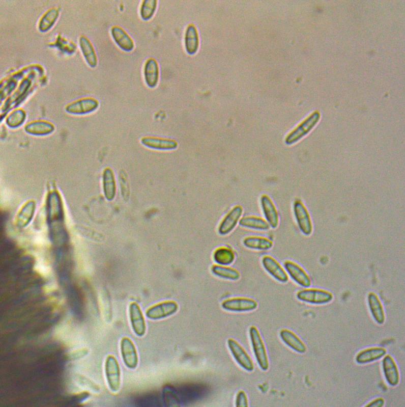
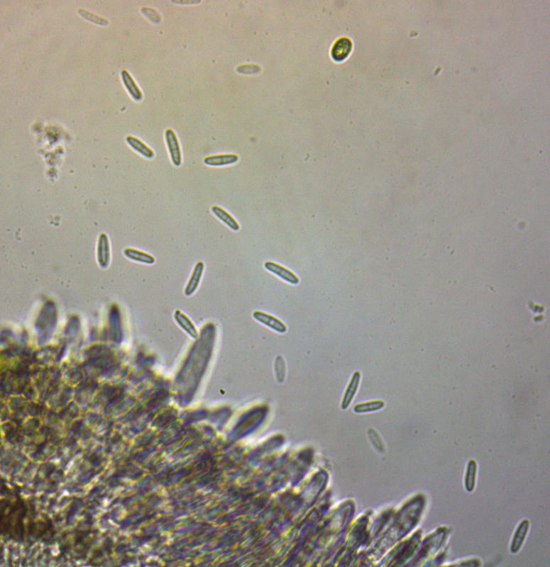
Microscopy (all dimensions in water): of fresh material
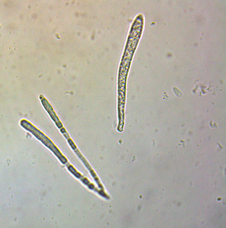
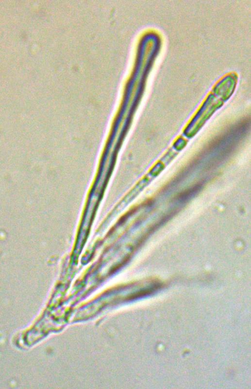
Asci: cylindrical, hyaline, pointy top, amyloid; with croziers, 67-73 x 7.35-7.61 µm.
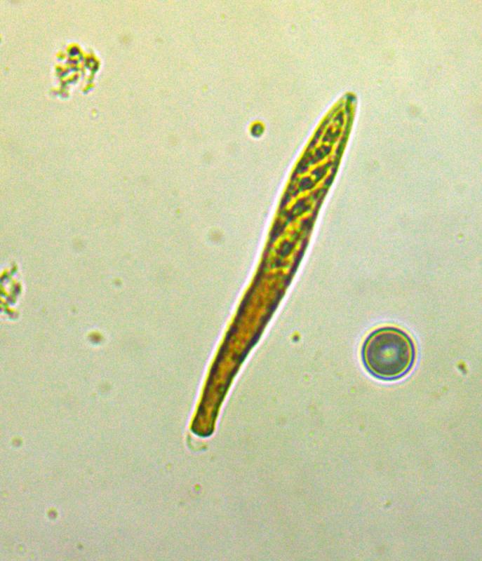
Spore: variable: cylindrical, amygdaloid to oblong with one septum; 7.58-12.08 x 2.62-3,68 µm; Q= 2,92
Paraphyse: with swollen top and refractive vacuole
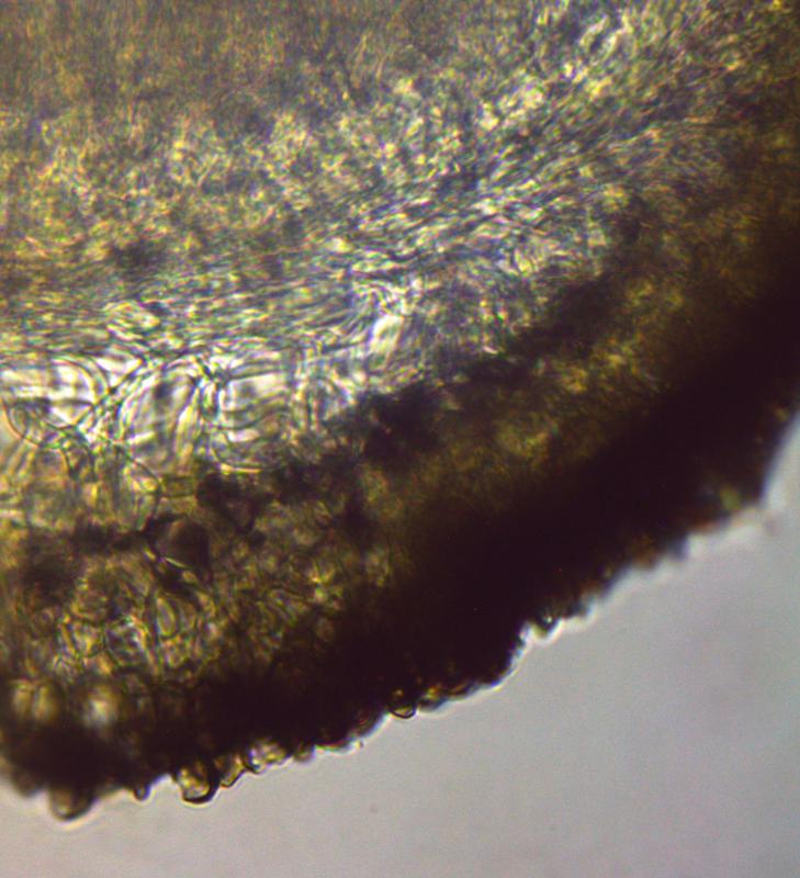
Texture:
- subhymenium texture intricata light brown hyphae
- ectal excipulum:dark brown, texture globulosa
- medullary excipulum: light brown, globulosa-angularis.
What is your opinion, can it be Mollisia lividofusca?
Thanks in advance!
Greetings,
François Bartholomeeusen

Hello,
I'm not too certain. The colour of the subhymenium is not to see in your foto, and I'm not sure wether you may be mixed up the subhymenium and the excipulum? The loosely arranged medulla below the subhymenium would be a good hint towards M. lividofusca, but the macroscopy is unusual pale. In this wet stage M. lividofusca would either show distinct brownish tinges in the hymenium or be dark blueish-grey. Also the septate spores are very unusual for M. lividofusca and last the spore width also in unusual broad.
What cocnerns the spores that may be an abnormal developement due to the winter temperatures may be?
Did you check the KOH-reation (it is most likely negative, but it's always worth a try ...)
And are you really certain about the colour of the subhymenial hyphae?
best regards,
Andreas
Thanks for your fast answer. You are right, due to the low temperature in the morning last week, the apothecia suffered probably from frostbite. I add a picture of spores with a more regular pattern which I measured again: 8,68-11,25 x 2,43-3 µm; Q= 4.05.
I also add two pictures of the frb, one by daylight and one through the binocular, and a picture of a new preparation. Would you be so kind te check the color of the hymenium and of the subhymenial hyphae. I am sorry to say that I am out of KOH.
Can this information shed some light on a determination?
Greetings,
François
Hello,
the daylight foto looks very much like M. lividofusca.
The other fotos I can not see too clear. A pitty that you don't have KOH, because if you have a thin cut through the apothecium in KOH, you can see the coloured subhymenium far better because all those refractive vacuoles are dissolved and don't mask the true colour of the hyphae.
best regards,
Andreas
Until the next time,
François
I have used KOH but not without problems! Cutting the apothecium was not easy, I couldn't get a nice sharp picture ... and I only saw a green discoloration. For me everything is still very unclear, what do you think of it?
Greetings,
François