
04-05-2026 16:39
 Stephen Martin Mifsud
ID request: This specimen was collected in Malta o
04-05-2026 09:50
 Castillo Joseba
Me mandan el material seco de Galicia,(España) re
28-04-2026 20:07
 Lothar Krieglsteiner
... on twig in the air at standing Ceratonia siliq
02-05-2026 12:42
Alain BRISSARDBonjour à tousJeuidi 30 avril dernier on m'a remi
02-05-2026 13:06
Pauline. PennaBonjour Please can someone help me with this id
01-05-2026 22:45
 Thierry Blondelle
Bonjour à tous, Une récolte sur bouse séchée d
14-04-2026 05:32
Ethan CrensonHi all, A few weeks back a friend pointed out som
28-04-2026 20:33
Vitus SchäfftleinHello, I found Trochila ilicina on Ilex aquifoliu
Orbilia sp. on Typha
Chris Yeates,
30-09-2014 21:49
 Bonsoir tous
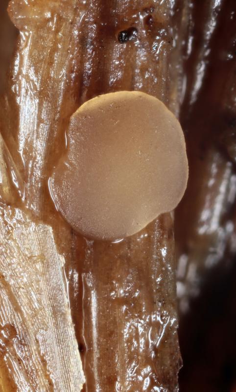
today I collected a few scattered apothecia of a discomycete which was difficult to see as it had the same pale brown colour as the surrounding stem of Typha latifolia.
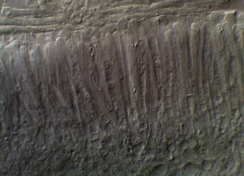
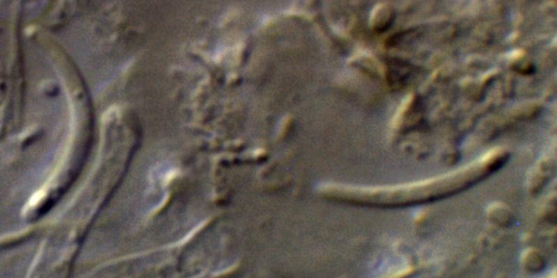
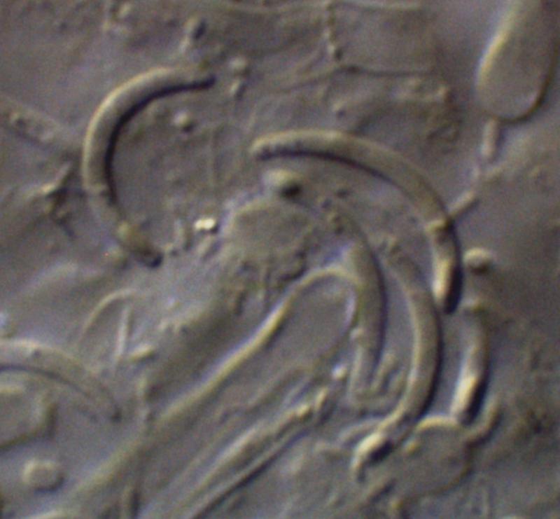
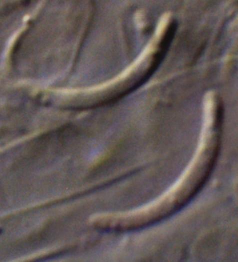
Under the microscope it was clear that it was an Orbilia (with the characteristic knob-topped paraphyses). Inside the asci the spores were clearly long-cylindric and curved with short spore bodies at one end (half the spores reversed). Unusually for an Orbilia, in my limited experience, in a water mount there were many free spores. These were very much curved, some almost into a semicircle - I wonder whether the tension to form this shape contributed to the ease in which so many were ejected in the water mount.
Here are some preliminary images - I can post more in due course, but these are typical. The spore measurements were made from spore tip to spore tip, so they are like the string on a bow, rather than an attempt to measure them along the curve. Measurements made so far are in the range 8.1-10.9 x 1.2-1.3(1.4)µm.
I note there is an Orbilia arundinacea listed as occurring on monocot's like Typha; but looking at the protologue in Velenovsky (1934) he says of the spores 'Sp. . . . rectae' and no spores seen could be described as anything near straight. I cannot find O. arundinacea in Zotto's images - maybe I have missed something.
As ever, help very much appreciated.
Cordialement
Chris
Hans-Otto Baral,
30-09-2014 22:16

Re : Orbilia sp. on Typha
Hi Chris
this would require a section of the margin, in order to see whether it is a nematode-trapping species or not, and conidia :-)
O. arundinacea is a synonym of O. rectispora - typical for Typha and the like, but, as you say, with rather straight spores.
It is sometimes that you have many free spores in Orbilia. Difficult to explain. But the spore curvature which is actually less so inside the ascus - logically - can hardly contribute to this. The spores are agglutinated in one packet prior to discharge, and the packet is ejectd as one entity.
Zotto
Zotto
Chris Yeates,
30-09-2014 22:28
Re : Orbilia sp. on Typha
Thanks Zotto for the quick response - and there was I thinking such distinctive spores might make it easy!
I have little material to work with but I shall have a go at the section, probably tomorrow. What am I looking for in the margin which would tell whether it is a nematode trapper?. I have seen no evidence of conidia but will have a very careful look.
Chris
I have little material to work with but I shall have a go at the section, probably tomorrow. What am I looking for in the margin which would tell whether it is a nematode trapper?. I have seen no evidence of conidia but will have a very careful look.
Chris
Hans-Otto Baral,
30-09-2014 22:34
Re : Orbilia sp. on Typha
Orbilia auricolor has just those spores, and it is a collective species with an Arthrobotrys anamorph, trapping nematodes by adhesive networks. However the spores are more tapered at the basal end in O. auricolor, so we are a bit reluctant.
Another option would be O. sambuci = O. fimicoloides, a lkewise plurivorous species with a Dactylella anamorph and no trapping capabilities. The Dactylella conidia are very characteristic: very large, fusiform, multiseptate.
The marginal excipulum forms distinct cell rows under a low angle in those with Dactylella, but roundish cells oriented under a high angle in those with Arthrobotrys.