
04-06-2026 11:36
Gernot FriebesHi,found on Vaccinium myrtillus.Asci: IKI –, 8-s
04-06-2026 07:02
 François Freléchoux
Bonjour, Voici la description d'une espèce qui p
04-06-2026 10:50
François Freléchoux
Bonjour, J'ai trouvé hier un petit asco observé
22-05-2026 13:29
Gernot FriebesHi,I am curious to hear your opinion on this mater
18-10-2022 00:12
Valencia Lopez Francisco JavierHola amigos/asRecientemente encontré esta colecci
03-06-2026 19:45
 Miguel Ángel Ribes
Good afternoonI'm completely baffled by this suppo
03-06-2026 14:39
Thomas FlammerApothecia yellow, glassy-transparent, 80 - 120 ymS

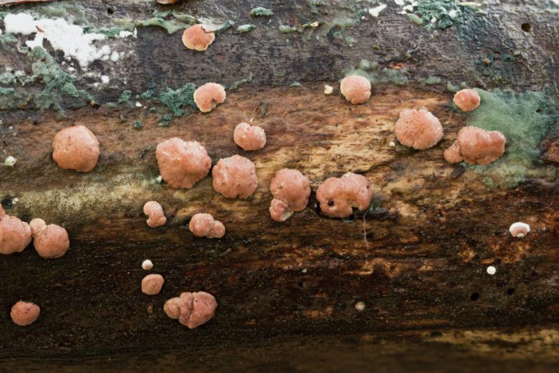
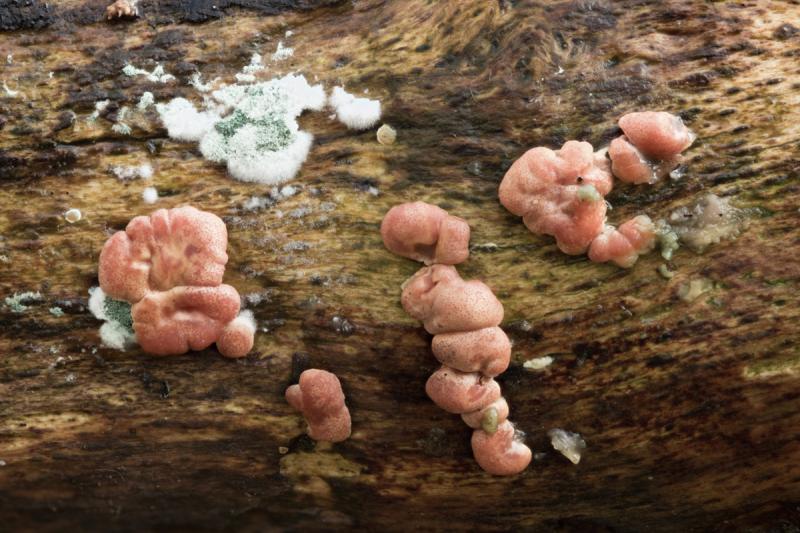
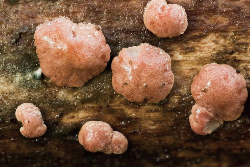
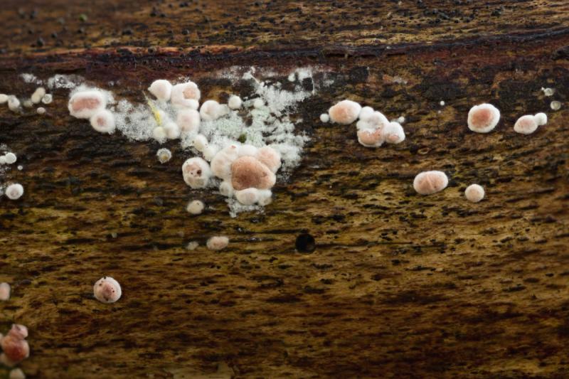
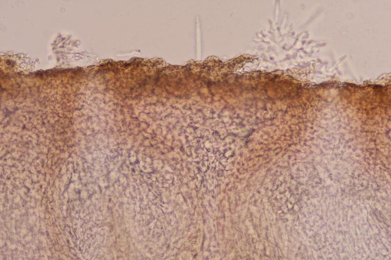
Here is what I believe is a Hypocrea found on Fagus branch on the forest floor. The stromata are not mature but the green anamorph was present which may help to identify it. The stromata were to 5mm and there were many of them. Attached to the substrate right up to the edge. They were a pinkish colour with visible darker ostiolar openings creating a pattern of dots on the surface. They dried to a pale brown.
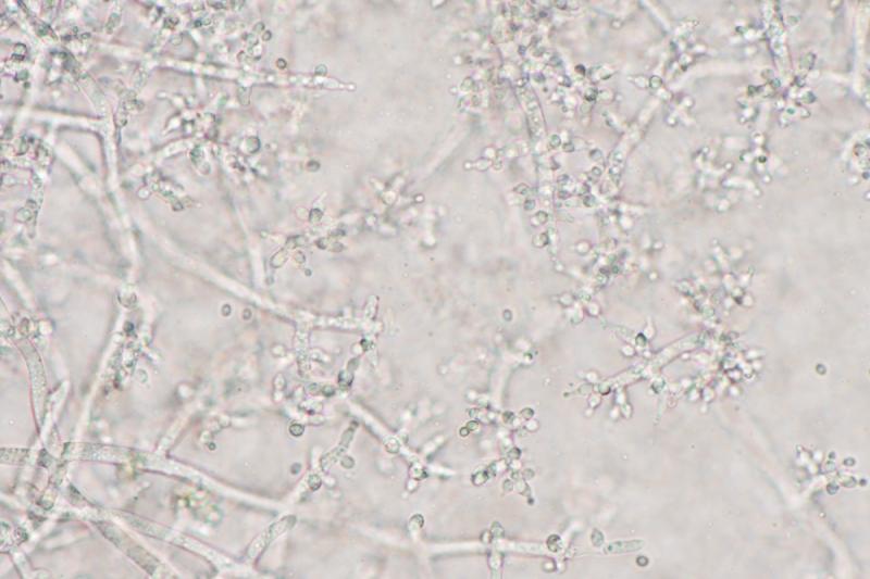
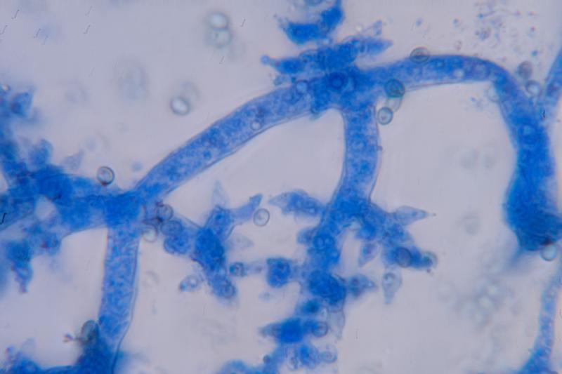
The anamorph was green and I don't have the language to describe the structure of the conidiophores and phialides, hopefully the pictures will help
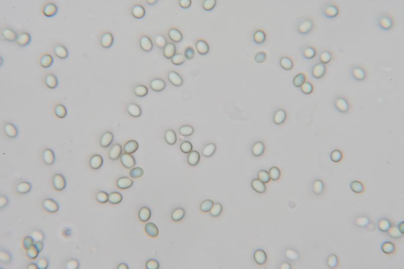
The conidia were smooth and ellipsoid 3.3 X 2.4 QE 1.4
My attempts to key it give Hypocrea minutispora and that does look OK with the structure of the conidiophores and phialides as described for that species in this paper although the conidia may be a bit on the large size.
If anyone can help I would appreciate it.
Many thanks
David

I think your fungus is H. minutispora but it would be necessary to compare it to H. pachybasidioides.
Christian
Thank you Christian,
I looked again at the paper I mentioned in my first post and it appears that H. pachybasidioides is in the "polysporum" clade and its anamorph, (Trichoderma Polysporum) has white/hyaline conidia where my specimen had green conidia. It looks like it also has infertile, corkscrew-like extensions of the conidiophores unlike my specimen. The green, smooth, ellipsoid conidia, along with the structure of the conidiophores pictured in the paper above, do seem to leave only T. minutisporum / H. minutispora.
Regards
David