
17-04-2026 19:16
 Enrique Rubio
Hi to everybodyI would appreciate any assistance r
14-04-2026 05:32
Ethan CrensonHi all, A few weeks back a friend pointed out som
17-04-2026 15:14
 Bruno Coué
Bonjour.Récoltes du 16/04/2026, sur feuilles mort
12-04-2026 15:52
Gernot FriebesHi,I'm looking for help with this anamorph collect
14-04-2026 21:52
Gernot FriebesHi,found on dead leaves of Carex elata. Conidia: 4
16-04-2026 22:09
Buckwheat PeteHello, I'd like to ask about this older specimen:
15-04-2026 19:33
Fátima Durán ManzanequeHi!! I need help, I found this Ascomycete but I d
14-04-2026 20:31
Gernot FriebesHi,can this be Psilachnum lateritioalbum on Phragm
12-04-2026 17:56
 Hardware Tony
Found on dead stems in February earlier this year
12-04-2026 12:22
 William Slosse
In a dune grassland in Oostduinkerke (Belgium), on
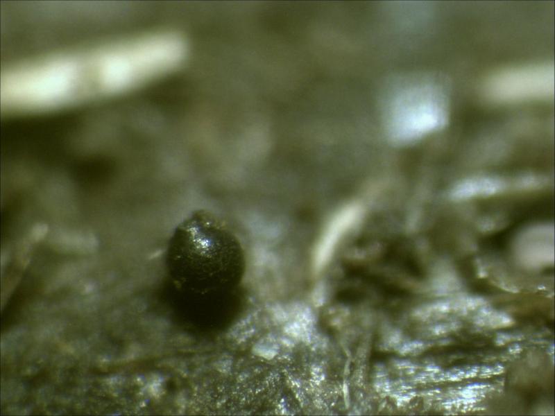
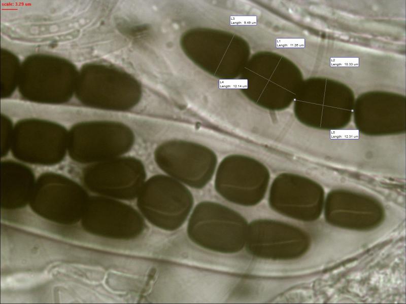
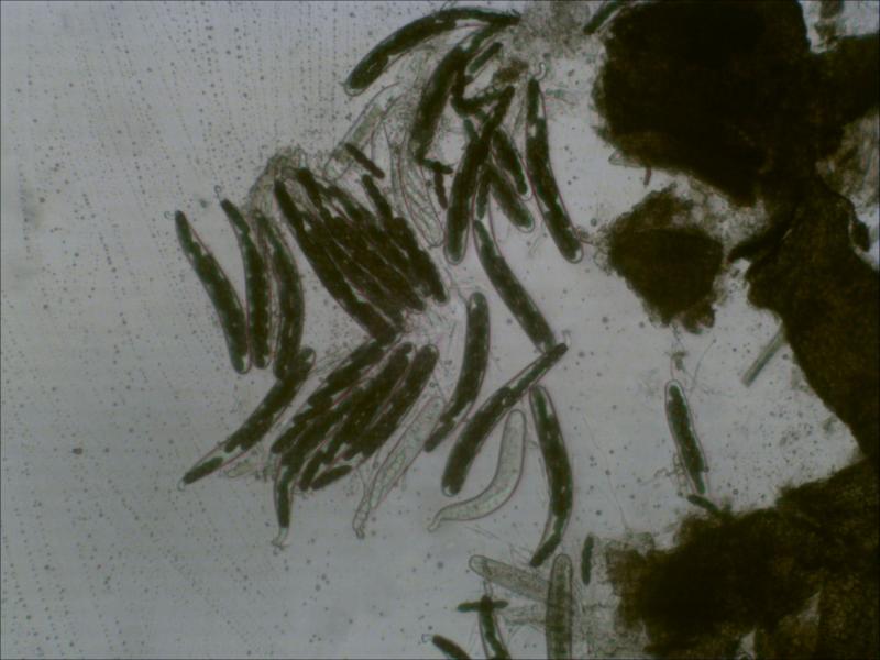
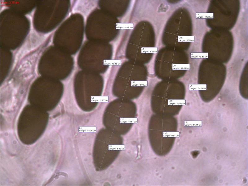
 Found on horse dung.
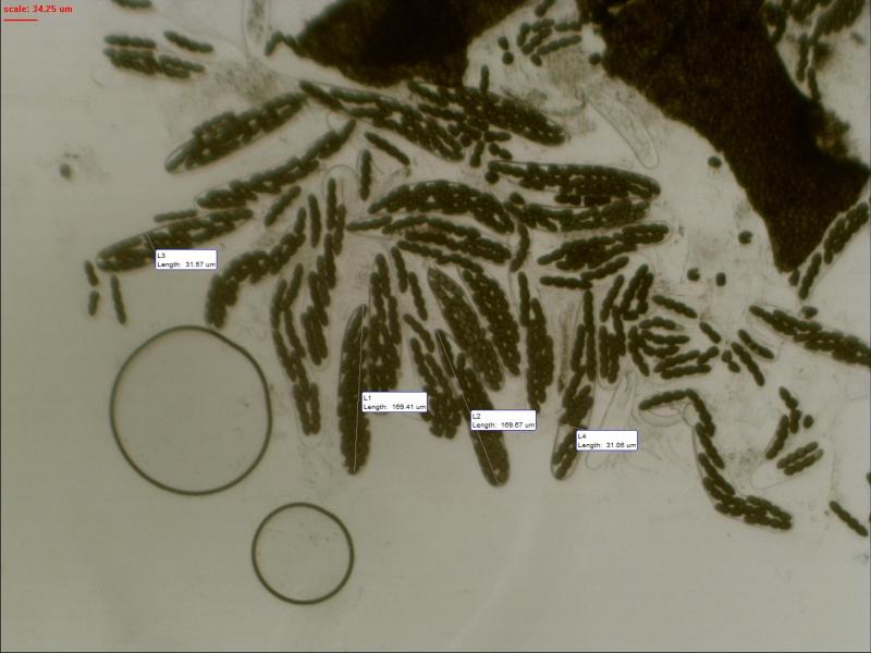
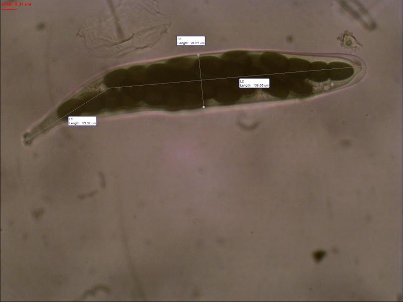
Asci: 169.41-169.67x31.06-31.57 um
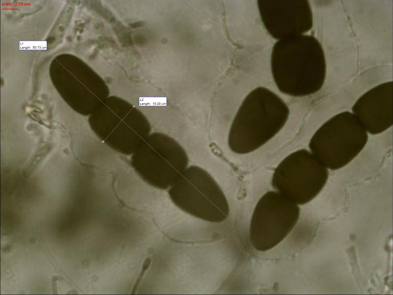
Spore: 50.73x10.20-11.26 um
De second cell slightly smaller than the third one 12.14 over 12.41 um
Germ slit: Parallel to slightly oblique whereby they bent near the septum

yes, this should be S. megalospora, compare with my finding from Austria last year:
Sporormiella megalospora
regards,
björn

Sporesize and germslit conforms better with Sporormiella capybarae.
The spores of Sporormiella grandispora have a parallel germslit and slightly larger spores.
And Sporormiella megalospora has much larger spores.
Norbert
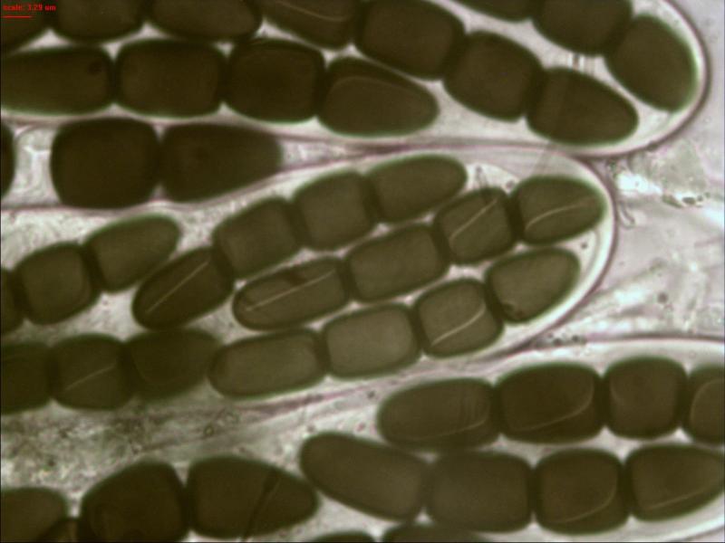
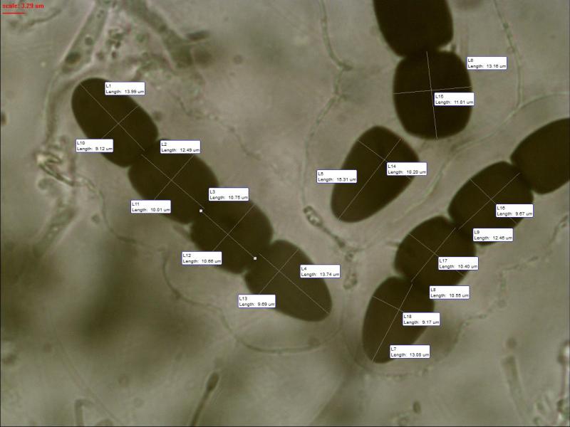
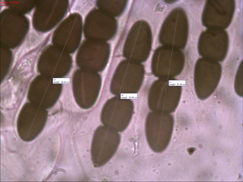
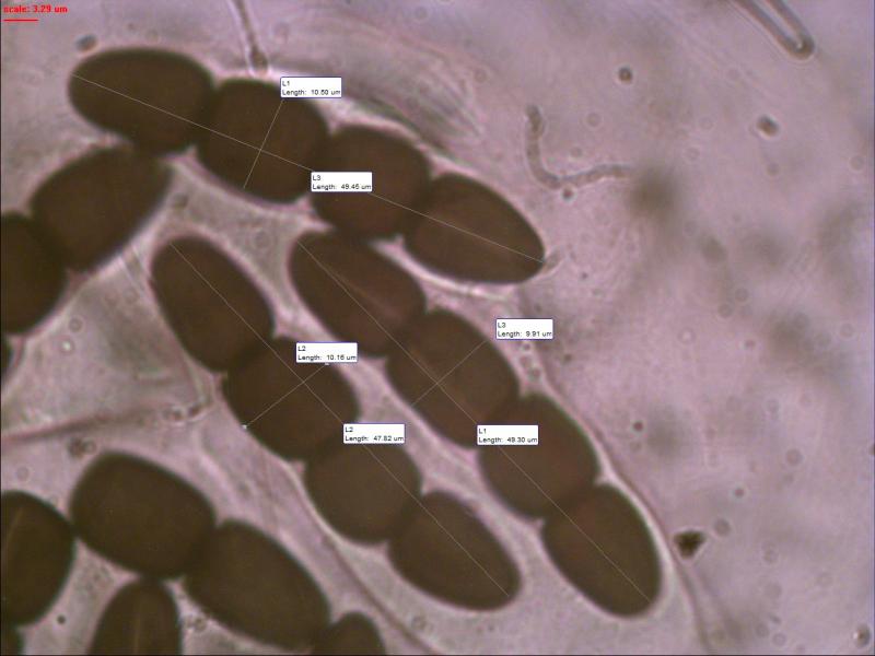
Hereby I send you a photo with measurements of each cell Norbert. Maybe this is a better confirmation.
Upper cell conical:
13.74x9.69; 13.08x9.17; 15.31x10.20 (extreme size)
Second cell barrel shaped:
10.55x10.40; 10.75x10.66; 13.16x11.81 (extreme size)
Third cell barrel shaped:
12.49x10.01; 12.46x9.67
Basel cell cylindrical rounded top:
13.99x9.12
Hopefully I can find more photo's of this one.
Do the germ slits also curve near the septum as they do with S. Grandispora Norbert?
In the article of Ahmed & Cain on page 442 about Sporormiella they also say for S. Grandispora that germ slits are usually parallel, occasionally slightly oblique, usually curved next to septum. Cells almost equal in size (in this case they ar not).

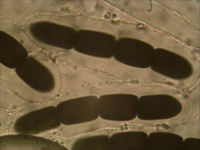
In the first photo, where we perceive the asci, they seem to have it as the base ends abruptly. Joop you can get a clearer picture? If this comfirme we would depart from S.capybarae which has asci gradually ending a long walk, and instead seek to S.intermedia.
Michel.
Michel
Michel.
Sorry, but in the moment I don't have enough time for an answer.
I'll write later.
Norbert
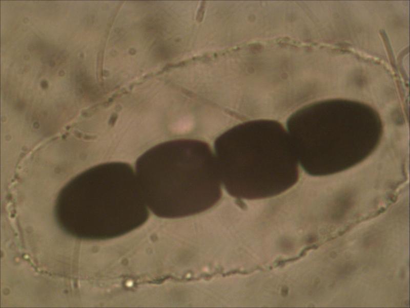
Is the shape of the gelatinous sheath a factor for determination?
They often differ in size and form, especially at the septa but they also can be fully cicular I will look up some photos to explain what I mean.
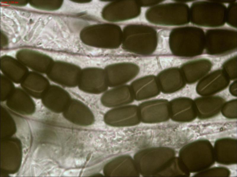
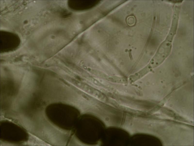
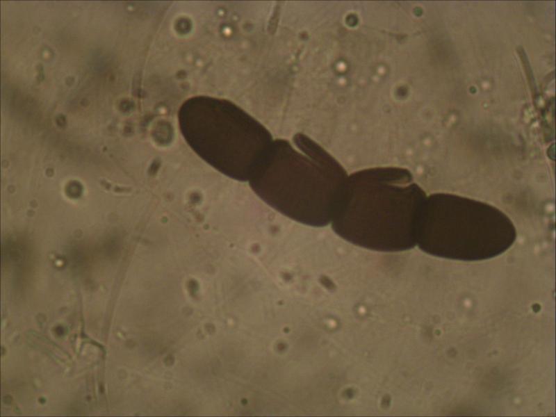
I also include a plhoto where you can see cells of sporormiella opening at the germ slit.

Michel.
Merci Michel
The other serie does not have short stalks for the asci I am still looking for better photo's that match.
In the first serie the upper cell is conical, the second barrel shaped the third and also the basal end cell cylindrical with a rounded end for the latter.