
22-04-2026 20:54
 Enrique Rubio
Hi to everybody.This Pyrenopeziza grew in moist le
24-04-2026 03:16
 David Chapados
Found while looking at something else from wood in
22-04-2026 01:06
 Richard VALERI
Bonjour à tous.Je vous présente cette Nectria s.
22-04-2026 20:17
 Marian Jagers
Is anyone familiar with the Hyphomycetes genus Pse

21-04-2026 13:36
Gernot FriebesHi,I am out of ideas for this one. I collected Sal
Mollisia on Carex
Marja Pennanen,
02-09-2010 16:04
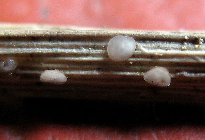
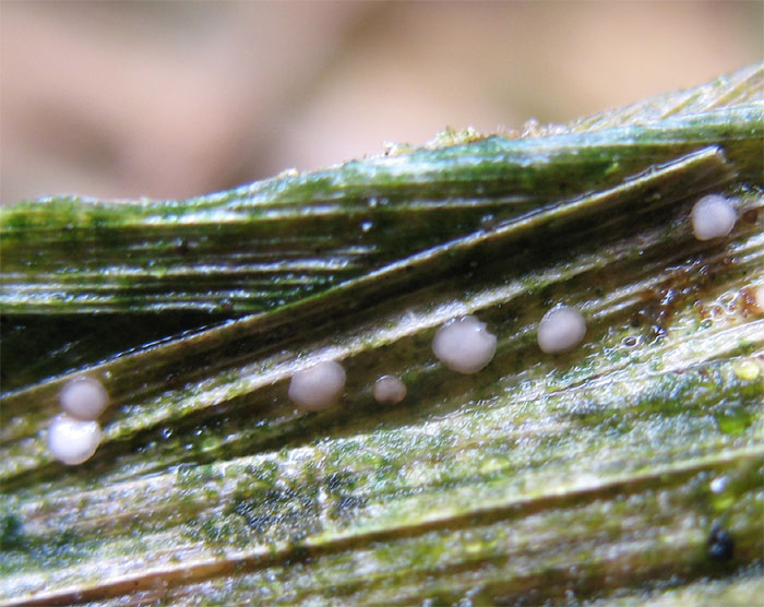
I found these on yesterday, too.
They were pink, but have dryed a little and turned paler, 0,5-0,8 mm wide.
Marja Pennanen,
02-09-2010 16:05
Re:Mollisia on Carex
This is a Mollisia species as far as I understand.
Marja Pennanen,
02-09-2010 16:07
Re:Mollisia on Carex
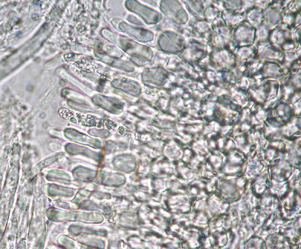
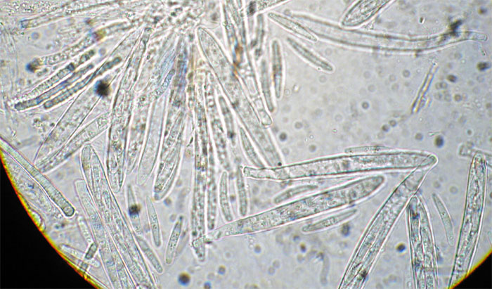
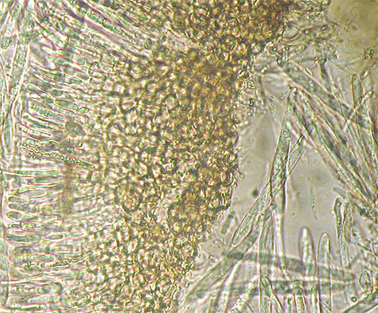
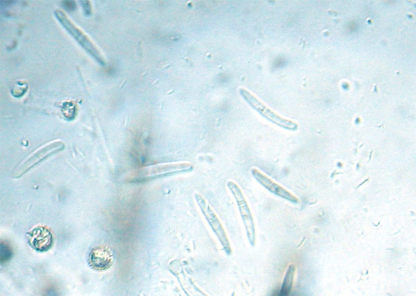
The spores were 13-18x1,5-2, often with one septa.
Asci 63-80x5-7, IKI blue and paraphyses 1,5-3 micrometers wide.
Marja
Asci 63-80x5-7, IKI blue and paraphyses 1,5-3 micrometers wide.
Marja
Hans-Otto Baral,
02-09-2010 16:56

Re:Mollisia on Carex
I see only the background pink, not the apothecia :-/
Due to the VBs in the paraphyses and the globose cells this is a Mollisia, yes. From the spores I am quite sure this is Mollisia submelaena. The only thin is that this species has rather dark, blackish apothecia due to a dark brown excipulum. Is the excipulum brownish near the base in your fungus?
Spores of submelaena are wider: 12-17 x 2.5-3(-4) µm. Are you sure with 1.5-2 µm? I think they could be a bit wider from your photo. Also considering the ascus width... What you also can do is to check the KOH-reaction. Quite a lot of Mollisias react yellow for a moment when coming in contact with KOH or NaOH. M. submelaena does not.
You can see the species taken by Enrique here:
http://www.ascofrance.fr/index.php?r=bdd&page=fiche&id=2743
Was it an acid bog?
Zotto
Marja Pennanen,
02-09-2010 18:06
Re:Mollisia on Carex
Hello Zotto,
The funus may be a little bit darker on the underside.
I saw nothing change color in KOH and I'm sure about the width of these, it may be a little over 2, but not much. Actually I saw only one spore narrower (14x1,5). all the rest were about 2 micrometers wide.
I measured only one ascus with the width 7, most of the asci were 5-6 micrometers wide.
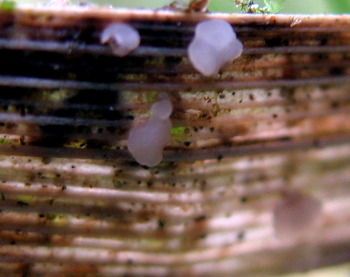
I attached here a poor insitu photo.
Marja
The funus may be a little bit darker on the underside.
I saw nothing change color in KOH and I'm sure about the width of these, it may be a little over 2, but not much. Actually I saw only one spore narrower (14x1,5). all the rest were about 2 micrometers wide.
I measured only one ascus with the width 7, most of the asci were 5-6 micrometers wide.
I attached here a poor insitu photo.
Marja
Hans-Otto Baral,
02-09-2010 18:22
Re:Mollisia on Carex
Thanks. Looks too white for me to be submelaena. Strange find! If you put an apo upside down on the glass and view with 400x on the excipulum, it would be good to know if there are brownish cells, perhaps also brownis anchoring hyphae.
was it in an acid bog?
Zotto
was it in an acid bog?
Zotto
Marja Pennanen,
02-09-2010 20:32
Re:Mollisia on Carex
Hi Otto,
I've got an ealier photo of the brown cells.
I didn't understand the word bog, checked it.
No it wasn't an acid bog, but a wet place (quagmire) in a coniferous woods (maybe an acid bog ;) )
Marja
I've got an ealier photo of the brown cells.
I didn't understand the word bog, checked it.
No it wasn't an acid bog, but a wet place (quagmire) in a coniferous woods (maybe an acid bog ;) )
Marja
Hans-Otto Baral,
02-09-2010 21:26
Re:Mollisia on Carex
Thanks, so only pale brownish. Another phantom, to be recollected in order to find out its variability.
Zotto
Zotto
Marja Pennanen,
07-09-2010 22:08
Re:Mollisia on Carex
Hello,
what do you mean by recollection? That someone else collects propably, because I found some more today on a different place, but maybe the same Carex. I don't know them very well.
I just assume this is the same, haven't checked it. I think, that I can fix the old Russian microscope (transformer broken) by using a led light.
I returned the loaned microscope to university yesterday.
Marja
Hans-Otto Baral,
07-09-2010 22:49
Re:Mollisia on Carex
Oh that's bad. I hope you will find a solution. I mean, the more collection one has studied of a species, the better one can identify or clarify it, because of variation, the next find will slightly differ in spore size etc. Curious whether your new find fits, i.e. has 1-septate spores.
Zotto
Zotto
Marja Pennanen,
08-09-2010 08:42
Re:Mollisia on Carex
Oh yes,
I used a led light and the septum was quite obvious and the cell shapes the same, I just didn't do any further measurements. As you may remember, I use myxomycete spores with the preparation and evaluate the sizes comparing to those now with the old microscope.
The place was propably a former (it's been a dry summer here) wet place (new to me) in a dell about 3 kilometers away from the first place.
This species is not very volumious, but I've got a feeling, that it may not be very rare. I'll check this kind of Carexes for this, when I meet them, if not in hurry. I live quite near the Russian border (under two hours driving), so there is a change, that the species is an easter one (or maybe not).
Dentist time in an hour, so with wishes of tolerable pain: Marja
I attach a photo here now a bit later.
Hans-Otto Baral,
08-09-2010 10:19
Re:Mollisia on Carex
Hope the dentist is a good one....
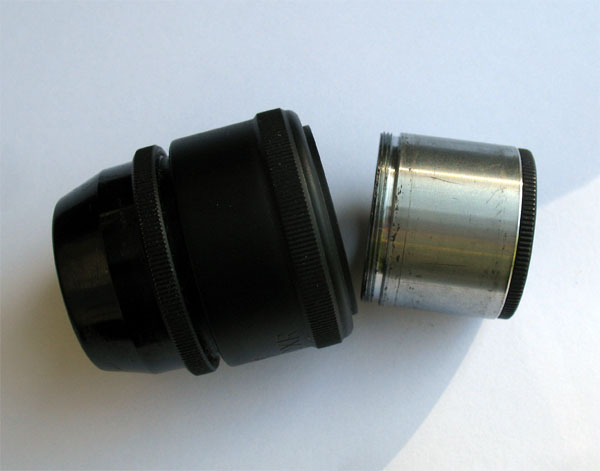
Is the problem that you do not have a measuring glass inside your eyepiece? Can you find out whether you can open the ocular and if there is an aperture on which such glass can be put?
Zotto
Is the problem that you do not have a measuring glass inside your eyepiece? Can you find out whether you can open the ocular and if there is an aperture on which such glass can be put?
Zotto
Marja Pennanen,
08-09-2010 11:16
Re:Mollisia on Carex
Hello,
no pain-no gain. Got a fixed tooth with very little pain.
The ocular was to be opened. I'll ask Seppo, weather he's got some broken ones kept with micrometers. Propably not and the sizes of new equipments are not the same.
There should be a simple measuring system on this one too. I just haven't been able to reveal that secret. I've even got the users guides, but in Russian and Spanih. Tried to translate from Spanish, but it's too slow job for me using the dictionary constantly. I'll apply for funding, but that will take months and may be useless. I can go to the Joensuu university and view there. It's one hour (sometimes more) in bus/side and costs 9,10 ¤/side, which is much for me now. But I'll manage somehow and after all I'm just a green amateur, what does those need ;)
Marja
Hans-Otto Baral,
08-09-2010 12:00
Re:Mollisia on Carex
It is really essential to be able to measure. When I had my first microscope (Kosmos) with single ocular, I already had a scale. And for the first years I avoided oil immersion what was a big fault...
Zotto
Zotto
Martin Bemmann,
08-09-2010 22:11

Re:Mollisia on Carex
Hi Marja,
you don't need an ocular-micrometer if you follow some rules. You just need a micrometer-slide (that you would need to calibrate an ocular-micrometer anyway), you can borrow it from someone since you need it just for an hour or so.
Restrict yourself for making photographs for measuring always with a certain adjustment of your camera (same resolution of pictures and same zoom-adjustment!, zero or full for instance). Then make pictures of the micrometer slide with the adjustment of your choice (mine is full zoomed with a Coolpix 5000 at highest resolution) of the micrometer with each of your objective lenses.
Next you download a measuring software such as ImageJ (free).
Load these photographs and measure the lines you have captured in your photograph: my camera gives 123 µm at 2560 pixels with the 100x lens.
Note down these values and when you load micros of your fungi done with your 100x, you just have to calibrate the software by telling it that 2560 pix are 123 µm (in my example) and all the measurements you will do are according to this calibration.
I hope this was not a description too confusing. Otherwise just contact me by PM.
Cheers,
Martin
Hans-Otto Baral,
08-09-2010 23:45
Re:Mollisia on Carex
Very good idea. But I do not use a software when measuring spores from photos. I think it is enough, or an alternative option, to simply put a scale bar (10 µm for instance) on the photos by the method described by Martin. Then everybody including yourself can evaluate spore size by a calculator (I still prefer using an old slide rule over an electronic calculator).
I have a small set of such scales which I rapidly put on my photos, macro or micro.
Zotto
I have a small set of such scales which I rapidly put on my photos, macro or micro.
Zotto