02-05-2026 13:06
Pauline. PennaBonjour Please can someone help me with this id
02-05-2026 12:42
Alain BRISSARDBonjour à tousJeuidi 30 avril dernier on m'a remi
01-05-2026 22:45
 Thierry Blondelle
Bonjour à tous, Une récolte sur bouse séchée d
28-04-2026 20:07
 Lothar Krieglsteiner
... on twig in the air at standing Ceratonia siliq
14-04-2026 05:32
Ethan CrensonHi all, A few weeks back a friend pointed out som
28-04-2026 20:33
Vitus SchäfftleinHello, I found Trochila ilicina on Ilex aquifoliu

30-04-2026 10:28
Rot BojanHello, by appearance I would say that I am dealing
27-04-2026 18:48
Tony MoverleyCollected 23rd April 2026, Norfolk, EnglandSwarms
27-04-2026 20:52
Lothar Krieglsteiner
Found on hanging tiwg of Olea europaea in dried-ou
 • Habitat and macro suggest Chlorociboria.
• P Bjork suggests C. aeruginascens and C. aeruginosa can usually be distinguished by phenology and macro (in https://www.svampar.se/smf/smt/SMT_2016_2.pdf).
• Earlier fruiting, darker colouring and disc, and concolorous core of the stipe, seem to suggest C. aeruginascens, although the stipe is not eccentric.
• C .aeruginascens confirmed by spores.
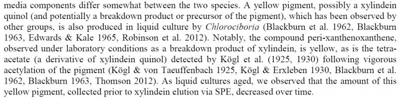
Habitat: Old and well decayed deciduous wood, decorticated, mainly in the cracks and crevices, some localised blackening, with extensive turquoise (blue-green) staining (not necessarily indicating areas of fruiting), some bryophytes, on the floor, shady area and usually damp, no undergrowth, under mature trees, mixed deciduous woodland, Low Weald, England, mid-September, after rain.
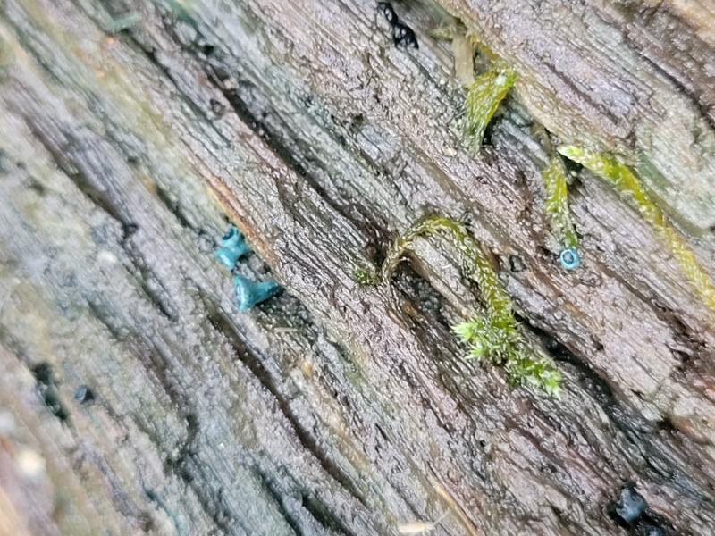
Apothecia: Several groups, solitary to caespitose in small groups, diameter < ~3 mm, initially globose-urceolate, then cupulate and eventually discoid, short and thick stipe, often with a common base, when collected receptacle and stipe with smooth appearance, dull, greyish-turquoise, mottled with blackish patches, after one week in damp box more vibrant due to covering of short whitish hairs (all stages of maturity), margin like receptacle, initially in-rolled, progressively opening and eventually flattened, disc always appears darker, flat colour, more smooth and gelatinous appearance, becoming shallower and paler in maturity, surface apparently becoming more irregular.
Associates: Some small blackish globose lumps on the wood with hyphae protruding, often close to the apothecia.
Asci: Cylindrical-clavate, croziers, rings bb, apex rounded to conical-truncate when turgid, usually more acute when flacid, more hemispherical when immature, usually biseriate, with spores becoming trapped at the apex when flaccid, slight thickening when flaccid but more noticeable when immature.
Spores: Narrow cylindrical, +/- homopolar with ends rounded, inequilateral and rarely slightly curved in profile view, ~2-4 small to medium LBs and some smaller ones, often in groups towards the poles.
Free living spores in water: (6.8) 7.1 - 8.6 (9.5) × 1.8 - 2.1 (2.2) µm, Q = (3.2) 3.6 - 4.5 (5.1), n = 30, Me = 7.7 × 2.0 µm, Qe = 3.9.
Paraphyses: Cylindrical, apex rounded, not noticeably inflated at the apex, multi-septate (3-4 counted), often branching at the base.
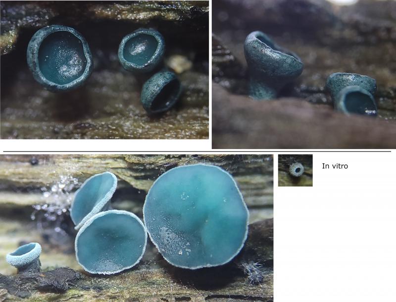
Exudate: Deep turquoise, in hymenium and ectal excipulum.
Medullary: Textura intricata, with little to no exudate/pigmentation.
Ectal excipulum: Difficult to view with heavy exudate, very dark in patches, hyphae appear irregular and dense, shortish hyphae protruding from the surface, sinuous to irregular and hyaline.
Some elongated crystal-like structures found around the hymenium of more outer section, crunching when the cover slip was squashed, may be visible as a strange patch on the disc of most mature apo, happened in vitro, uncertain of explanation.
 Sections-0006.jpeg
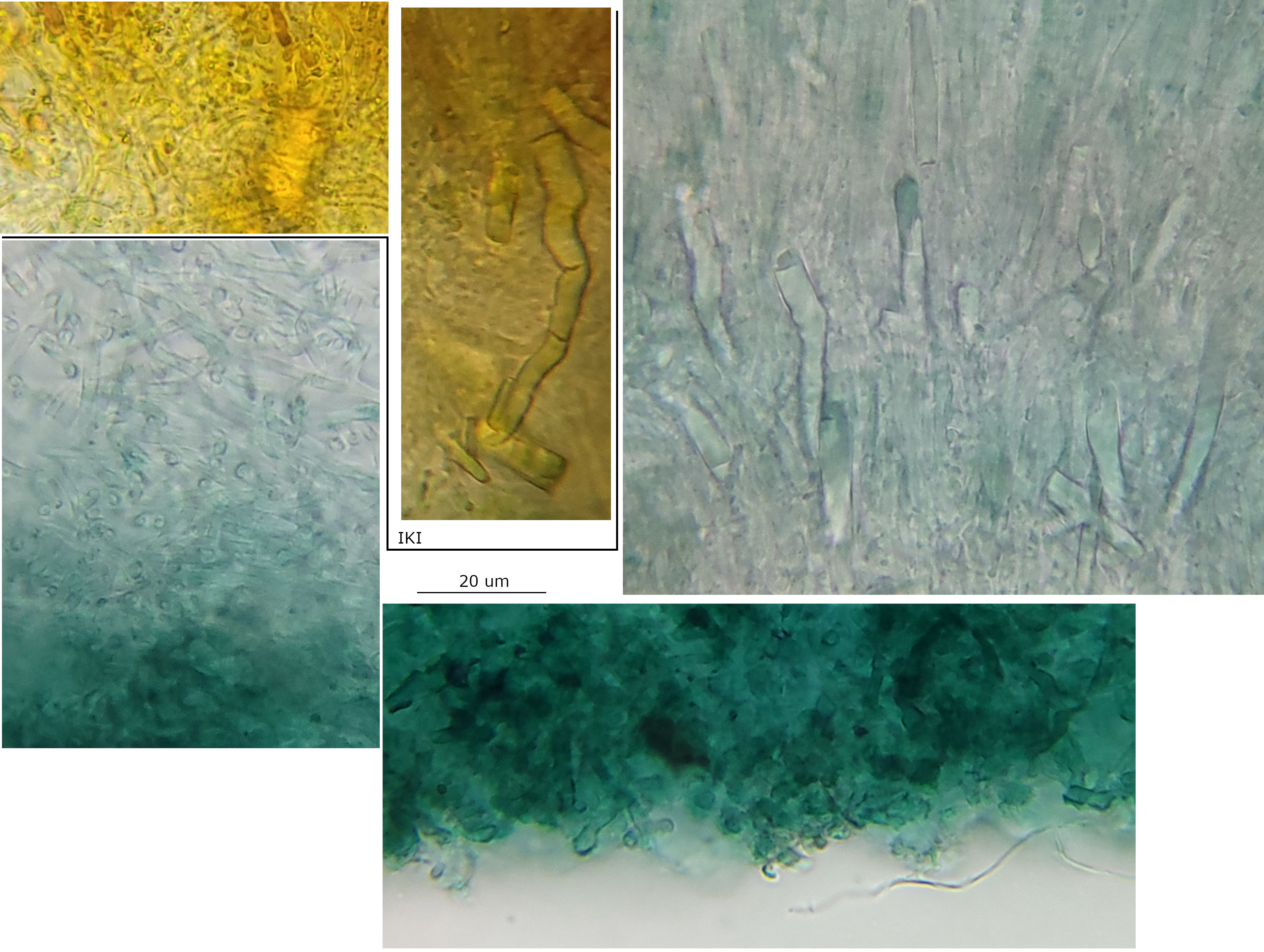 Excipulum-0009.jpeg
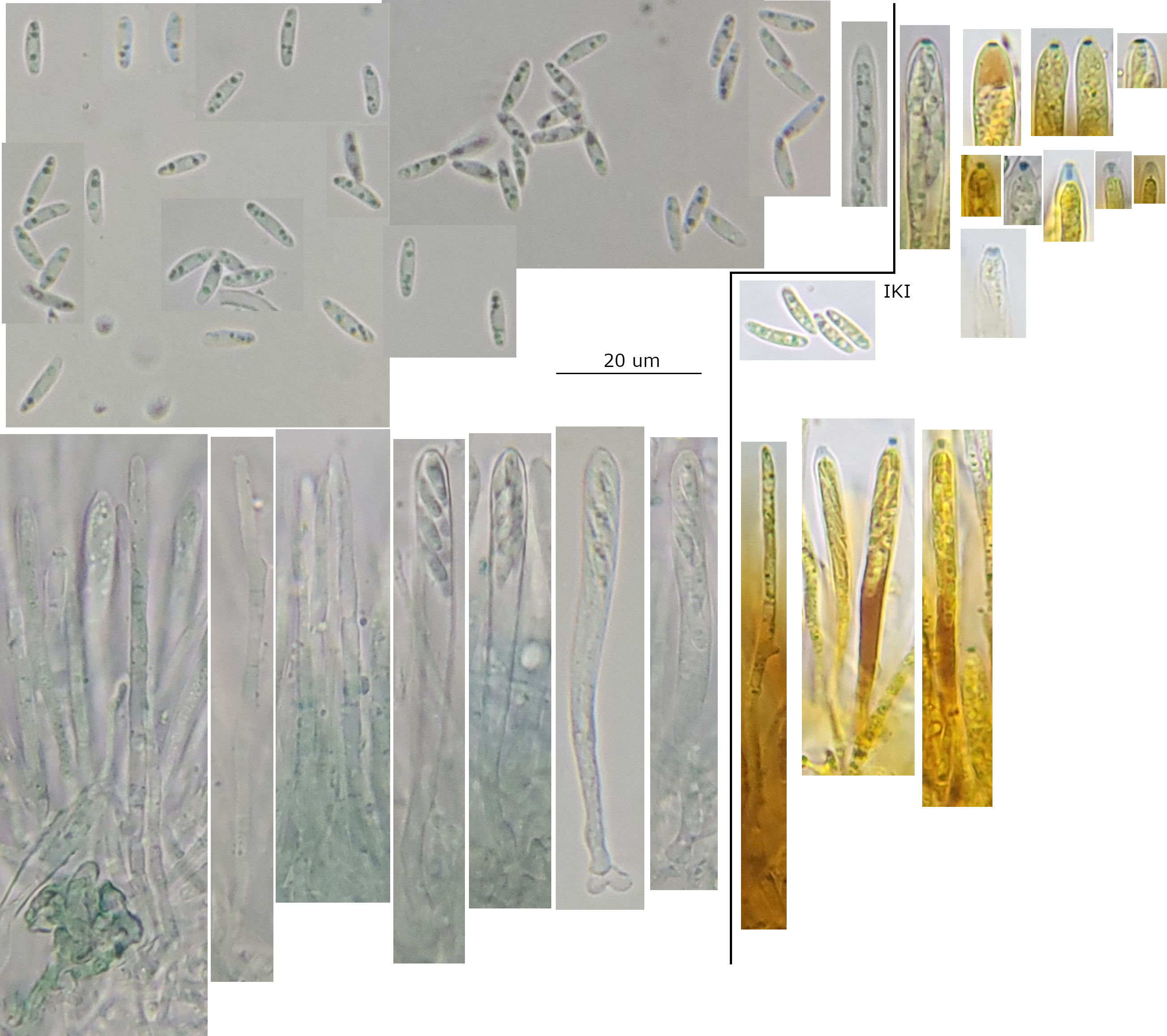 Hymenium-0016.jpeg
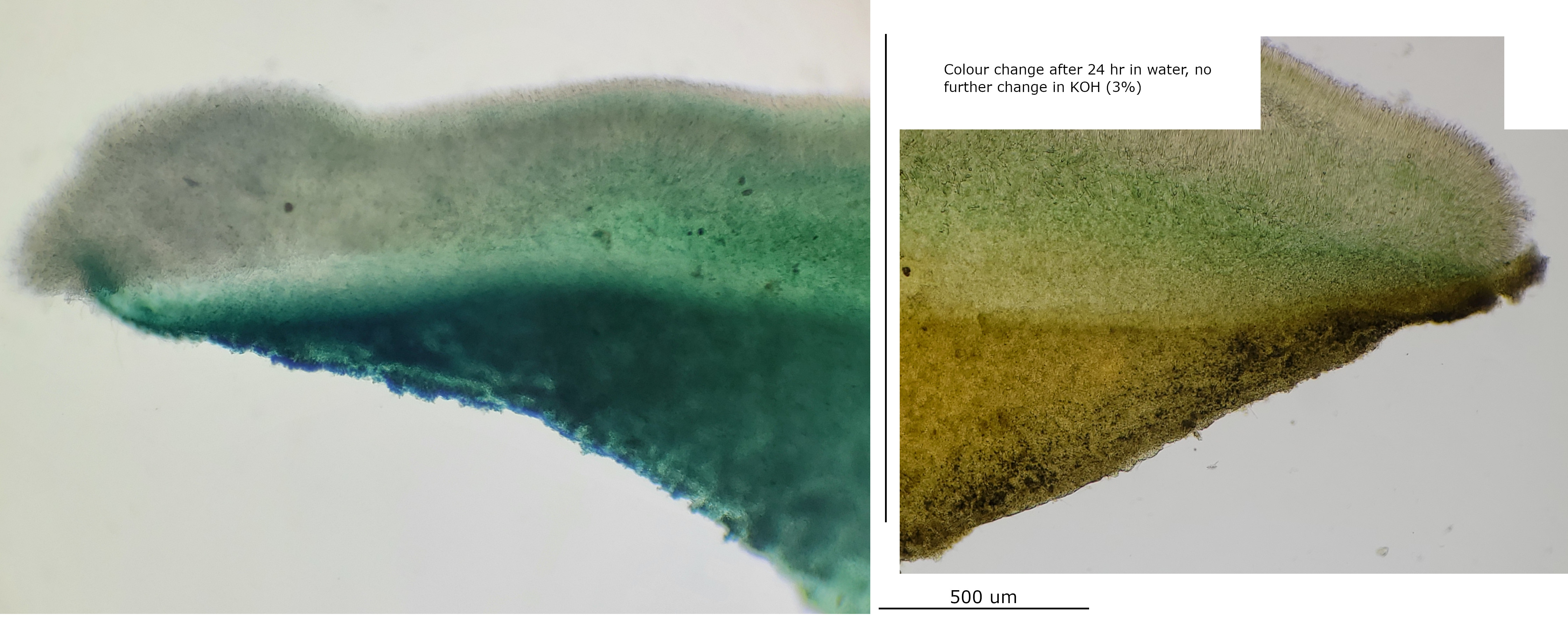
Good question about the exudate, that was my interpretation of the gelatinised appearance. I should test with KOH but unfortunately I didn't do it at the time.
I left a water mount for ~24 hours, and the colour faded to a brownish-yellow in the exposed parts (hymenium and ectal but not so much medullary), it is less opaque in the ectal but still appears gelatinised. Copious bacteria and some protists also developed in the mount.
Adding 3% KOH (only % I have) to this several times (to ensure %) did not have any visible effect on the pigments or gelatinised appearance, even after waiting several minutes and then agitating the cover slip and waiting again.
I have attached some photos. I think you can just about make out the textura epidermoidea in the second set.
 Section-24Hr-water-0001.jpeg
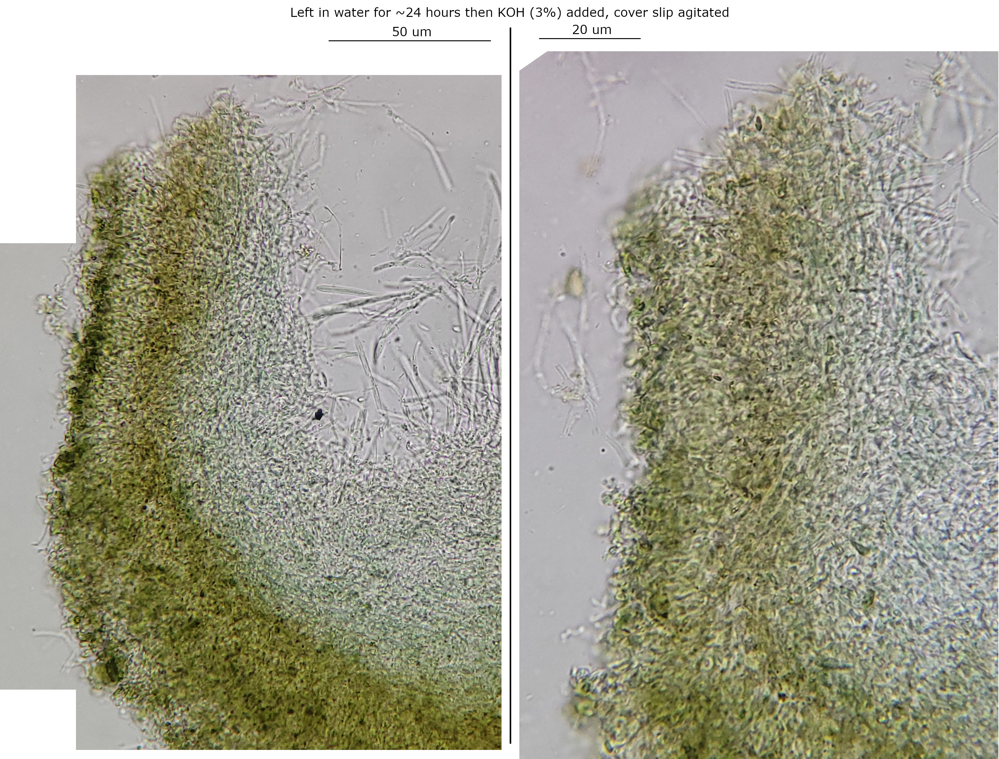 Excipulum-24hr-water-0001.jpeg
The pigment, xylindein, seems to be relatively well studied due to its historic and commercial value in staining wood, and there is no known synthesis. The Tudor et al. (2014) paper, linking the anamorphs, says that Rommier (1868) first extracted the pigment, named it, and found it was water soluble.
It is reported to be produced in the hyphae and gradually diffuses through the substrate. A yellow pigment (or xylindein quinol) is also reported as occasional.