
12-07-2015 00:05
 Nedim Jukic
This one from the same locality as the previous on
06-06-2026 17:44
Steve ClementsBonjour, This disco was on planed wood 3 x 1.5 cm
14-08-2016 23:15
 Alex Akulov
Dear friendsCan you help me to find the descriptio

05-06-2026 11:02
Thomas Læssøehttps://svampe.databasen.org/observations/10596691
04-06-2026 11:36
Gernot FriebesHi,found on Vaccinium myrtillus.Asci: IKI –, 8-s
05-06-2026 12:10
 François Freléchoux
Capitotricha sp. sur Lonicea caerulea Caractères
19-05-2026 10:27
Patrice TANCHAUDBonjour, récolte récente sur terre retournée i
04-06-2026 18:39
Gernot FriebesHi,I collected this species in two different locat
 Files included for resolution loss in attached photos.
• Looks like Hyaloscyphaceae (without VBs, s.l.), with exudate maybe Hyaloscypha or Hyphodiscus.
• Hyaloscypha rather than Hyphodiscus: IKI bb, large spores/asci, no warty hairs.
• Doesn't fit in Huhtinen's monograph: IKI bb, croziers, large spores, substrate, and no (true) hairs.
• Seems H. minuta-like, but differs in IKI bb, croziers, and larger spores. Possibly a similar observation in Baral's Hyaloscypha folder.
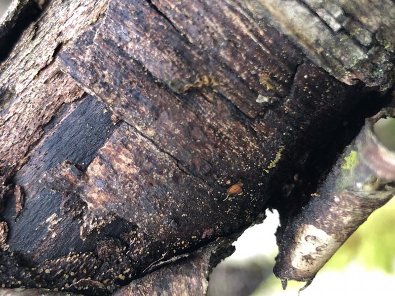
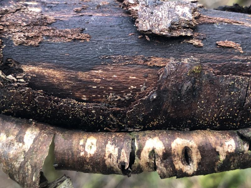
Habitat: On exposed inner-bark of Betula pendula, ~2 m from the ground, on area of dead branch with diameter ~10 cm, seemingly xeric, branch attached to dead trunk on the ground, generally damp area but no bodies of water in immediate vicinity, mixed deciduous woodland, southern England, early March, after wet weather.
Associates: Old-looking stromatic dematiaceous fungus (possibly a common associate), Hyalorbilia inflatula on similar parts lower down on the same branch, bryophytes. Black synnemata? developing in a damp box, not investigated yet.
Preparation and methods: Stored in the fridge for two days, sections examined from two mature and yellowish apothecia, mounted in tap water, gentle pressure required to squash without immediately killing the asci, IKI added to water mount.
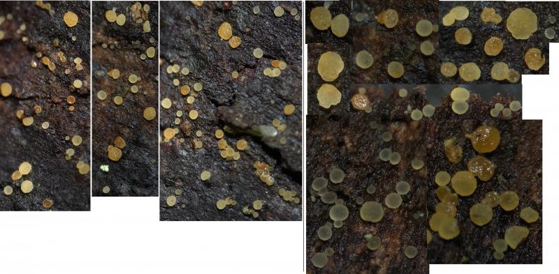
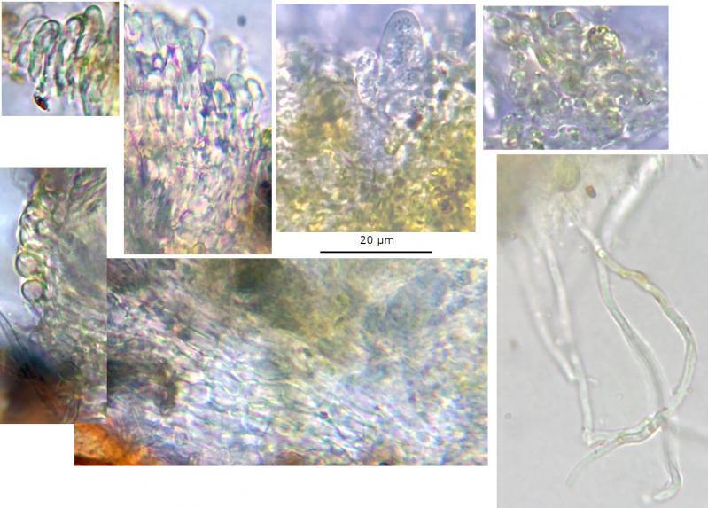
Apothecia: Gregarious to clustered, often 2-10 caespitose, very small, whitish-greyish going yellowish, orange-brown when over mature, translucent, opaque when over mature, initially discoid-turbinate to pulvinate, becoming pulvinate and broadly appressed, sessile, superficial, relatively thick, uneven edge to slightly lobate in maturity, strongly gelatinous appearance, texture soft-gelatinous, margin indistinct, no hairs visible; disc concolorous, translucent, greyish in the centre, usually concave, surface becoming more uneven and occasionally convex when over mature; no subiculum visible.
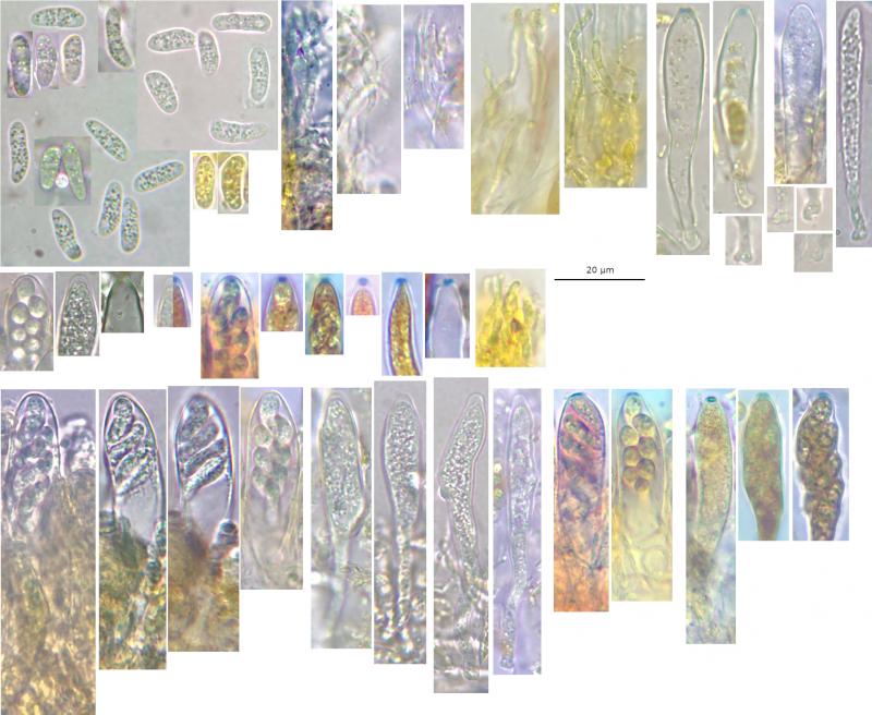
IKI: Ascus rings bb calycina-type, ascoplasm sometimes dextrinoid, paraphyses and spores yellow.
Asci: Relatively large, 8-spored, cylindrical-clavate, apex conical to obtuse, calycina-type rings, upper ring sometimes more laterally spread, sometimes small apical canal, croziers, some perforations observed, medium-walled?, 1-2-seriate, one spore at the apex, no inversely oriented spores observed; discharge via poricidal opening, thickening sometimes not retracted, ring material still present, still relatively turgid after (width ~9 µm).
Mature vital (turgid): ~50-70 × 10-12 µm, clavate, mostly 2-seriate, spores obliquely oriented often close to horizontal, apex more rounded to obtuse (profile to front?), mostly no apical thickening evident, rarely with very slight thickening, rings very reduced, pars sporifera ~30-40%.
Dead (flaccid): ~40-65 x 4.5-9.5 µm, some smaller ones may be immature, cylindrical to narrowly-clavate, occasionally more broadly-clavate, often irregularly shaped due to protruding spores, apical thickening ~1-2.5 µm, thickening tapering to ~50% of length, apex more conical, spores more disorganised, pars sporifera ~60-90%.
Spores: Cylindrical to clavate, inequilateral and occasionally slightly curved in profile view, poles rounded to obtuse, mostly 1(-2)-septate predischarge, OCI (3) 4-5 with many smallish greenish LBs, nuclei often visible as a clearer area often with nucleolus, often heteropolar with the apex wider and often more obtuse, apical cell usually larger ~50-70% of length, no budding observed. When over mature, LBs in a cell merge into one large LB, no dead cells observed.
Measurements of mature vital spores mostly discharged in water mount and a few in asci:
(8.3) 10-13.8 (14.6) × (3.3) 3.6-4.7 (5.1) µm, Q = (2.1) 2.5-3.8 (4), N = 25, Me = 12 × 4.1 µm, Qe = 3.
Paraphyses: Cylindrical-filiform, apex rounded and uninflated to slightly inflated or slightly lanceolate, 1-3 septate, apical cell more sinuous/irregular, occasionally penultimate cell also inflated.
Exudate: Bright yellow, more orange in concentration, lumpy/cloddy, oily residue in water mount, forming epithecium extending into the hymenium, coating ectal excipulum.
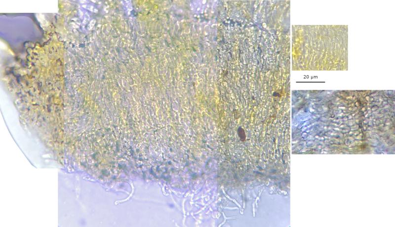
Subhymenium and medullary: Weakly developed, difficult to distinguish, hyaline.
Ectal: Textura prismatica, external cells occasionally more globose (appearing to form rings around receptacle?), oriented parallel to the surface; marginal cells slightly elongated and gently clavate-capitate, occasionally more acute and slightly-capitate or hair-like but not protruding, unornamented, hyaline (without exudate).
Subiculum: Anchoring hyphae around the base, hyaline.
 Hymenium-0003.jpeg
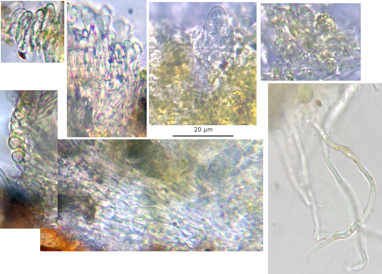 Excipulum-0001.jpeg
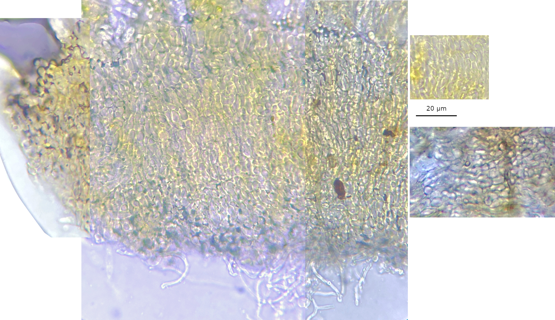 Excipulum-60-0001.jpeg
The macro does look similar to Leptodontidium, or Arachnopezziza without hairs. I saw a description of Polydesmia as downy in FoTE and a picture of P. pruniosa, but unfortunately I did not connect that with this appearance.
The form of the paraphyses, as you suggested, and also the highly guttulate and septate spores, and association with a stromatic pyrenomycete seem fitting for Polydesmia. Looking at your observation of P. 'sorbi', I agree, there are no measurements but everything certainly looks the same.
There appears to be some host specificity in Polydesmia, perhaps this species is characterised by the presence on certain pyrenomycetes on deciduous bark. Having a very brief look, the indistinct margin, excipullar cells, and spore size (ascus size), could also be distinguishing from the other species.
In the phylogenies of the Kosenen et al. (2021) paper, I saw that Arachnoscypha and Polydesmia grouped outside Arachnopezizaceae. It would be interesting to get more coverage of Polydesmia and also describe this species.