
12-11-2019 10:32
 Miguel Ángel Ribes
Hi againExactly at the same place than my previous
25-12-2019 17:54
Valencia Lopez Francisco JavierHola a todos/asEstas supuestas pezizas estaban en
28-07-2011 18:31
 Alex Akulov
Dear FriendsToday I made the pdf file of Velenovsk
12-07-2015 00:05
 Nedim Jukic
This one from the same locality as the previous on


30-05-2026 21:12
Philippe PELLICIERSur branche de mélèze (Larix) près de la neige,
31-05-2026 10:35
Hulda Caroline HolteHello,I collected this species growing on a rather
25-05-2026 16:35
 Bernard CLESSE
Bonjour à toutes et tous,J'ai trouvé récemment,
29-05-2026 15:35
daniel FERREBonjour à tous,Je voudrais votre aide pour cette
Ascobolus cf. foliicola
Zuzana Sochorová (Egertová),
28-06-2022 20:12
 Hello,
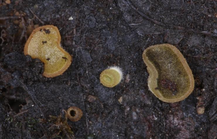
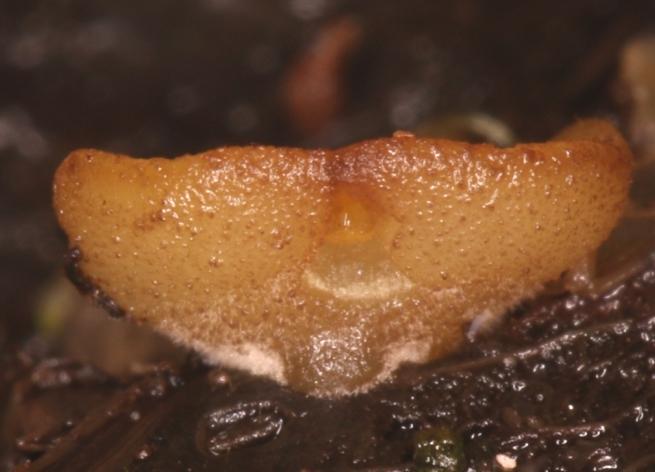
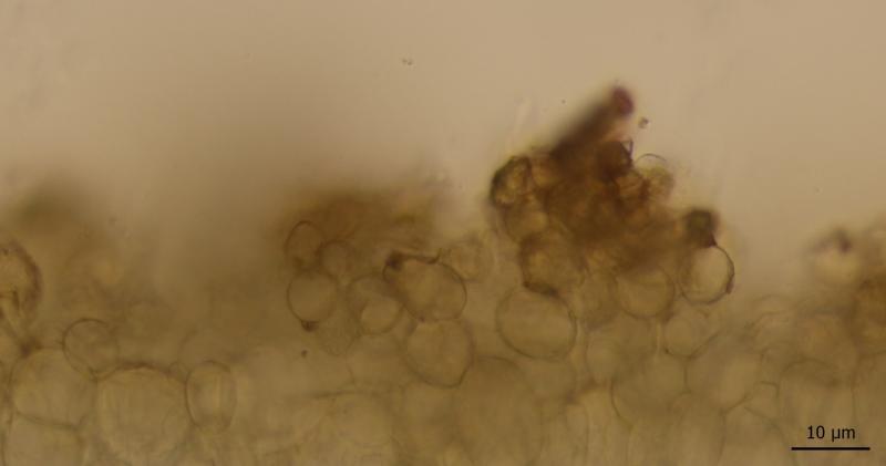
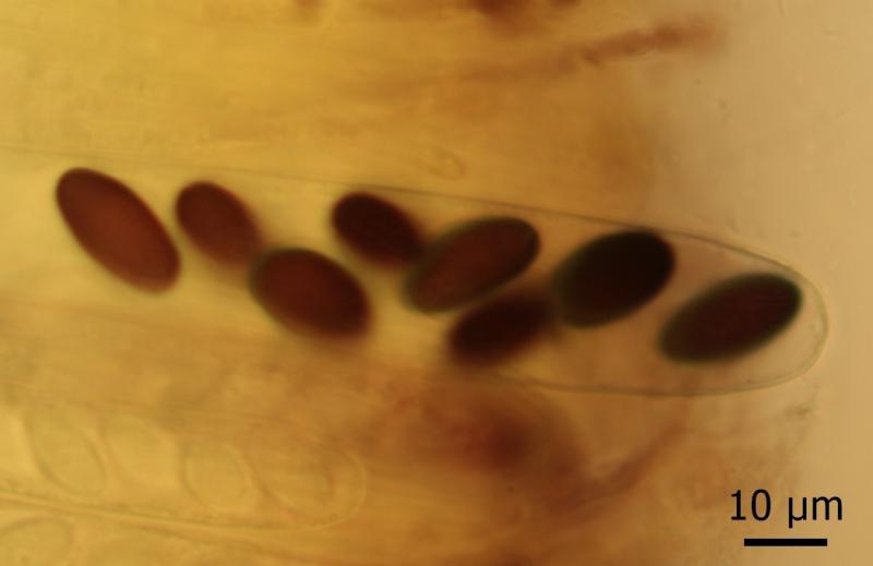
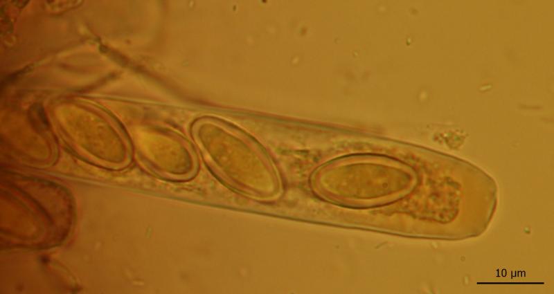
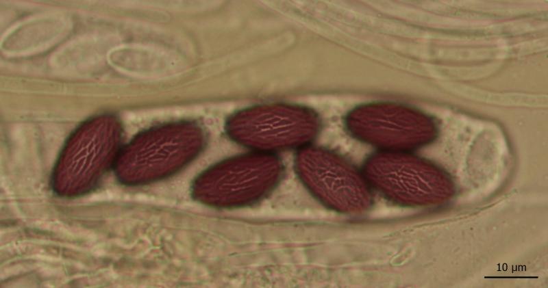
I repeatedly collect a foliicolous Ascobolus in one locality in Moravia (200 m a.s.l., on decaying, very wet leaves of Alnus and Salix, close to the Morava river).
First I identified it as Ascobolus foliicola, but now I am in doubts whether the identification is correct.
The features highlighted by van Brummelen as important for distinguishing A. foliicola and A. denudatus are the following ones:
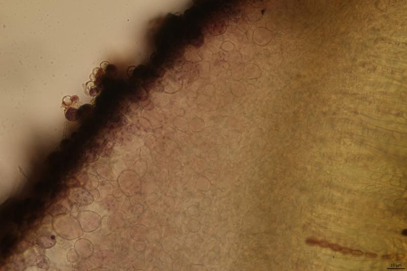
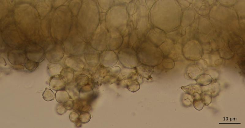
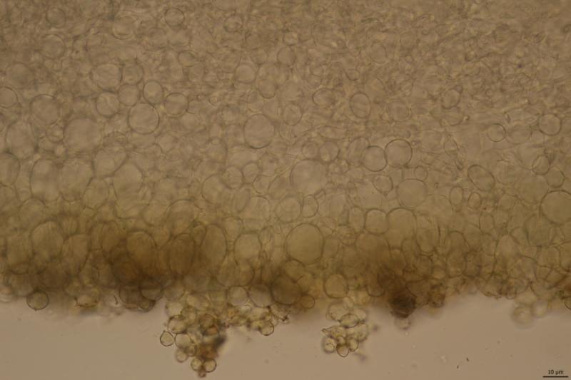
* stalked receptacle (foliicola) vs. sessile receptacle (denudatus) - I would consider my apothecia rather sessile or only shortly stipitate
* margin permanently furfuraceous (foliicola) vs. soon smooth (denudatus) - here my find fits better A. denudatus
* excipular warts consisting of subglobose and pyriform cells (foliicola) vs. only subglobose cells (denudatus) - I can see a few pyriform cells, but the majority is subglobose
Ornamentation of ascospores clearly fits better A. foliicola, at least when judging from van Brummelen´s illustrations.. Their dimensions are (18.1)18.5-20 (20.4) × 9.7-10.9) µm
Q = 1.8 -1.9
Me = 19.2 × 10.3 µm
How would you determine it?
Zuzana

Zuzana Sochorová (Egertová),
28-06-2022 20:13
Re : Ascobolus cf. foliicola
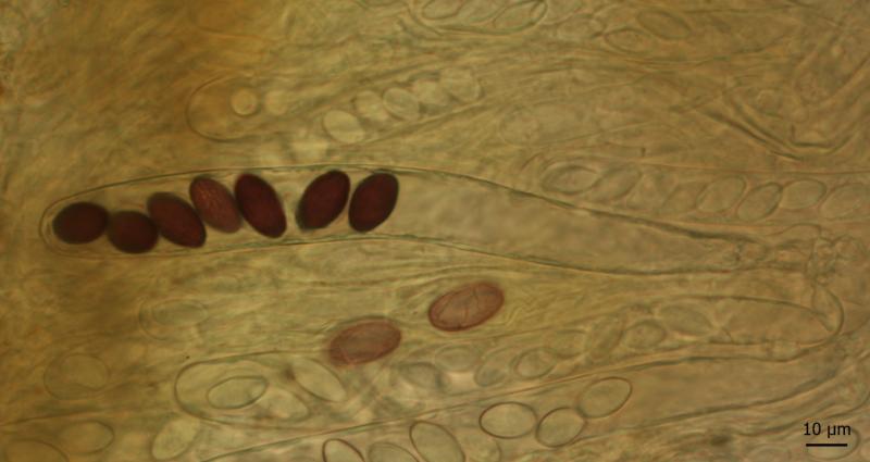
Wall of asci is not blue neither in IKI nor in MLZ, but the content of immature asci as well as cells of ectal excipulum turns violet in IKI.
Zuzana Sochorová (Egertová),
28-06-2022 20:17
Re : Ascobolus cf. foliicola
more

Till Lohmeyer,
29-06-2022 10:05
Re : Ascobolus cf. foliicola
Good morning, Zuzana -
comparing van Brummelen's illustrations 40 c (foliicola) and 41 (denudatus) you will note a remarkable difference: The spores of foliicola look like "drawn by a trembling hand", those of denudatus don't. Thus, I'm pretty sure, your fungus should be A. foliicola.
Best regards from Bavaria
Till
comparing van Brummelen's illustrations 40 c (foliicola) and 41 (denudatus) you will note a remarkable difference: The spores of foliicola look like "drawn by a trembling hand", those of denudatus don't. Thus, I'm pretty sure, your fungus should be A. foliicola.
Best regards from Bavaria
Till
Zuzana Sochorová (Egertová),
29-06-2022 17:48
Re : Ascobolus cf. foliicola
Hello Till,
thank you very much!
Zuzana
thank you very much!
Zuzana
Nicolas VAN VOOREN,
30-06-2022 16:00

Re : Ascobolus cf. foliicola
Hello.
A. denudatus can be excluded because its ascospores always show some pustules.
In the other hand, I do not recognize A. foliicola I used to collect, but I do not have any other idea.
A. denudatus can be excluded because its ascospores always show some pustules.
In the other hand, I do not recognize A. foliicola I used to collect, but I do not have any other idea.
Zuzana Sochorová (Egertová),
01-07-2022 18:14
Re : Ascobolus cf. foliicola
Thank you :-)