17-04-2026 19:16
 Enrique Rubio
Hi to everybodyI would appreciate any assistance r
14-04-2026 05:32
Ethan CrensonHi all, A few weeks back a friend pointed out som
17-04-2026 15:14
 Bruno Coué
Bonjour.Récoltes du 16/04/2026, sur feuilles mort
12-04-2026 15:52
Gernot FriebesHi,I'm looking for help with this anamorph collect
14-04-2026 21:52
Gernot FriebesHi,found on dead leaves of Carex elata. Conidia: 4
16-04-2026 22:09
Buckwheat PeteHello, I'd like to ask about this older specimen:
15-04-2026 19:33
Fátima Durán ManzanequeHi!! I need help, I found this Ascomycete but I d
14-04-2026 20:31
Gernot FriebesHi,can this be Psilachnum lateritioalbum on Phragm
12-04-2026 17:56
 Hardware Tony
Found on dead stems in February earlier this year
12-04-2026 12:22
 William Slosse
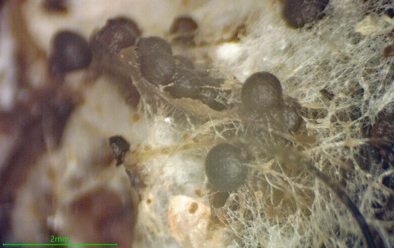
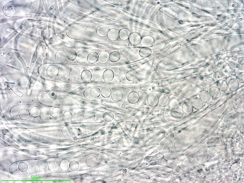
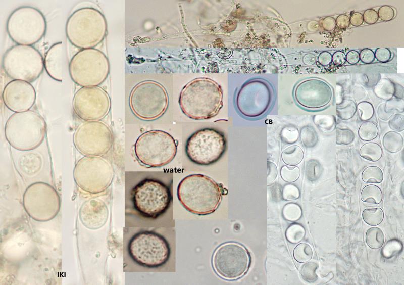
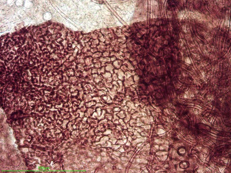
In a dune grassland in Oostduinkerke (Belgium), on
The asci are thin-walled and looong, up to 200 µm, IKI-. Most of them are eight-spored but many six-spored with one spore aborted. The spores are (sub)globose, on average 12.3 x 11.1 µm, hyaline (wrong colour balance in the large water spore images). The spore wall is two layered, calyptrate, in water the surface looks ornamented with short crests but in unheated CB the spores are perfectly smooth. The outer layer is cyanophilous. The spores also look slightly dextrinoid in IKI.
In another corner of the culture (without mould) there were some Preussia funiculata, which externally look exactly the same. Also the ascus length matches but the spores are, of course, completely different.
I have no idea what animal this is. Anyone?