
27-04-2026 18:48
Tony MoverleyCollected 23rd April 2026, Norfolk, EnglandSwarms
14-04-2026 05:32
Ethan CrensonHi all, A few weeks back a friend pointed out som
27-04-2026 20:52
 Lothar Krieglsteiner
Found on hanging tiwg of Olea europaea in dried-ou
28-04-2026 22:51
 Bernard CLESSE
Bonsoir à toutes et tous,Pourriez-vous m'aider à
29-04-2026 08:01
Lothar Krieglsteiner
... on twig attached to small tree of Citrus auran
29-04-2026 10:44
Lothar Krieglsteiner
growing at moist, drying-out soil at the side of a
28-04-2026 20:33
Vitus SchäfftleinHello, I found Trochila ilicina on Ilex aquifoliu

28-04-2026 21:50
 Pablo Sandoval
Hola a todos,Espero se encuentren bien. Hace mucho
27-04-2026 18:05
Lothar Krieglsteiner
... still attached at standing tree. The green con
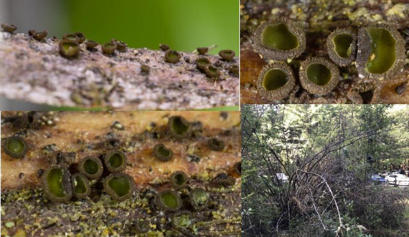
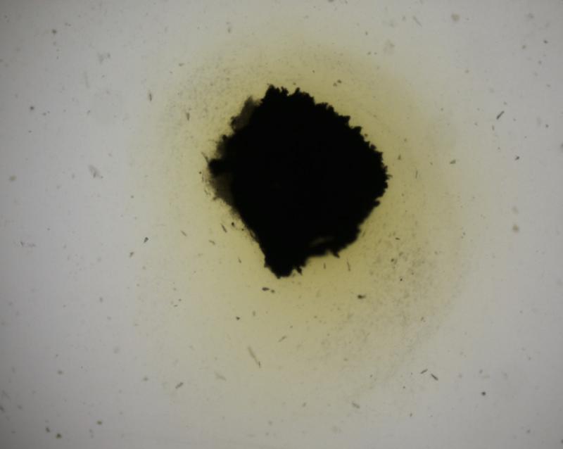
 Cups about 1 mm across, growing in large numbers on branches of a tall (2.5 m) dead shrub.
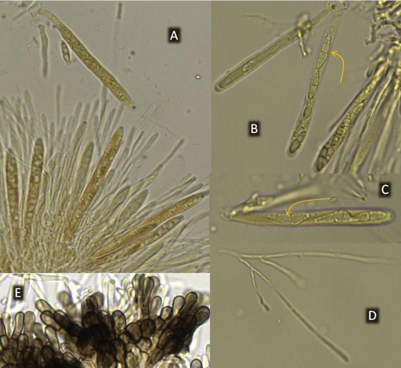
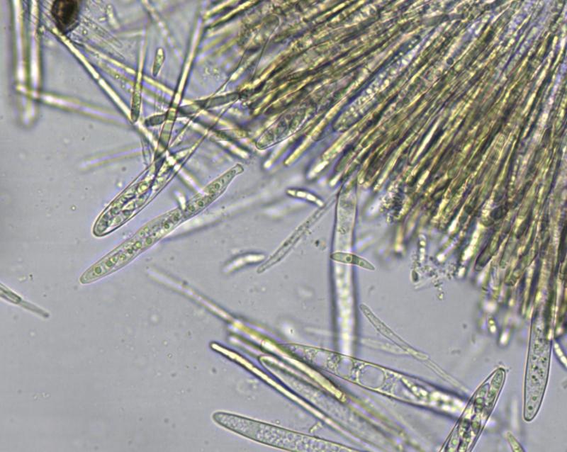
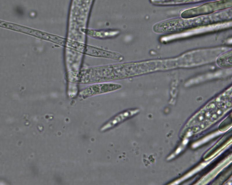
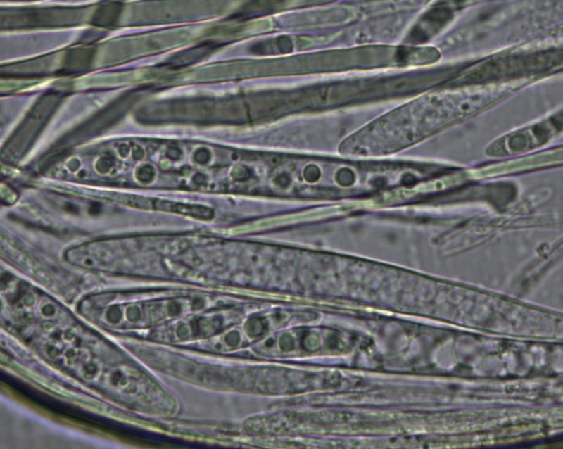
 Asci about 80x8, with amyloid tips. Dozens of spores I could see were all with 3 septa and measured 13-15 x 3.5-4. But in two asci (figs. B, C) there were 5-septate spores, measuring 23x5 and 18x3.5. Forking paraphyses (D) were common.
My only guess is Patellariopsis, but if the top ascus in B is bitunicate (as it looks to me), then this genus should be eliminated.
Thanks for any help you might have.

I am not sure if the branch was still attached and the fungus drought-tolerant.
Regrettably you did not mount in water. It is a recent collection? It is important to know the contents of the living spores and paraphyses.
The ectal excipulum at the flanks os of globose cells?
I don't know about drought-tolerance of these. There has been a lot of rain this winter. In any case, they were growing on free standing sticks/branhes, at the height of about 150-180 cm. It's a deciduous shrub and I'll check later if some of it grows leaves, so we can identify it.
Would be great to identify the shrub. Yes, when these grew on standing dead branches, they must withstand drought for a couple of weeks.
Your area is mediterranean, is it? It may be that these cups do not occur during summer but show only a limited tolerance.
Zotto
Yes, the climate in the area is kind of Mediterranean, with rain in the winter and cool and dry summers, with lot of fog.
Sava
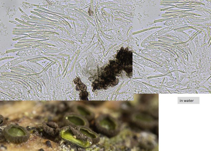
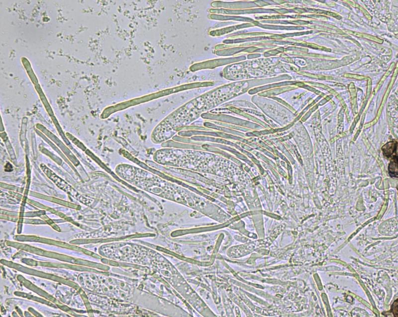
So we can be quite sure that it is a Mollisia. Good would be to observe living asci and free spores. It is quite probable that the spores are septate when still in the living asci, but this should be proved. Also the oil drop pattern in the mature, freshly ejected spores is important.
Septate spores are rather rare in wood-inhabiting Mollisias.
This weekend I'm going to revisit the place and try to learn something about the shrub, and also check how the discos are doing.


I hope I'll find more of these cups soon and then I'll look at samples from serveral of them. I will take any advice you might have about the procedure. I printed a copy of your 1992 paper, perhaps it's all said there...
And if they are truely 5-septate, it can still be that these are overmature ones.
If I had a scale to your photos I could measure more than 3 in the living asci. Often the spores are a little smaller in this case, but in the present species I do not see much difference to outside.
Fortunately, the shrub is alive. Most of it is dead, but there are buds at enough branch tips and soon we will see its leaves. For experts, the photos I took yesterday would probably be enough to tell the shrub genus.
It was interesting to find an asexual form growing alongside the cups. It has round, olive-green conidia, about 4 microns. A likely anamorph, I guess.
The photos are in the attached pdf. The second file contains measurements and microshots from which I selected asci and spores to measure. I used only one cup for this, mounting one slice of it in water and another one in Melzer's. If you think more measurement would be worthwhile, please let me know what you would do.
Thanks again, it's been a nice learning experience.
 MollisiaBigBasin-1-0001.pdf
See http://www.gbif-mycology.de/HostedSites/Baral/
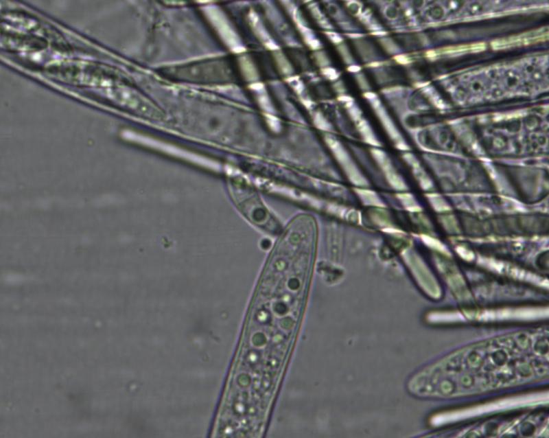
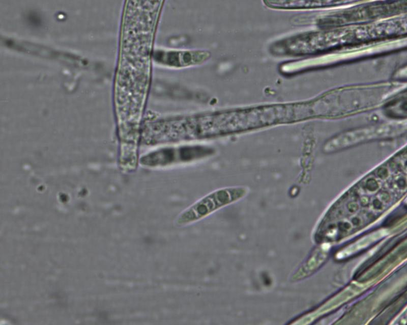
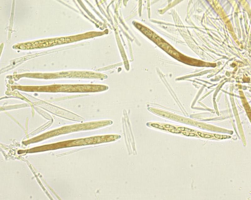
This third image shows above right a spore with a thick gel sheath!
Indeed spores are 3-septate, one can see it in free living spores by slight constrictions. Asci arise from croziers.
What I still miss is a median section to show the excipulm at margin and flanks.
Zotto
In the attached file, the first three photos show excipular cell from near the cup margin. The next three are from the flanks or near cup base. If you don't see what you want in these photos, please let me know what you'd like to be done differently.
The last three photos show some branching 'binding' hyphae that looked unusual to me. I tried to find them on another mount, but couldn't.
Lastly, a macro feature that I haven't mentioned so far: these cups are quite sticky (resinous?). When I'm making sections, they stubbornly get themselves attached to needles and tweezers.
In any case, with a razor blade it should be possible to make a median (vertical) section, at least at the flanks, with yome luck also at the margin. In addition, the anchoring hyphae (subiculum), usually also brown in Mollisia, might be of interest.
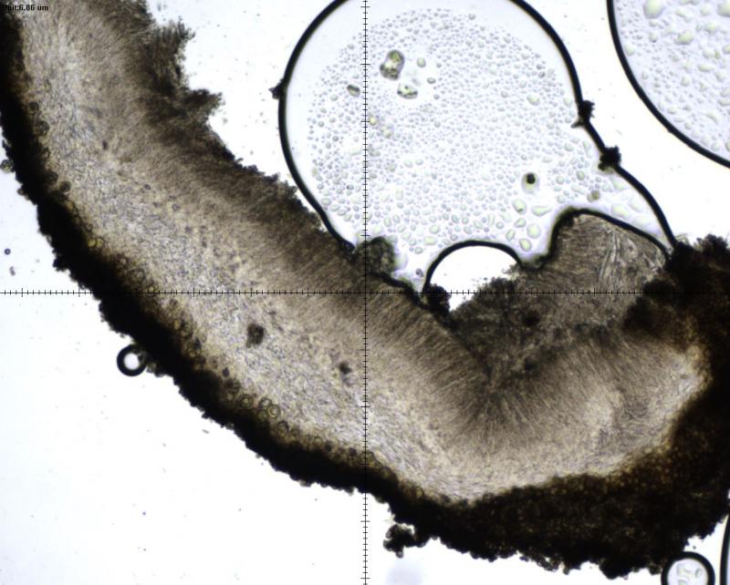
The remaining photos are an attempt to show the anchoring hyphae. I took a vertical section down to the substrate and including a piece of it. You can see it in the photo that begins this sequence (page 6). In the remaining photos I tried to capture the tissue between the wood and excipulum. There was no subiculum that I could discern through the dissecting scope.
If there is something more that I can do, it will need to wait for a week because I'll be out of the country. When I'm back, I'll revisit the location to get a photo of the shrub with its leaves.
Many thanks!
On the beforelast pic I see some brown hyphae which are anchoring hyphae. So there seems to be no abundant subiculum.
I've only seen herbarium specimens, which still showed the nice green hymenium but the ectal excipulum was darker. It is nice to see this one fresh, I'll have to keep an eye out for this here in British Columbia.
Cash, E. K. (1936). Some ascomycetes new to California. Mycologia, 28(3), 247-252.
Again a case where the yellow KOH-reaction seems to have been overlooked.
No sequence seems to exist in GenBank. I suspect that placement in Nipterella is not justified, depending on where the type N. duplex belongs. N. duplex reminds me somewhat of Mollisia atlantica ined., which has a bit smaller spores.
It seems curious that Dennis and also Müller & Defago still laid so much stress on spore septation or did they see more distinguishing features against Mollisia?
Nipterella parksii definitely in Mollisiaceae but it would be great to have sequences of N. duplex to see if they form a clade or not. The bright hymenium, scurfy ectal excipulum comprised of monilioid cells, and ascospores are quite distinct. I realize now that I have access to N. tsugae specimens including the holotype; I'll have to take a look at these soon and see if it belongs in this family or not.
I almost forgot about these species but they are very striking, I'll keep an eye out for them.
I also wonder if there is a scutum-like structure in parksii, judging from the present micro-pics.
And I wonder if the gel sheath around the spores is real and if it stains lilaceous in Cresyl Blue as I observed in Nimbomollisia.
Looking through my pictures of N. parksii, I don't see any gel sheath in cresyl blue or water, but Sava's pictures do seem to show some apparent ascospore sheaths.