
28-04-2026 20:07
 Lothar Krieglsteiner
... on twig in the air at standing Ceratonia siliq
14-04-2026 05:32
Ethan CrensonHi all, A few weeks back a friend pointed out som
28-04-2026 20:33
Vitus SchäfftleinHello, I found Trochila ilicina on Ilex aquifoliu

30-04-2026 10:28
Rot BojanHello, by appearance I would say that I am dealing
27-04-2026 18:48
Tony MoverleyCollected 23rd April 2026, Norfolk, EnglandSwarms
27-04-2026 20:52
Lothar Krieglsteiner
Found on hanging tiwg of Olea europaea in dried-ou
28-04-2026 22:51
 Bernard CLESSE
Bonsoir à toutes et tous,Pourriez-vous m'aider à
29-04-2026 08:01
Lothar Krieglsteiner
... on twig attached to small tree of Citrus auran
29-04-2026 10:44
Lothar Krieglsteiner
growing at moist, drying-out soil at the side of a
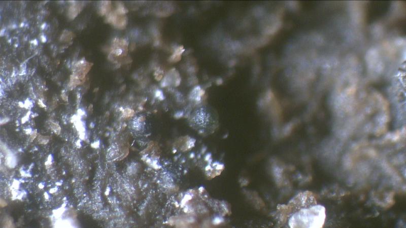
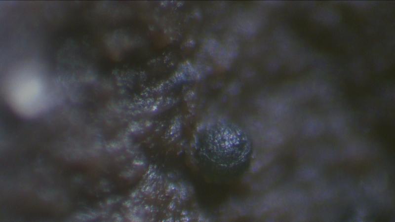
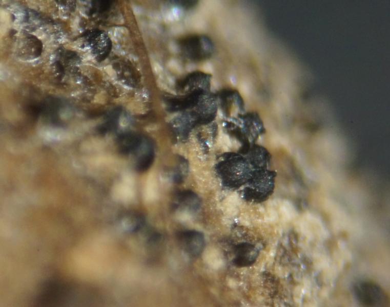
 Found on cow dung, most of the time in the visinity of Schizothecium species.
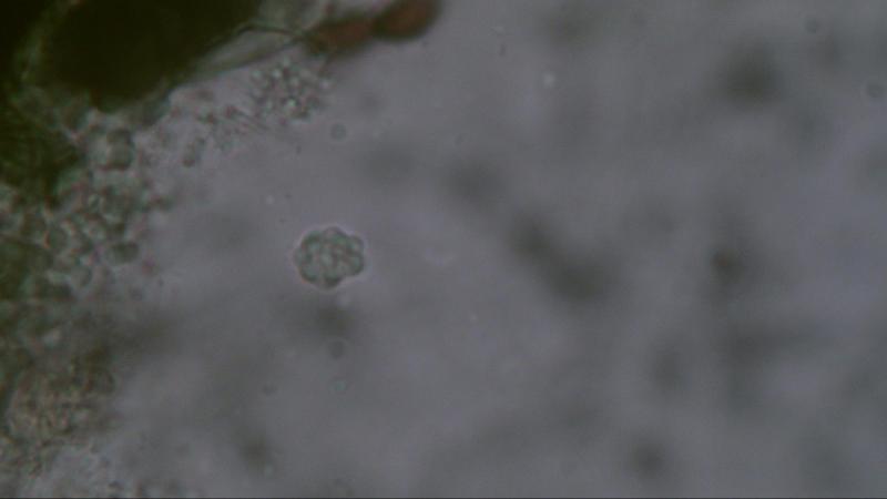
Fruitbody: Round 170,2-173 um in diameter, surrounded by a gelatinous layer approx. 60 um thick, dark green in colour.
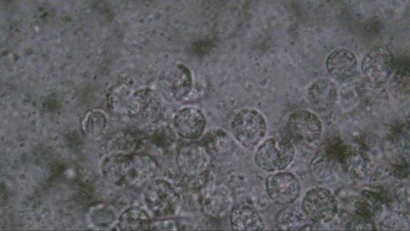
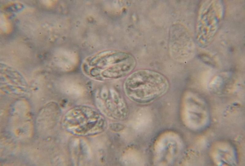
Spores: Round and/or pointed 6.8-7.3 um covered with round warts 2.2-2.5 um.
The first time spores were measured when in water but missing the warts so the second time measurement was performed in Melzer.
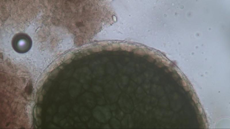

That is a very interesting fungus. Do the fruiting bodies have an opening of some kind? The round asci look like the kind you find in a cleistothecium but your pictures suggest that maybe there is an ostiole. Also, do the asci contain 8 ascospores or are there more than that?
Regards,
David

What you show on all your photos are in my opinion asci and it is very difficult to see the spores alone. Are there any hairs? Afraid to be could you look for the genera Lophotrichus, Kernia. There is also the genus Orbicula but in the latter the asci are cylindrical.
Michel.


Can you provide me the following article.
Pithoascus nidicola (Massee & E.S. Salmon) Arx, Proceedings van de Koninklijke Nederlandse Akademie van Wetenschappen Section C 76 (3): 292 (1973) [MB#320551]
Regards,
Joop

Hello Sven,
I will try to get the article in the library of Naturalis when I can find the time to do so.
If it is succesful I will send you a copy.
Joop
Joop
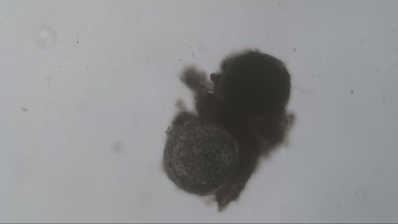
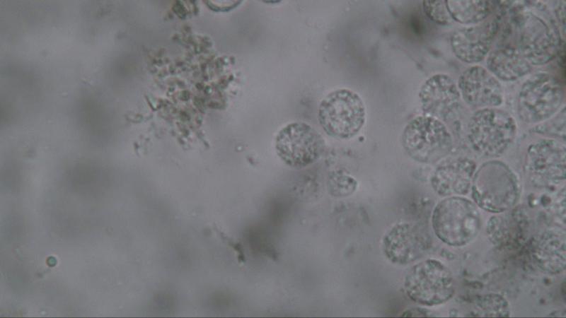
In my opinion it is a single ascus species like Thelebolus stercoreus containing hundreds of spores.
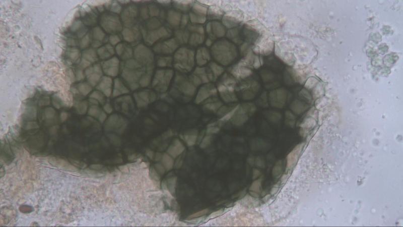
I did not find any ostiole but maybe that is possible when these species are ripe.
It is typical that these species were found together with Schizothecium conicum species.
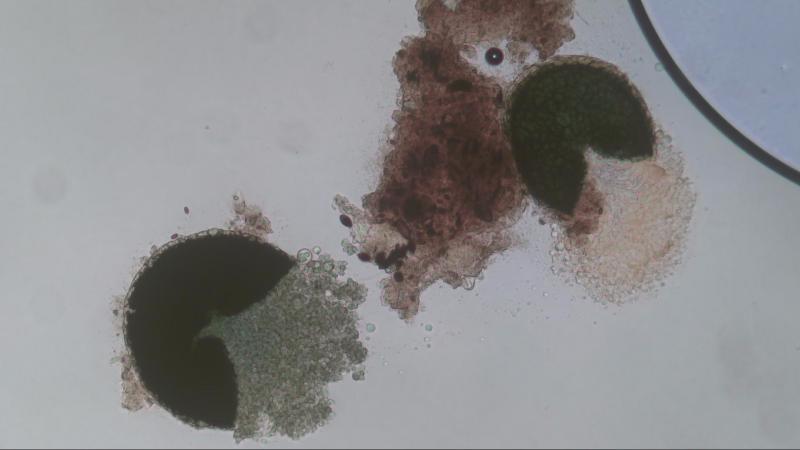
When putting pressure on the cover glass the species bursts open (photo #4).
Joop
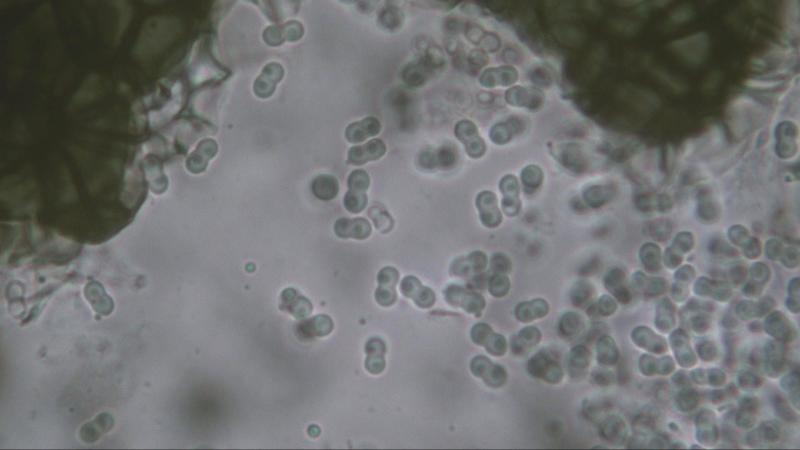
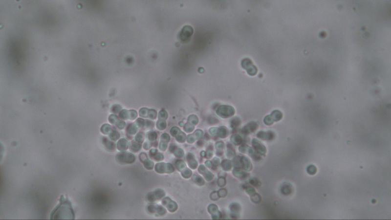
I collected some more information about this species and it will present different shapes of spores when using different fluids.
Photo #1 when using water.
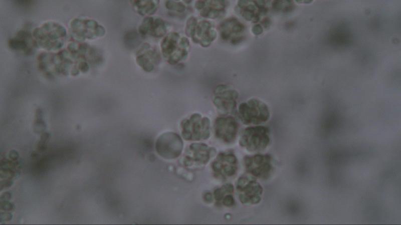
Photo #2 when using Melzer
Photo #3&4 when using Congo Red.
It seems to me that the presentation on #2 show the spores as seen in #3&4 clinging to each other. Whereby at first I thought that they were warts.
The spores consist of 2 cells, each cell measures 2.45 um in diameter, the total of 2 cells combined is 4.9 um.
Each cell is filled with a "the bary bubble".
When measuring the cells as presented in #2 (or other photos I made) the result will be the same as in #3&4 namely 2.45 um.
Greetings,
Joop

I believe this fungus is Mycoarachis inversa, a species characterized by two-celled peanut-shaped spores and a cleistothecial peridium with the hyaline layers on the outside (hence "inversa").
David
Joop
