06-02-2026 15:40
 Robin Isaksson
dear all,anyone that have this one? and can send i

05-02-2026 15:07
Found on a fallen needle of Pinus halepensis, diam

05-02-2026 06:43
Stefan BlaserHello everybody, Any help on this one would be mu

18-08-2025 15:07
 Lothar Krieglsteiner
.. 20.7.25, in subarctic habital. The liverwort i

02-02-2026 21:46
Margot en Geert VullingsOn a barkless poplar branch, we found hairy discs

02-02-2026 14:55
 Andgelo Mombert
Bonjour,Sur thalle de Lobaria pulmonaria.Conidiome

02-02-2026 14:33
 Andgelo Mombert
Bonjour,Sur le thalle de Peltigera praetextata, ne

31-01-2026 10:22
 Michel Hairaud
Bonjour, Cette hypocreale parasite en nombre les
Mollisiaceae sp. ?
Nicolas VAN VOOREN,
22-07-2008 00:37
 Je sèche sur une espèce récoltée ce week-end dans la tourbière du Grand Leyat (Savoie), alt. 1660 m, sur branche morte d'Alnus viridis.
J'ai un peu cherché du côté des Mollisiaceae mais sans succès...
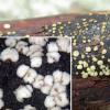
Macroscopical description: apothecia sessile, pulvinulate, at first yellowish then pale cream to whitish; outer surface furfuraceous, ochraceous. Ø 0.3–1.5 mm
No subiculum. Reaction to KOH 4%: none.
Nicolas VAN VOOREN,
22-07-2008 00:47

Re:Mollisiaceae sp. ?
Microscopical features: asci 82-87 x 7-9 µm, with crozier, containing 8 sp. (± biseriate) ; pars sporifera as long as the ascus length; apical ring IKI bb.
Paraphyses sublanceolate, large to 3.2-4 µm, hyalin, septate, with a great VB in the upper part.
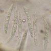
Spores fusiform elongated, 25-33 x (2.7) 3-3.2 µm, hyalin, with a lot of LB, and sometimes with a septum ("very mature" spores) but never in ascus.
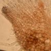
Excipulum at the base of textura globulosa, with brown cells, Ø 8-20 µm, becoming more elongated on the lateral part.
Any suggestion?
René Dougoud,
22-07-2008 07:07
Re:Mollisiaceae sp. ?
Bien Cher,
Je te réponds depuis le job et donc de mémoire, pour te dire que cela me fait penser à Mollisia ramealis !
Amitiés
René
Je te réponds depuis le job et donc de mémoire, pour te dire que cela me fait penser à Mollisia ramealis !
Amitiés
René
Hans-Otto Baral,
22-07-2008 12:34
Nicolas VAN VOOREN,
22-07-2008 21:44

Re:Mollisiaceae sp. ?
Merci beaucoup.
Raúl Tena Lahoz,
24-07-2008 11:02

Re:Mollisiaceae sp. ?
Spores of this species can be seen as these of my photo? Rest of features of my collection fit with those of Nicolas (except for colour, always yellowish). The explanation could be a fusion of the oil drops? Also spores like those Zotto sends are seen. Medium: distilled water.
Thanks!
Thanks!
Hans-Otto Baral,
24-07-2008 12:16

Re:Mollisiaceae sp. ?
Hi Raul
yes, in the free spores the LBs have fused and these spores are dead. There is a central small area which is unclear whether it is a septum or perhaps the nucleus.
Zotto
yes, in the free spores the LBs have fused and these spores are dead. There is a central small area which is unclear whether it is a septum or perhaps the nucleus.
Zotto
Raúl Tena Lahoz,
24-07-2008 12:38

Re:Mollisiaceae sp. ?
Hi Zotto
I think that, in this case, it´s the nucleus. So better to use tap water? I don´t use it because I live in a very calcareous place, so I think it´s hard water. Maybe water of springs in acid soils could be better?
Thaks a lot!
¡Muchas gracias!
I think that, in this case, it´s the nucleus. So better to use tap water? I don´t use it because I live in a very calcareous place, so I think it´s hard water. Maybe water of springs in acid soils could be better?
Thaks a lot!
¡Muchas gracias!
Hans-Otto Baral,
24-07-2008 12:43

Re:Mollisiaceae sp. ?
I am sure it makes no difference to use distilled water, or hard tap water. Visible osmotic effects need higher concentrations. To see living spores depends on various matters: the fungus must be in good condition, it must be kept moist if it is xerointolerant as in the Mollisia here, you may only slightly press on the cover slip, and you may not apply heat or lethal chemicals.
Zotto
Zotto
Raúl Tena Lahoz,
24-07-2008 12:52

Re:Mollisiaceae sp. ?
Ok, Zotto. I wasn´t sure about that. Thank you for your advices.

