
11-04-2026 15:45
 Zuzana Sochorová (Egertová)
Please, could anyone send me this paper?Moyne G.,
11-04-2026 13:34
Artem PtukhaHello, I am seeking assistance with the identific

11-04-2026 10:19
 Michel Hairaud
Chers amis d'Ascofrance , voici une très bonne no
11-04-2026 10:10
Michel Hairaud
Dear Ascofrance members, here is some very good ne
10-04-2026 23:22
Gernot FriebesHi,ascospores are 1- to 3-septate, approximately
10-04-2026 15:51
 William Slosse
Hello everyone, On 08/04/26, I found a growth sit
09-04-2026 15:25
Jac GelderblomOn bare soil between mosses Ifound an asco I deter
09-04-2026 13:55
Thomas Læssøehttps://svampe.databasen.org/observations/10589176
09-04-2026 10:12
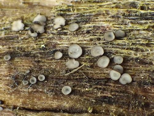
Thomas Læssøehttps://svampe.databasen.org/observations/10587061
These specimens were found in a public garden with diverse vegetation, growing on the wood a young tree (maybe of the family of palm trees).
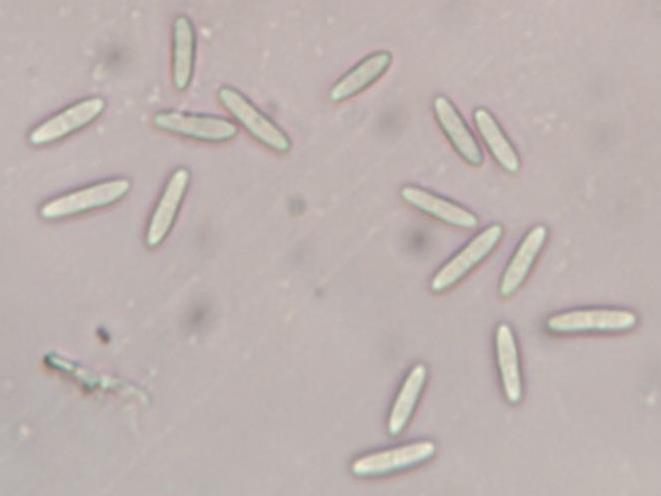
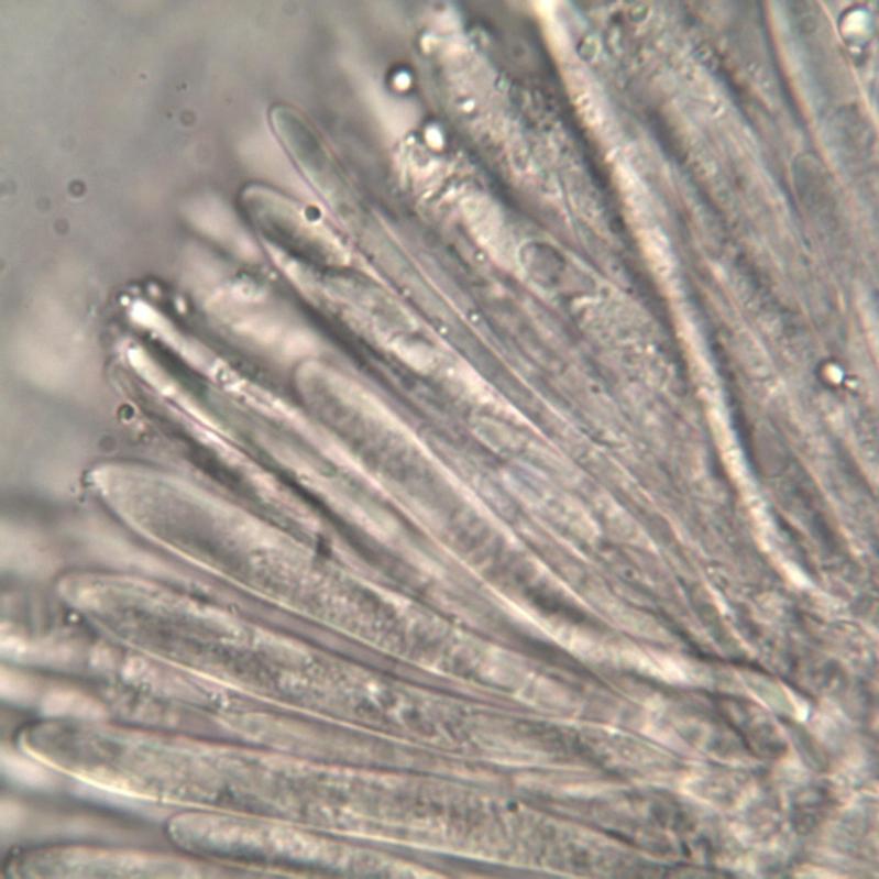
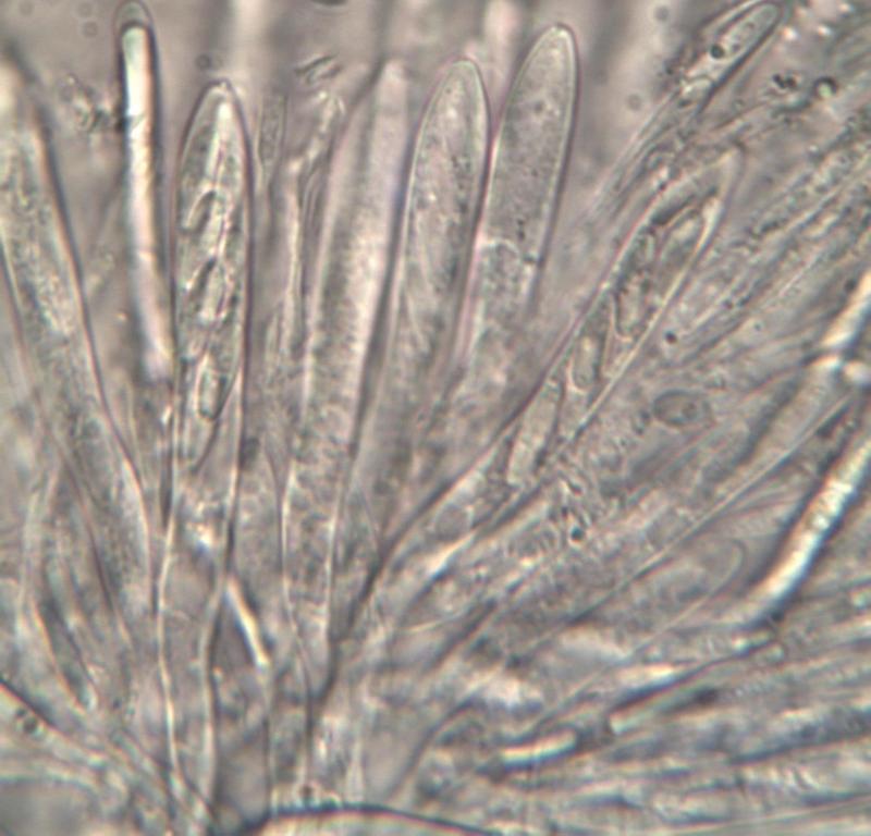
The spores were measured from the free found at magnification 400 and inside the asci at magnification 1000, with values in the ranges 8-11 x 2.2-2.8 µm. I had doubts about the reaction in lugol of the asci apices and, at the end, I observed the reaction in Melzer.
I will appreciate any hint about its classification.
Thanks in advance,
zaca


this is likely a Mollisia. This is very difficult to determine this species. If has grown in wood still more difficultly. Here a link, unfortunately only into German, but at the moment the single possibility to read so much about these species.
Greetings Peter.
http://www.mollisia.de/
Mollisia was my first thought, but then Orbilia comes into the scene and I hesitated.
I will look for the link you provided, though I can't read German.
Best regards,
zaca
an Orbilia is not this. Here a collage of Mollisia-species, this must not be already correct any more.
Greetings Peter.

Yes, there are some very similar.
Thanks again,
zaca


Hi Zaca,
definitely a Mollisia, as Peter said.
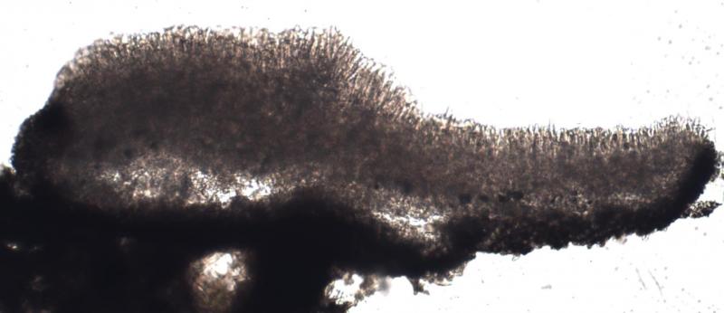
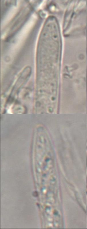
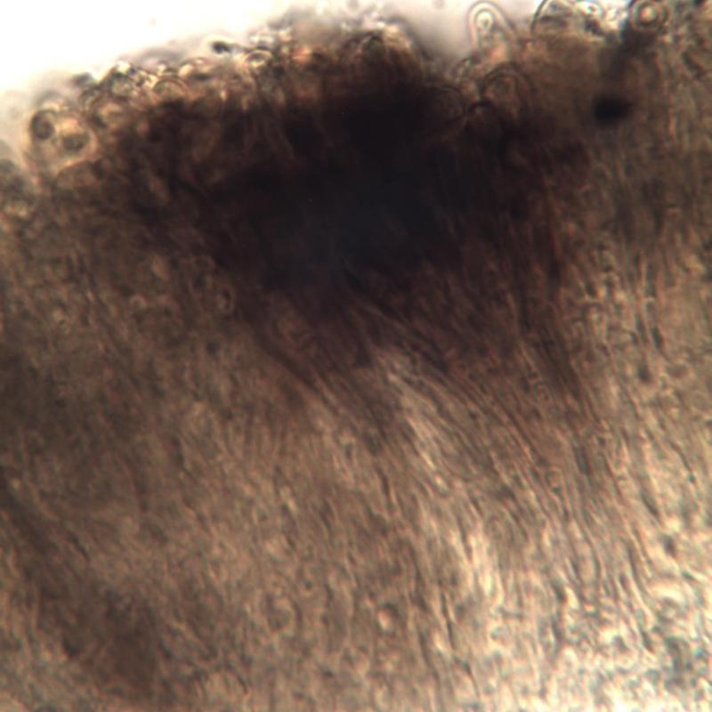
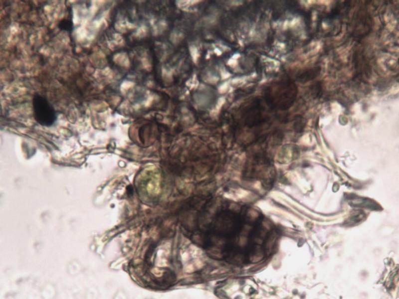
You see the vacuolar body in the paraphyses in your picture(s), this is diagnostic. As is the dark textura globulosa.
About the species I do not ponder - but looks rather normal ...
Best regards from Lothar
Let's see if anyone will p+ropose a name for it,
Best regards,
zaca

for Mollisia please always check the reaction on KOH. It guess it should be colourless in this case. Please also use Lugol's instead of Melzers. Some species show a red reaction of the ascus porus before addition of KOH (and after addition of KOH red or blue).
Please also check the Homepage of Ingo Wagner: http://asco-sonneberg.de/pages/ueberblick/ascomycetes/helotiales/mollisiaceae/mollisia.php and compare your collection especially to the ones that are named "Mollisia cinera". This could fit to your species.
Best regards,
Florian
Hi together,
cinerea - yes, sure ...
But: nobody knows at the moment, which species is meant by this name. I talked with Andreas Gminder some weeks ago, and I remember that there are different species in this complex (the most common of them is named olivaceocinerea by Andreas at the moment, provisionally).
If somebody, then he could give a trustable name to your collection.
"Cinerea" is only a quite vague determination in the moment.
Best regards, Lothar
i know that the "clear" M. cinereas on Ingos page fit to the concept of Andreas. I just tried to give some (general) help for the determination. Spore size and shape, the lack of oil in the spores (this is hard to see on the pictures, so Zaca should check this "live") and shape of the cells of the ectal excipulum fit quite well for M. cinerea. There are some important information missing, so as i wrote and underlined it could be M. cinerea.
Beste Grüße,
Florian
Quoting Lothar in both of his last comments:
"definitely a Mollisia, as Peter said" and "cinerea - yes, sure ..."
So, the question is more: which infrataxon in the M. cinerea complex is this?
Some comment to the helpfull messages by Florian:
- the reaction to KOH was negative, as you presumed;
- I started the analysis of the amyloidity with lugol and only because the reaction was doubtful I add a drop of melzer (that is, in absolute values when looking to a photo take in lugol is hard to say that the result is positive; the result seems more clear after comparing with the same in water; anyway far from being conclusive);
- I gave a look the the link you provided, and after looking to the "cinerea" there I found that the closer to what I observe is from "Mollisia-cinerea-110305" (but there are many similar) except for my doubtful reaction in lugol.
Summarizing, entering to the universe of a collective species I have no means to go further, due to the obvious lack of quality of my equipment, but I'm happy with the result anyway.
Thanks to all who participated in this forum,
Best wishes,
zaca
Zaca - Some of your photos (particularly the 9th and 10th) show spores inside asci already with septae. I can't be 100% sure but the asci look live and I wonder whether Mollisia 'atlantica' could be a good fit?
The picture of the free spores is a little unclear, but perhaps many of these are septate too? What does everyone think - Am I seeing things?
Amitiés,
Nick
You must have an eagle-eye and this is a case to say: "if the equipment is bad the observer is still worse"!
Damn, you're right! I looked to all my photos from the microscopy and was about to quit. But then two sucessive photos appeared, confirming that you are not dreaming and, on the contrary, seeing the right things. I upload part of these photos, clearly exhibiting septate spores. This means M. atlantica? What are the features of this species?
Thank you, Nick, for bringing us in the right direction.
Best regards,
zaca

