31-03-2026 08:19
 Bernard CLESSE
Bonjour à toutes et tous,Pourriez-vous m'aider à
30-03-2026 12:03
 William Slosse
Hello all,On 27/03/26, in Kraaiveld in Wingene (Be
25-03-2026 10:35
Hulda Caroline HolteHello,I collected this species growing on a dead b
30-03-2026 09:53
Yanick BOULANGERBonjourVoici des petites fructifications poilues s
27-03-2026 10:47
Åge OterhalsI have tentatively identified this Stictis to S. f
28-03-2026 07:55
 Marc Detollenaere
Hello everybody,Yesterday I found a number of whit
26-03-2026 15:31
 Åke Widgren
Hello,I found this one in October last year, on r
27-03-2026 15:23
Gernot FriebesHi,this Trichopezizella deviates from typical T. b
 Hi, I wonder if this is interesting to someone here.
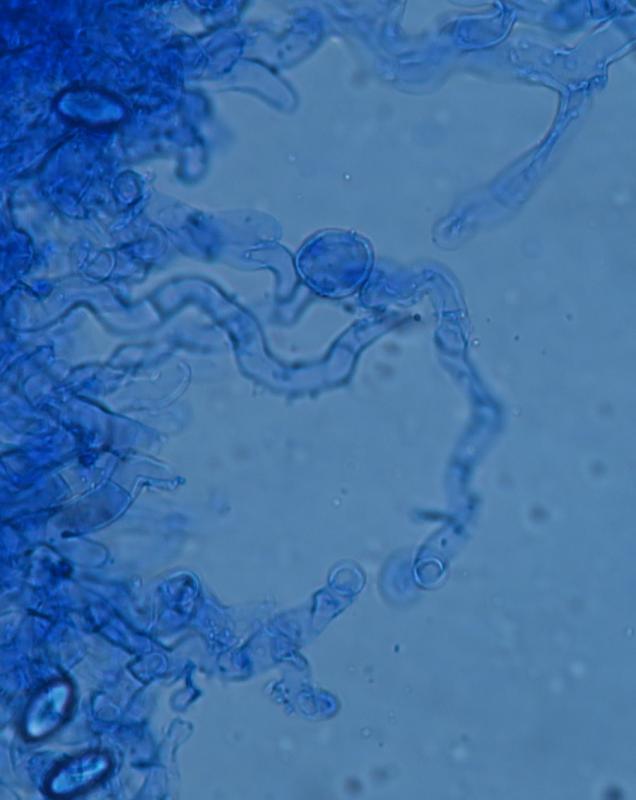
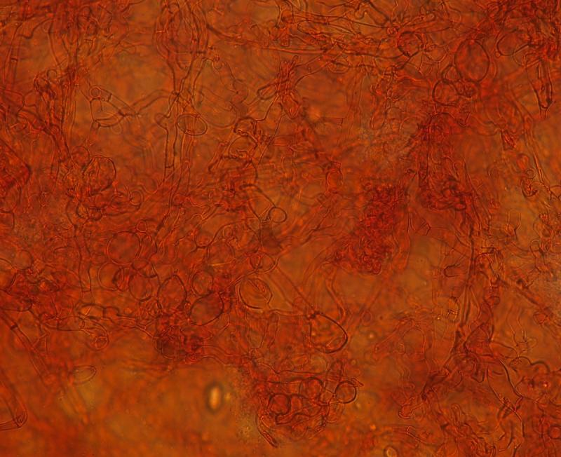
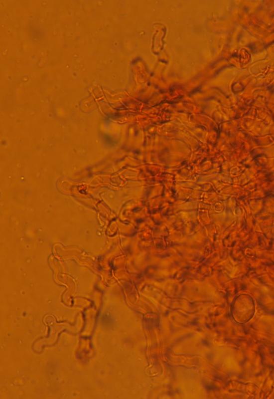
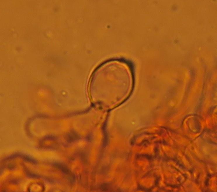
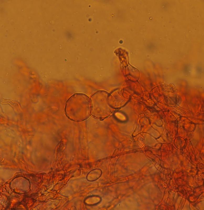
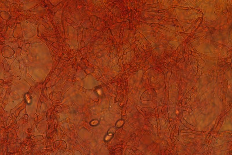
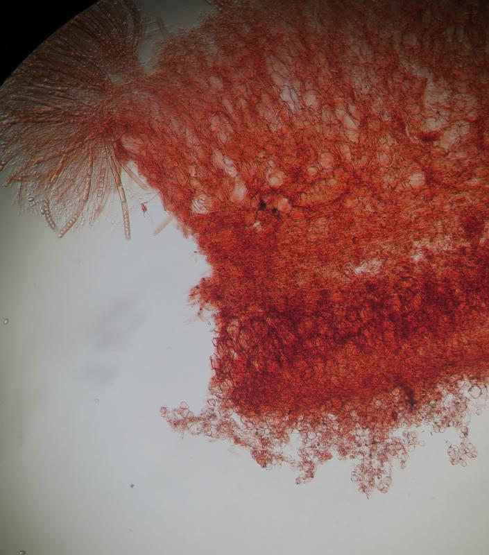
Recently I have collected a large population of clustered Peziza domiciliana on a limestone wall. I have placed this in the fridge and went to examine it today. I noticed that on the abaxial surface, there was a layer of another mould-like fungus, quite hard and compact, almost as if encrusted that it literally pealed off from the surface of the Peziza
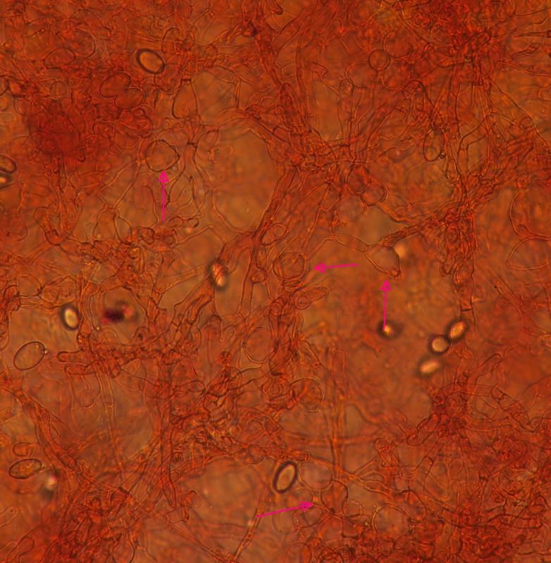
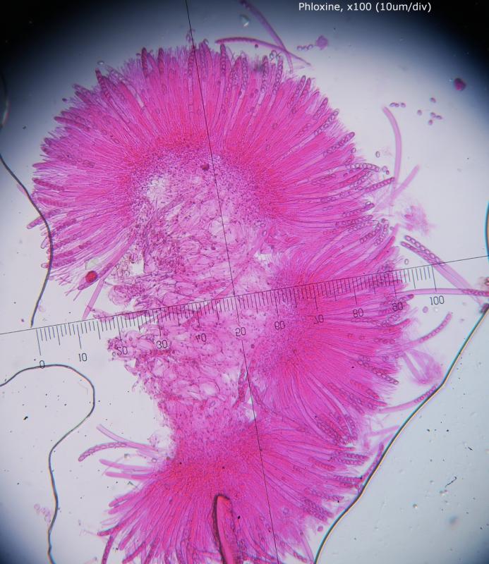
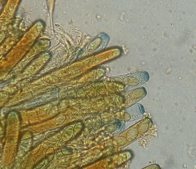
I gave it a quick shot under the microscope and it was obvious to be a parasitic fungus. It is difficult to describe but it consisted of tightly intertwined curled hyphae which here and there, it produced swellings (glubular hyphae) which I think has a sexual function.
I am dropping all images here, and if you have some comments or perhaps a genus (or family) I would love to learn
Thanks in advance


Hi Stephen and others,
I cannot say much about the possible parasite - so I don`t.
The Peziza I would possibly have called P. cerea (muralis) by pure macroscopy and ecologal condtions, but I don`t know much about the taxonomy today and about the limits to similar species (varia, domiciliana?).
Best regards from Lothar
Oui, il s'agit très probablement de Peziza varia (= P. cerea, P. micropus, et de très probablement de P. muralis). Cette espèce est assez communément récoltée sur des murs humides. S'agissant des hyphes, je ne pense pas qu'il s'agit d'un parasite, mais plutôt d'une prolifération anormalement abondante d'hyphes de l'excipulum ectal, peut être la conséquence d'un stress ou des conditions locales.
Cordialement
René
Yes, it is most likely Peziza varia (= P. cerea, P. micropus, and most likely P. muralis). This species is commonly harvested on damp walls. As for the hyphae, I do not think it is a parasite, but rather an abnormally abundant proliferation of hyphae of ectal excipulum, may be the result of stress or local conditions .
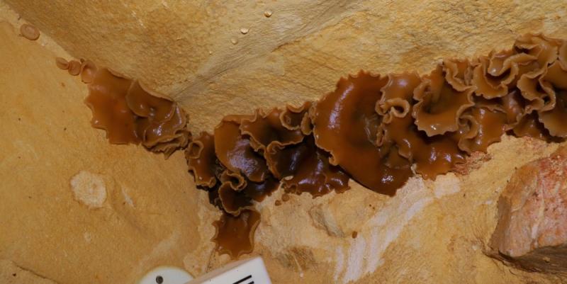
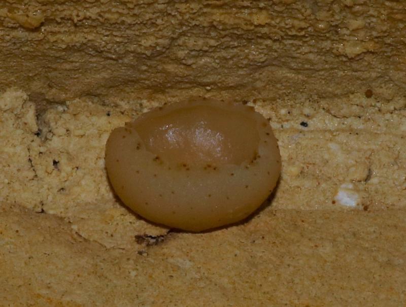
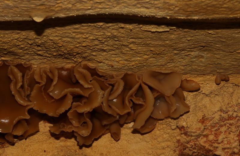
Habitat: Wet Limestone wall (Globigerina Limestone)
Habitat notes: Room with water literally dripping from the wall
Remarks: Numerous clustered specimens of various sizes
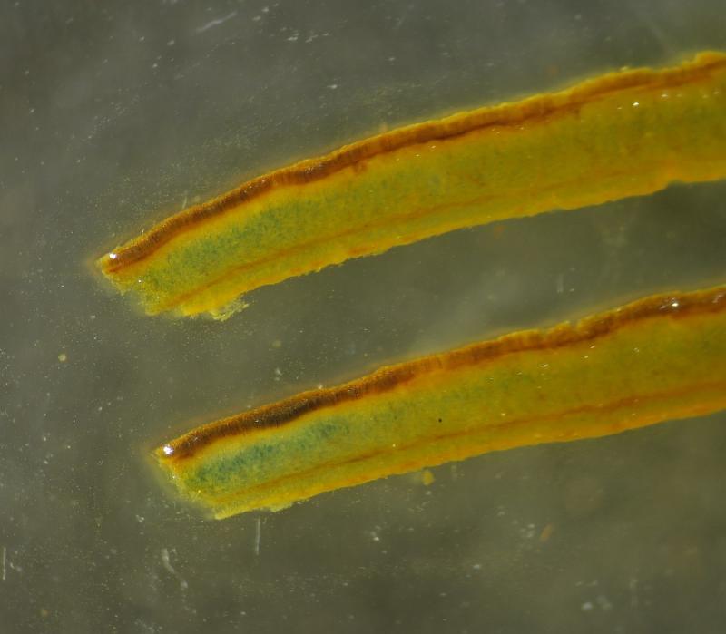
Acocarp colour (adaxial): Young specimens are pale buff, semitranslucent then gradually turning toffee brown when fully mature
Acocarp colour (abaxial): Beige to pale buff when wet but ccustose cream or almost white when dry
Ascocarp diameter: 30 – 95 mm (mean: 60.42 mm)
Ascocarp rim: Irregularly lobed and sinusoidal especially in adult specimens
Ascocarp shape: Initially bowl shaped when young and rather regular then flattening out to a saucer- shape with an irregular border, ocassionally trumpet shaped.
Stipe length: Stipe absent
Stipe colour: Stipe absent
Texture Smooth: to finely granulose
Flesh colour: Buff, semi-hyaline
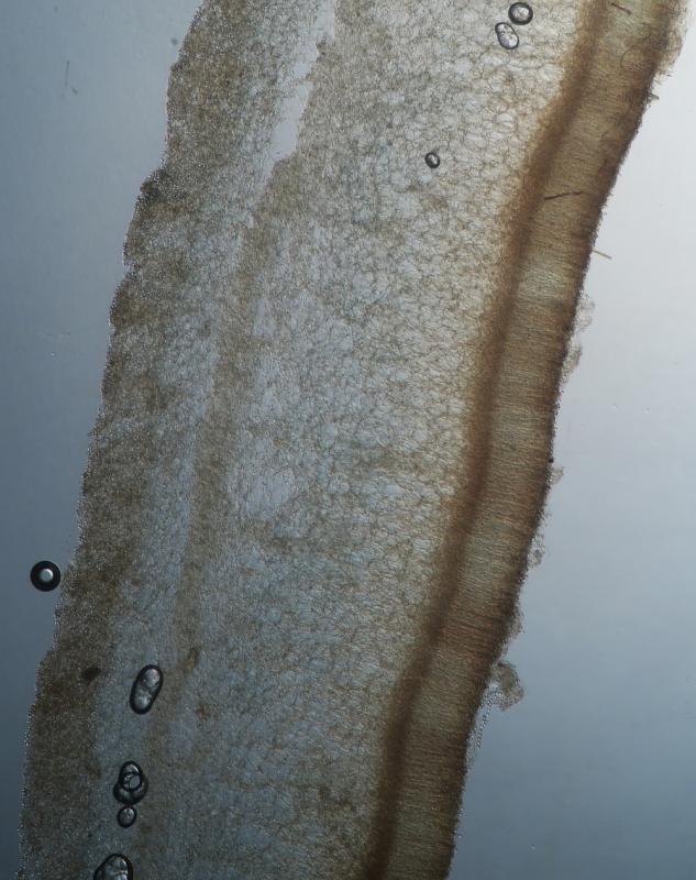
Additional notes: Five layers detected; hymenium, a thin subhymenium, medullary excipulum, ectal excipulum and an outer abaxial epithilial layer
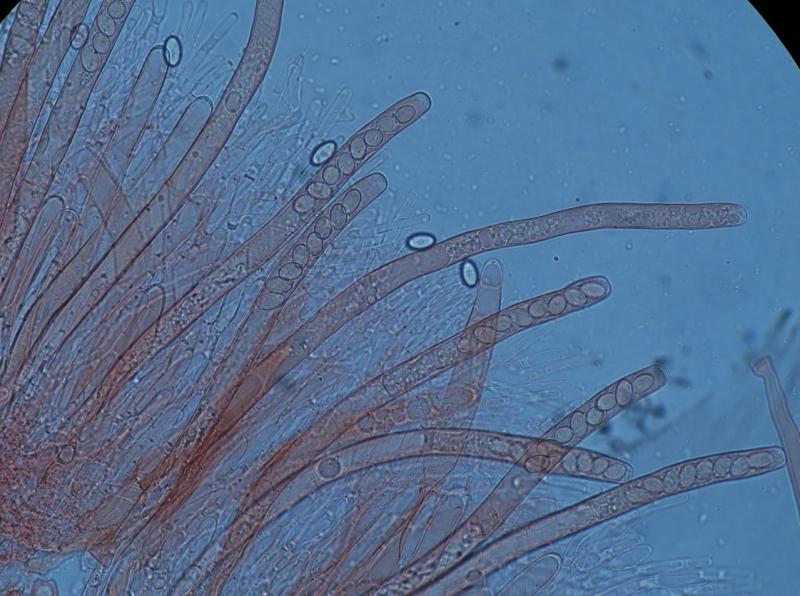
PARAPHYSES.
Paraphyses: Numerous in dense clusters
Paraphyses shape (apex): Not differentiated, or very slightly inflated
Paraphyses width: 5-7 µm, ocassionally inflated to 10-15um at the middle
Paraphyses length: 70–80 µm, distinctly shorter from the asci
Paraphyses septation: Two or more septa, usually up to five
Few paraphyses are inflated about the middle part, wile others have conspicuous vacuoles that do not stain in congo red, melzer or Cotton blue
ASCI
Ascospore release: Apical orifice without operculum
Shape: Cylindrical, slender, arcuate
No. of Spores: 8
Operculum: Not observed
Tunic (Wall): Unitruncate
Ascum length (range): 205.49 - 286.82 µm
Ascum length (mean): 254 µm
Ascum width (range): 10 - 14.28 µm
Ascum width (mean): 12.1 µm
Ascum L:W ratio: 21.1
Iodine reaction (J +/J -): Strongly J+ve at the tips. Apart from the asci tips, the subhymenium and young asci also stain blue in Melzer
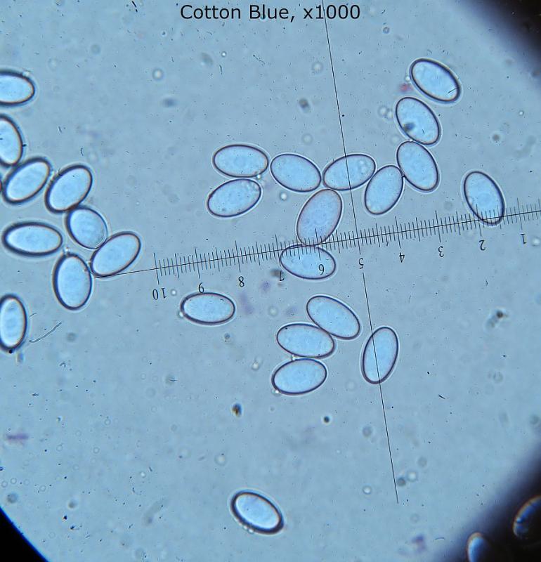
ASCOSPORES
Spore length (range): 12.29 - 14.85 µm
Spore length (mean): 13.3 µm
Spore width (range): 7.11 - 8.92 µm
Spore width (mean): 8.0 µm
Spore Q factor (range): 1.52 - 2.04 µm
Spore Q factor (mean): 1.7
Spore shape: Oval
Spore septa: 0
Spore surface: Finely rough, appearing as cloudiness under x 1000
Oil bodies Not observed
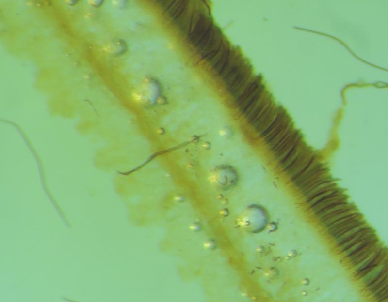
Ascocarp Layers 6: A whitish hymenium, sitting on a narrow and darker layer subhymenium composed small pigmented hyphae (and possily giving the brownish colour of the ascocarp). Below is the spongy medullary excipulum and then the ectal excipulum separated by a thin pigmented layer of tubular hyaphae. An epithilial tissue is finally found at the abaxial surface and is composed of small globular cells
Excipulum layers: 2 , separated at the lower quarter by a thin layer of textura intricata of short tubular hyphae 7-10um wide.
Medullary Excipulum: Spongy thick layer of textura angulosa, 40-70um wide
Ectal Excipulum: Narrow layer of tessuta angulosa, 20-50um wide.
Abaxial epithilium Thin crustose layer of inticated hyphae with several swellings
Impressively, when I blew on the ascocarp collection placed in a container, I could see a puff of white spores - FEW seconds later! Its like there is a mechanism involving the paraphyses. Also when I dropped some Iodine (or water) on the surface, the fruiting body ejected spores which could be seen as cloudy puffs floating in the air!!! Incredible!
The habitat of varia is quite diverse but mostly on the ground not walls, where P. domiciliana is a wall specialist.
http://www.mycocharentes.fr/pdf1/489.pdf