18-05-2026 12:43
Sylvie Le GoffBonjour à tousPuis je avoir votre aide sur ce que
18-05-2026 10:13
 Lieve Deceuninck
Dear forum members,I identified this as the teleom

17-05-2026 19:05
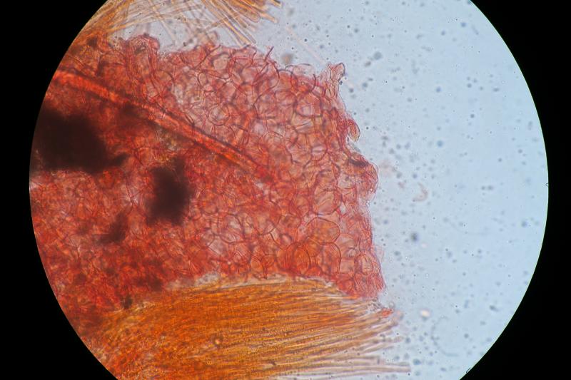
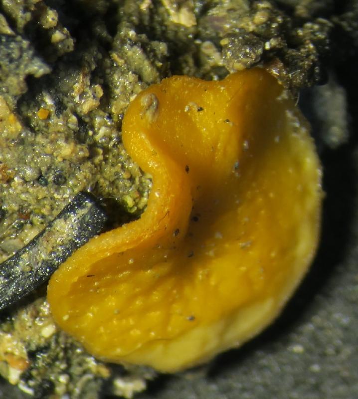
Thomas FlammerI have found this tiny 200 ym cup shaped apothecia
17-05-2026 16:41
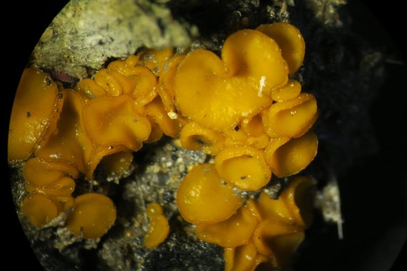
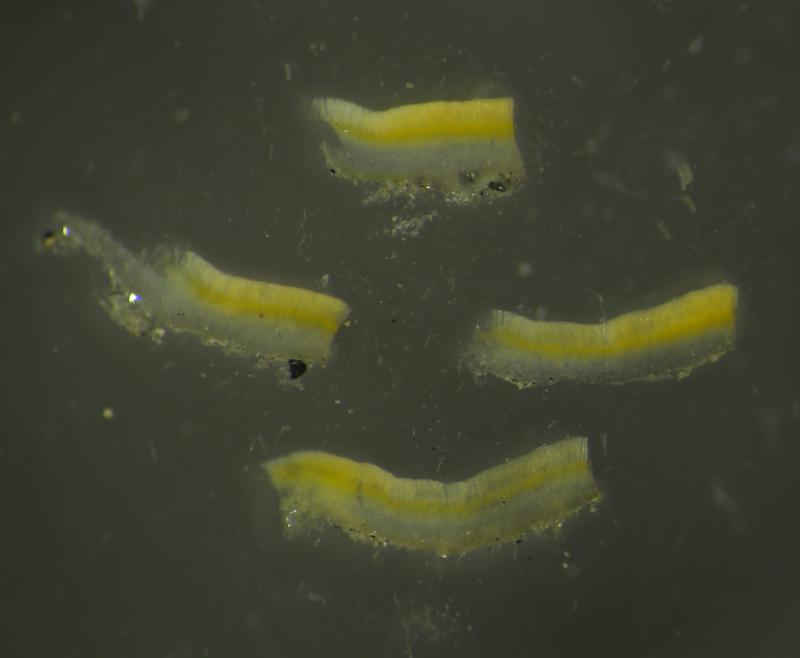
Margot en Geert VullingsWe found this Lachnum on an old Rubus stem.Fruitbo
05-04-2026 22:46
 Lothar Krieglsteiner
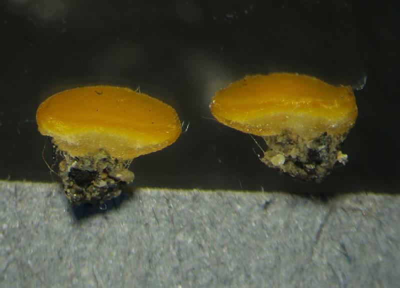
on wood of Ceratonia, Algarve, 3.4.2026.The color
15-05-2026 13:33
Sylvie Le GoffBonjour à tousJe serais très reconnaissante enve
16-03-2011 14:31
roman vargas albertoHi. I would like some opinion about this Peziza
14-05-2026 05:36
Ethan CrensonHi all, I haven't paid much attention to Lachnu
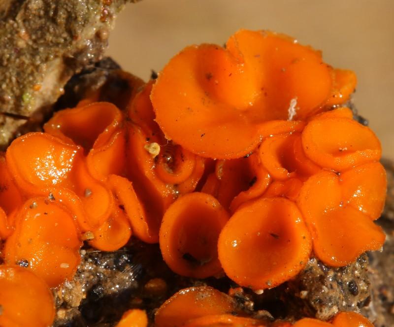
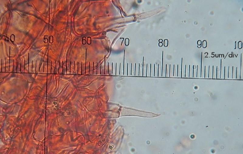
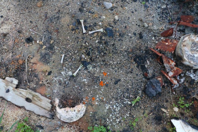
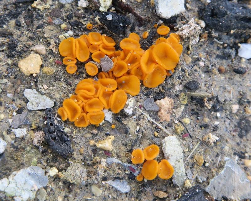
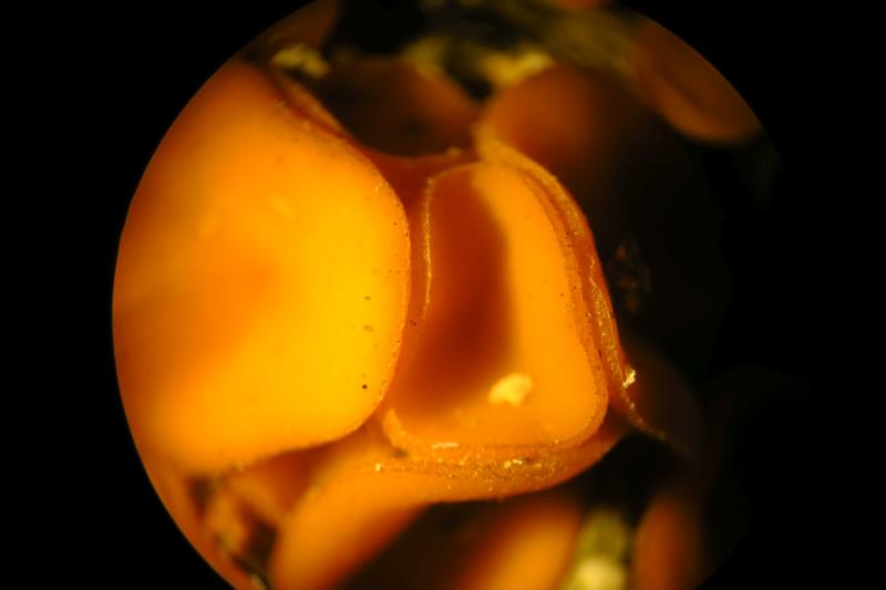
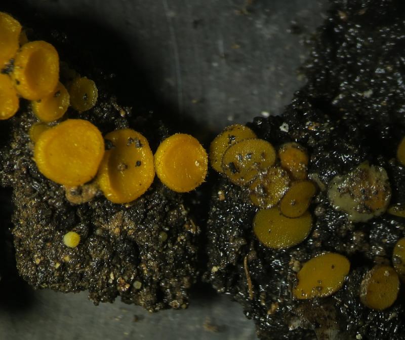
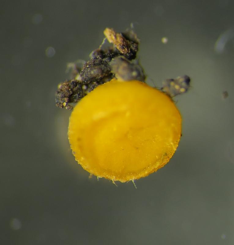
 Hi, I found another rubber-orange coloured Ascomycete on burnt wooden debris on the soil in an open field. My thoughts went on Anthracobia, but definitely this species had no visible hair on the rim. With the microscope I have only a suspect if I have observed "hair?" at 40um length.
 Here's some important data.
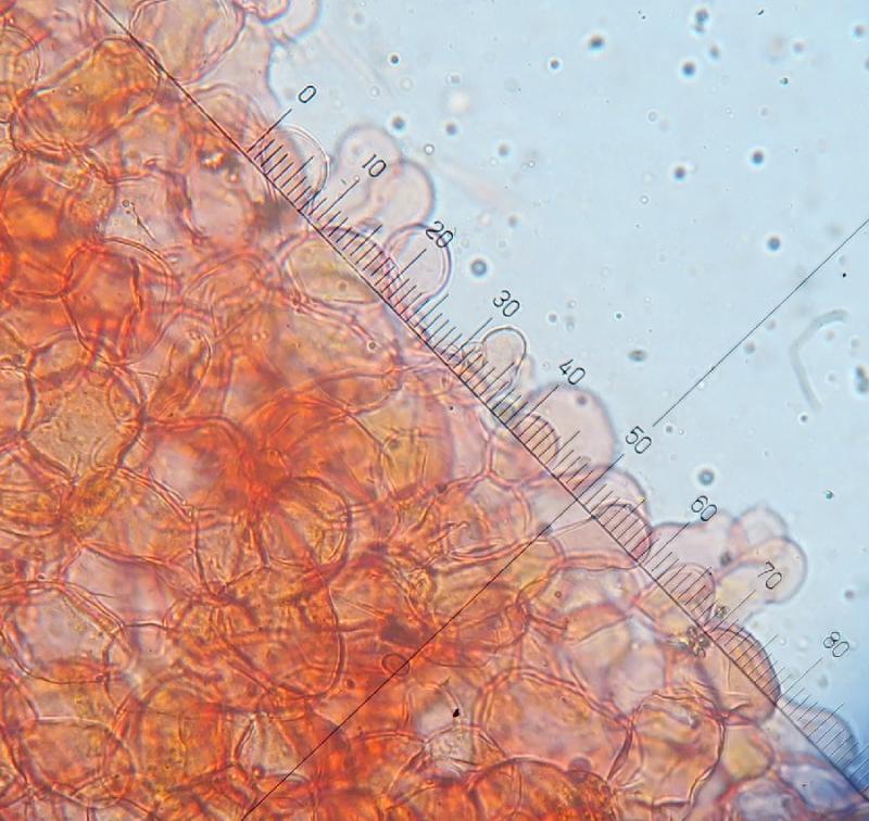
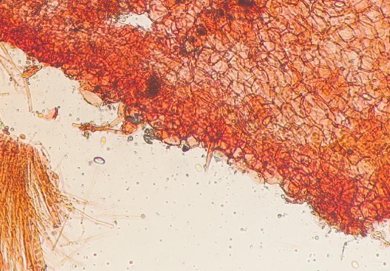
Sessile saucer-shaped Ascocarp, vivid apricot-orange (like industrial rubber), abaxial surface same but more opaque-greyish and rough; diameter 3 – 9 µm (mean: 5.5 µm) with a concolourous rim having a very narrow and barely visible hyaline border.
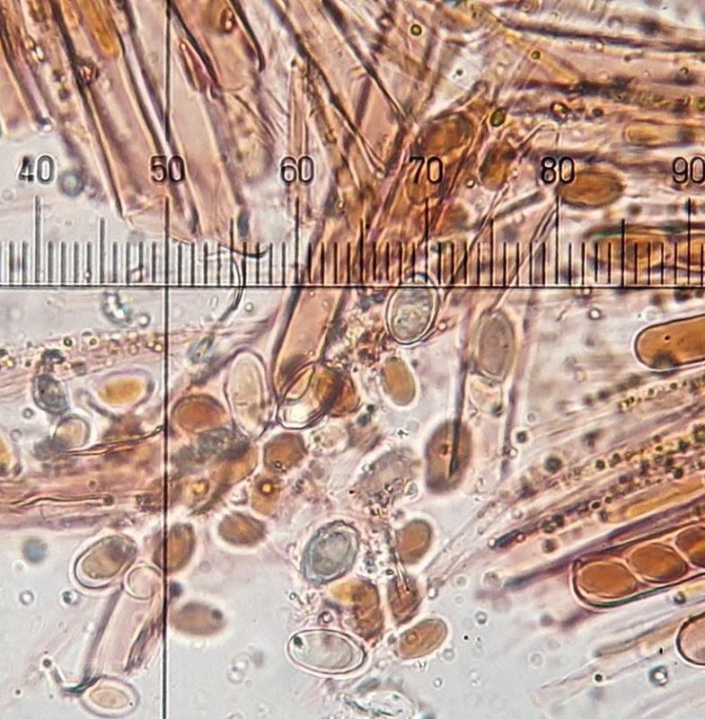
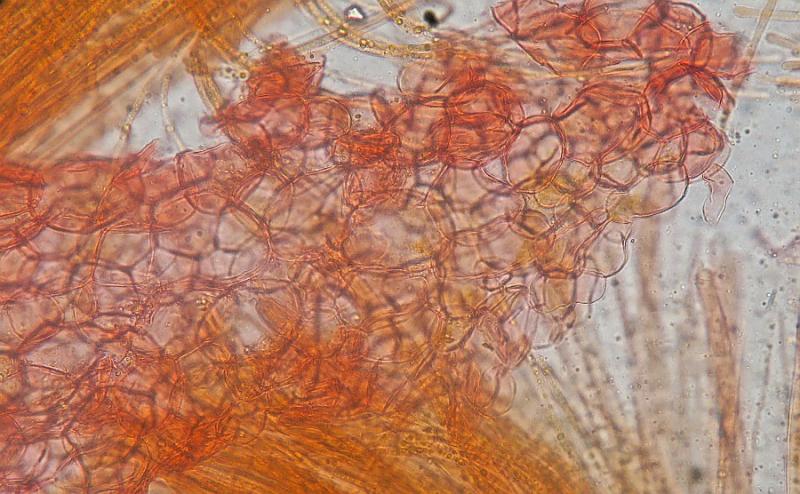
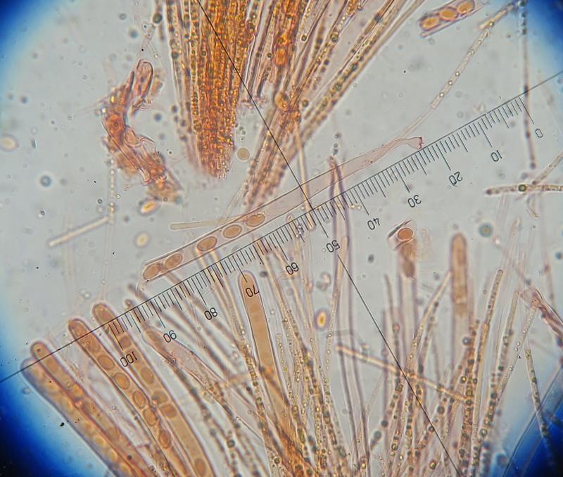
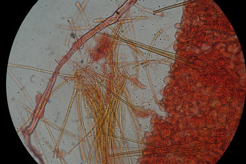
Flesh colour yellowish just below the hymenium then greyish-beige further down. Ascocarp marcescent; Lower surface of ascocarp with small white-hyaline hair-like rhizoidsParaphyses Numerous often in clusters of few numbers outnumbering the asci
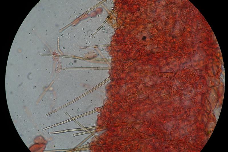
Paraphyses narrow and filiform (3um wide), terminating with a slightly swollen, hence sub-clavate apex, up to 7um wide. Paraphyses contain many vacuoles which becomes readily pigmented in Lugol's Iodine and Cong red. Paraphyses length 210-240um, same length or slightly overtopping the asci
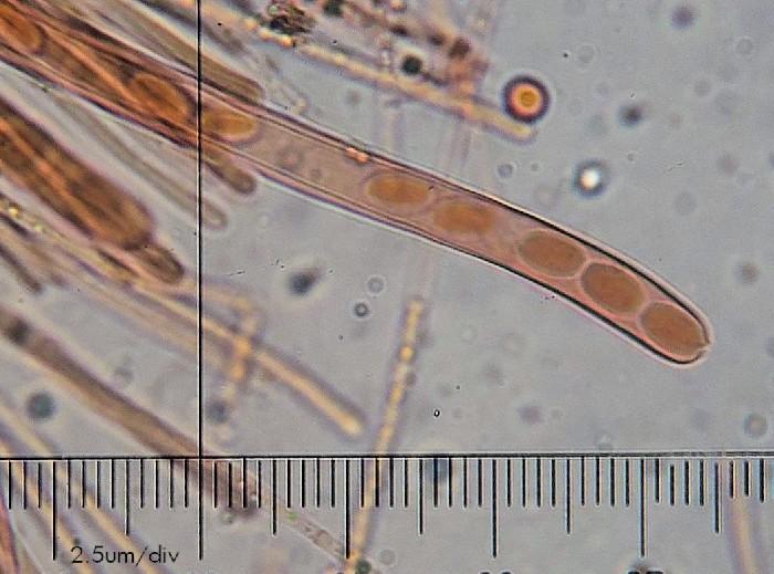
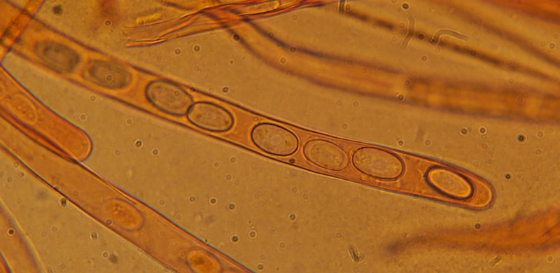
Asci inoperculate, release of ascospores likely by splitting of apex; cylindrical in shape, slender, rounded obtuse apex, often with an abrupt bent or curve at the base. 8 spores per ascum, unitunicat, smooth; 167 - 245 µm x 9.5 - 13.5 µm (L:W ratio 17.5), J -ve
Ascospores 11.8 - 14.69 (mean 13.1 µm) x 6.49 - 9.58 (mean) 7.6 µm), Q. 1.49 - 2.12 µm (mean) 1.7); fusoid-elliptical, widest at the centre with two identical rounded poles, aseptate, smooth surface, oil bodies absent (unless it consists of a large oil body occupying most of the spore space.
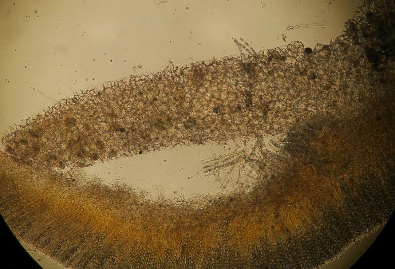
Excipulum (medullary) Spherical to broadly elliptical usually with obtuse angles forming an isohedral , 30-48 µm wide
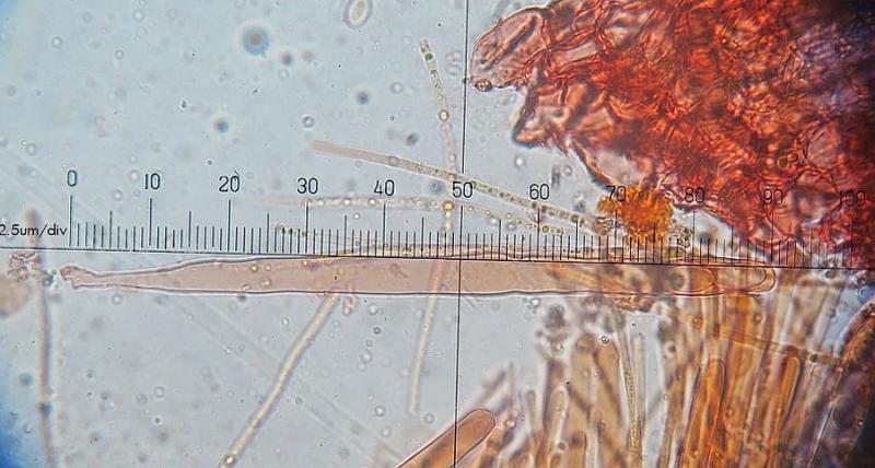
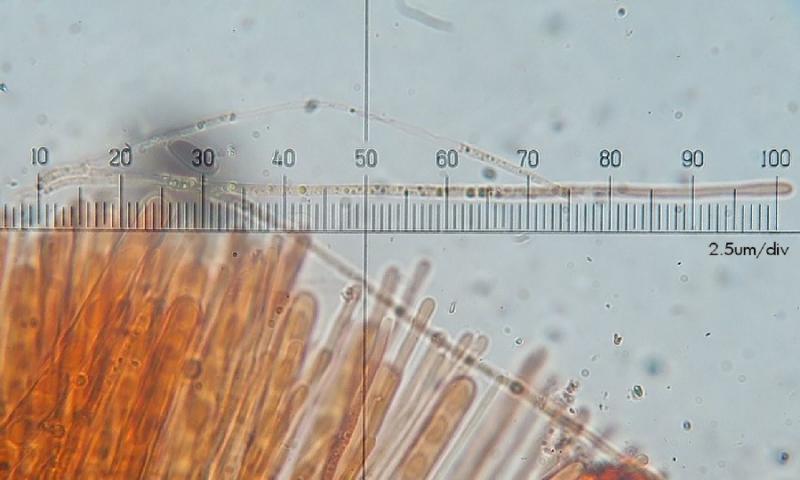
No distinct hairs on rim, although some slides showed a filamentous projection of about 40um in length, cylindrical often in two layers of hyphae.

What other options ??? Tricharina spp.
I shall check again for the hairs, but I don't see anything distinct from the macro and from the micro!
Hmm.... any help please ?
Key to genera to Pyromycetes would be lovely!

it reminds me most at cheilymenia. there are some taxa with quite inconspicious hairs. and some terrestric ones. why not on burnt ground? and there could be dung-infiltration even there ...
perhaps you find some more significant hairs?
possible ornamentation would also be interesting. striate?
best
dirk
So here are more photos showing the microscopy and the ascocarps

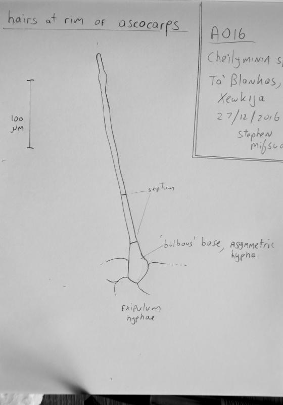
Note 29/Dec B: Yesterday I have found another population in a different locality. I can confirm now that the habitat is burnt ground and it was sharing the habitat with an Anthracobia (cf nitida) sp. Interestingly, this populations had some individuals with hyaline hairs. These were detected by the naked eye and hand lens. In my opinion, mature specimens lost the hair. I have managed to photograph them - they are over 100um long, usually 1 or 2 septate, one septum when present, about half length of the hair, the other always present at the base below the basal rounded cell.
I wonder why the two specimens I studied, there was a low number of asci... possibly because the ascocarp was young,
 Spores do not show striations.

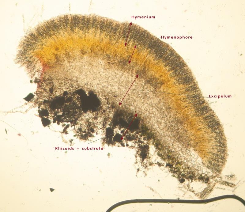
In my non-expert opinion, the excipulum is one layer (Spherical hyphae) lying below a hymenophore of tubular elements and then the hymenium of asci.
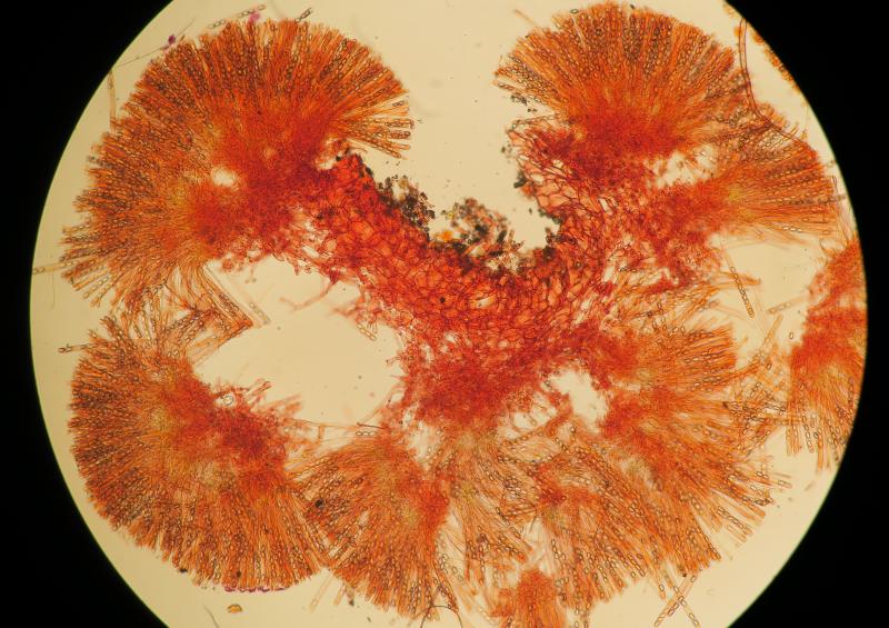
Note that the hymenium (asci + paraphyses) were strongly dextrinoid in IKI
BUT
another interpretation is that there is both ectal and medullary layers, the latter corresponding to the tubular hyphae, and the ectal composed of spherical cells
I have reported the second Cheilymenia from another site (see Note 29/Dec B above) with burnt ground and assumed that both are the same species. Now I think they are different species, or at least, it cannot be assumed so before checking well. So the hair I depicted from the second population for the moment should not be attributed with the first population to avoid confusion. Let's stick to the first posted population.
I've also reported that this population was also found close to animal bones and other sort of debris. Marginal hairs very rare or caduacous or (absent?). Now I think that this is Cheilomenia cadavarina, reported on necrotic material and peat and superficially have a good similarity.
Look here:
http://www.biodiversidadvirtual.org/hongos/Cheilymenia-cadaverina-(Velen.)-Svr-269-ek-img25066.html
I shall collect, yet another specimen and check immediatelly for the presence of hair on fresh material to compare. Some more photos are found here.
Regards the second Cheilymenia, I will submit a different post