05-05-2026 22:40
Gernot FriebesHi,I believe this is a Plagiostoma growing on a Sa
04-05-2026 18:13
 Stephen Martin Mifsud
ID request for what seems to be a true aquatic fun
04-05-2026 16:39
Stephen Martin Mifsud
ID request: This specimen was collected in Malta o
28-07-2011 18:31
 Alex Akulov
Dear FriendsToday I made the pdf file of Velenovsk
28-04-2026 20:07
 Lothar Krieglsteiner
... on twig in the air at standing Ceratonia siliq
04-05-2026 09:50
 Castillo Joseba
Me mandan el material seco de Galicia,(España) re
02-05-2026 12:42
Alain BRISSARDBonjour à tousJeuidi 30 avril dernier on m'a remi
02-05-2026 13:06
Pauline. PennaBonjour Please can someone help me with this id
01-05-2026 22:45
 Thierry Blondelle
Bonjour à tous, Une récolte sur bouse séchée d
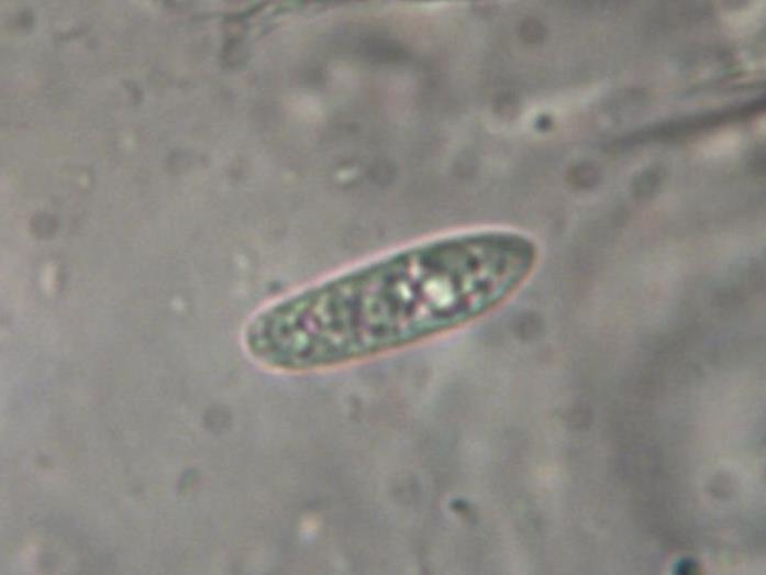
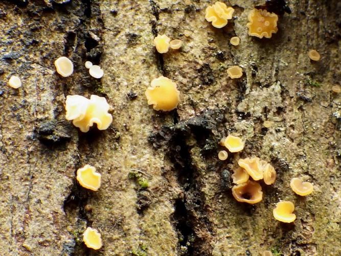
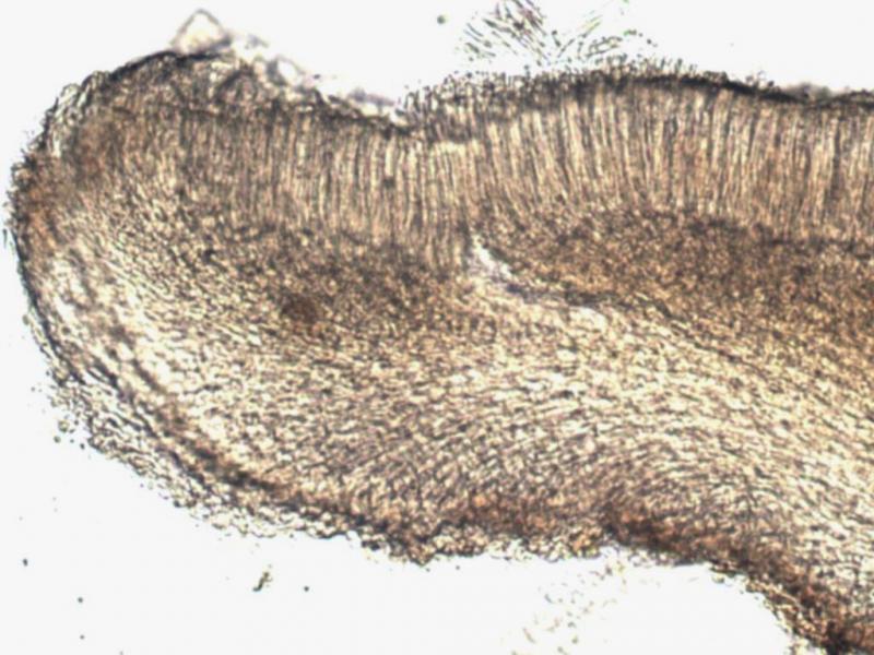
I found this cups growing on the bark of Eucalyptus. It seems to be Mollisia or close. However, the spores are a bit to large. I got the following dimensions for them:
(9.7) 9.8 - 14.1 (14.6) × (2.2) 2.4 - 3.7 (3.9) µm
Q = (2.8) 3.1 - 4.4 (4.6) ; N = 23
Me = 11.9 × 3.3 µm ; Qe = 3.7 .
I attach a set of photos with its features.
I would like to have some help for its classification.
Thanks in advance,
zaca

Hello,
the spores wouldn't rule out Mollisia, but all the rest doesn't fit.
I think this is a Hymenoscyphus or a related genus.
best regards,
Andreas
Let's move the target genus.
Best regards,
zaca

Zotto
Hello,
may be it's worth comparing with Hymenoscyphus phialea.
best regards,
Andreas

Bonjour,
I would suggest a comparison with Bisporella subpallida, at least the macro would fit and also the excipulum ?
Amitiés
Michel
Many thanks for your comments, opinions and proposals.
I'm a bit confused by now: I could not find any information about Hymenoscyphus phialea, suggested by Andreas, and regarding Bisporella subpallida, mentioned by Michel and Zotto, that species has spores much smaller than my specimen, despite of the similar macro appearance, as I could see in several places. I checked again the dimensions of the spores.
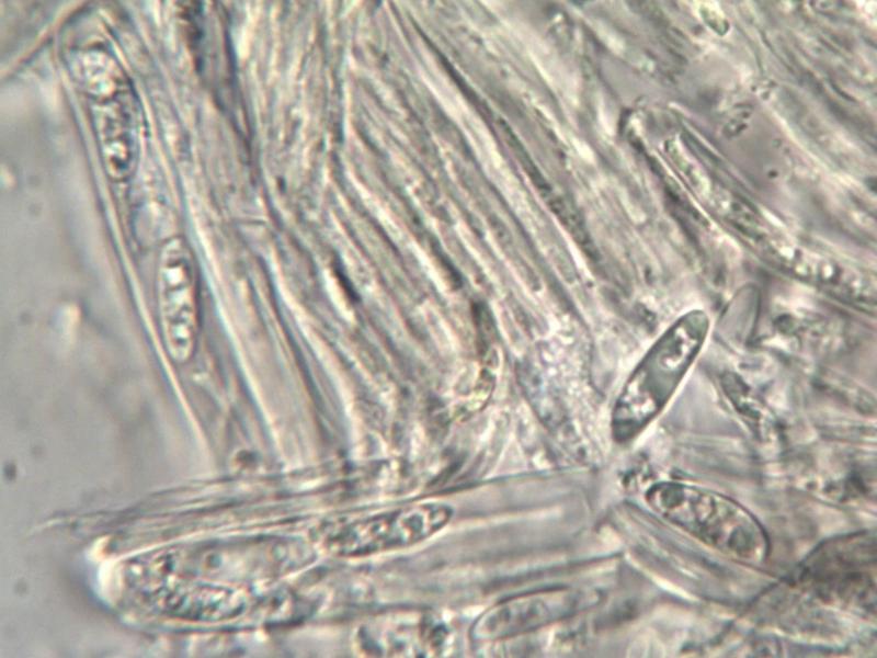
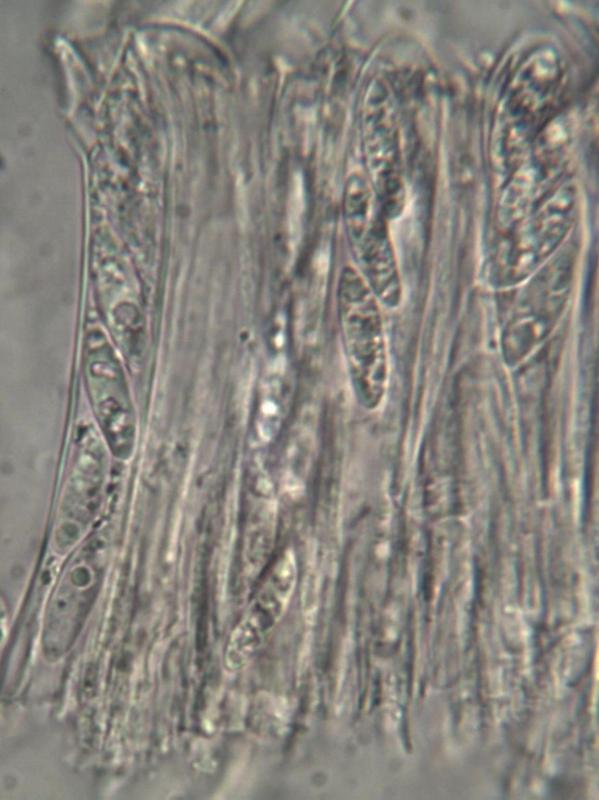
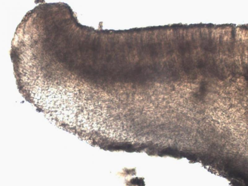
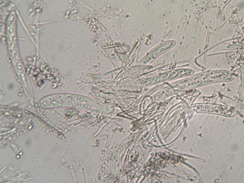
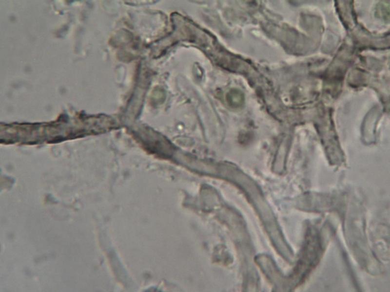
I take the oportunity to upload two photos, one of the ectal exciple and the other with some strange hyphae seeming incrusted.
Regards,
zaca
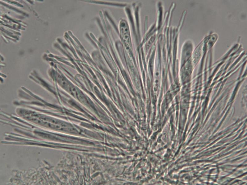
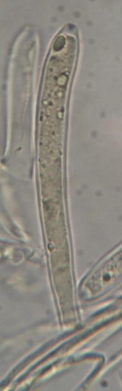
The asci look amyloid, do they? The shape of ring would really be helpful.
The correct name for Andreas' suggestion is Helotium phiala, but that species has inamyloid asci.
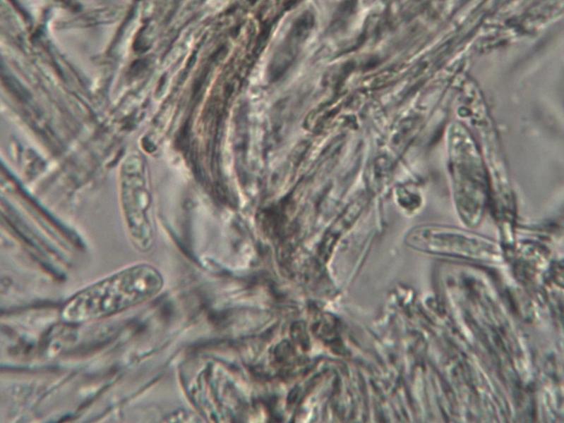
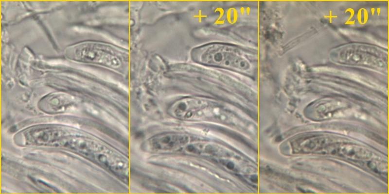
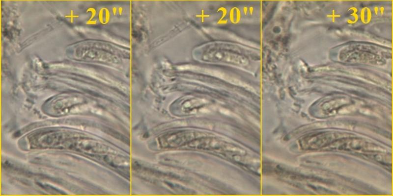
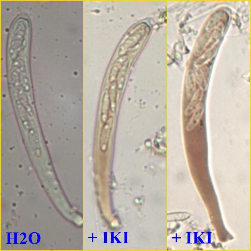
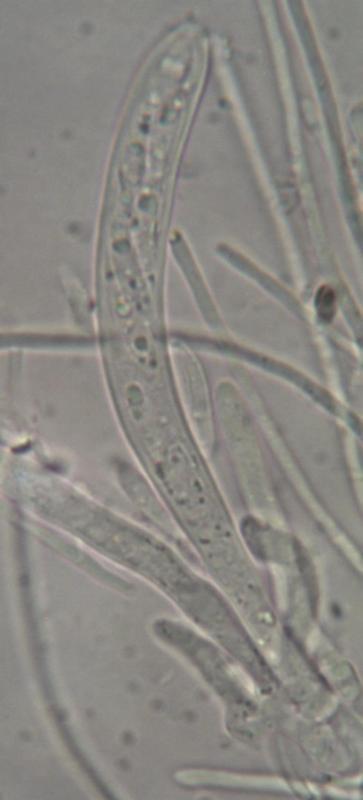
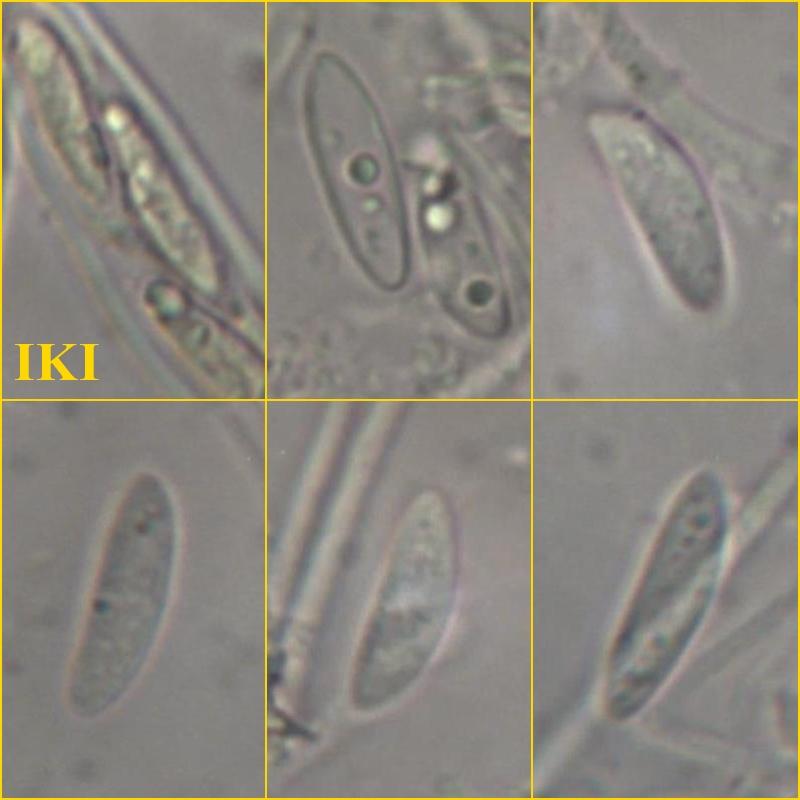
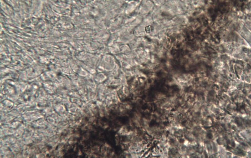
I will repeat the microscopy and if something new I will post it here. As far as I can see from the previous micro, it is difficult to classify the asci as amyloid since there is a very diffuse bluish tone at the apex and that's all, but if you see the photo in water the appex already seems a bit blue. I also took a photo, that I upload now, of the Lugol reaction in a mount with KOH and that gave blue apex.
Best regards, zaca
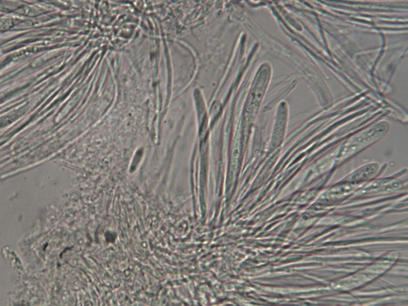
If you find the fungus again, please make preparations in water with only slight pressure on the cover slip. maybe we can then proceed in finding a genus.
Following the recommendations of Zotto I remade the microscopic observation, using a mount in water to observe the spores; After joined IKI and analyzed the evolution of the reaction, taking photos at regular intervals. As expected from the previous observation, the bluish apex reaction of asci is very discrete. I attach some photos, hoping that these can be useful.