
28-04-2026 20:07
 Lothar Krieglsteiner
... on twig in the air at standing Ceratonia siliq
12-05-2026 15:41
 Nicolas VAN VOOREN
Dear Ascolovers, especially interested in Pezizale
27-04-2026 20:52
Lothar Krieglsteiner
Found on hanging tiwg of Olea europaea in dried-ou
11-05-2026 20:22
Lothar Krieglsteiner
on attached twig of standing Ficus caricaquite uns
11-05-2026 12:32
 Bernard CLESSE
Pourriez-vous m'aider à identifier cette héloti
29-04-2026 10:44
Lothar Krieglsteiner
growing at moist, drying-out soil at the side of a
05-04-2026 22:46
Lothar Krieglsteiner
on wood of Ceratonia, Algarve, 3.4.2026.The color
10-05-2026 16:18
brigitte vignotbonjour trouvée en Ariège sur bois une petite
 Greetings AscoFrance!
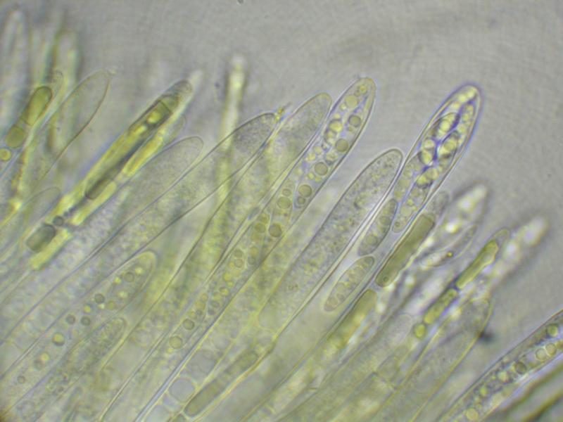
Here is a curious discomycete, field IDed to Ionomidotis by Paula DeSanto on the recent Peck Foray in Watkins Glen, New York. This particular find is from a mixed, predominantly hardwood forest within the Meads Creek State Forest. When I got a look at the dried material and field photos, I saw enough resemblance to my own Ionomidotis collection from North Carolina (http://mushroomobserver.org/174774) to consider the possibility, but upon preparing the material for microscopy we noticed that KOH extractable pigments (3% solution) were conspicuously absent. Can it still be Ionomidotis without this reaction? Perhaps this is a member of some other genus in the Encoelioideae?
Ascus tips inamyloid, despite appearing somewhat bluish in the micrographs. No paraphyses observed.
Spores:
9.5-14×=2.5-4.5?m (x=12.25×3.325?m, Q= 2.44-5.2?m, Qm=3.781?m, m=20, s=1)
13.5 x 3 ; 4.5
13 x 4 ; 3.25
13 x 2.5 ; 5.2
12.5 x 3.5 ; 3.57
14 x 4 ; 3.5
13 x 3 ; 4.33
9.5 x 3 ; 3.17
12.5 x 4 ; 3.13
13 x 3.5 ; 3.71
11 x 4.5 ; 2.44
13.5 x 3 ; 4.5
13.5 x 3 ; 4.5
10.5 x 4 ; 2.65
12 x 3 ; 4
12.5 x 2.5 ; 5
12.5 x 3 ; 4.17
9.5 x 3 ; 3.17
14 x 4 ; 3.5
11.5 x 3 ; 3.83
10.5 x 3 ; 3.5
Many thanks!
-Danny N.
PS: The images are all apparently too large for the site :( Please find them on Mushroom Observer here: http://mushroomobserver.org/218595

I am reminded of a Chlorencoelia, but the two species for which I have images, C. versiformis and C. torta) have distinctly amyloid asci. The spores would fit.
I am sure that the paraphyses would be seen when squashing the hymenium. If you had pictures from fresh material the genus Chlorencoelia would show a striking feature in the paraphyses (vacuolar bodies, see attach).
Zotto
Also, I believe the fact that the bottle of Melzer's used was labelled "Melzer's Replacement" may have something to do with the lack of observed blueing. Will use a more reliable reagent for the second set of micrographs.
These vacuolar bodies are a useful character at the family level. They are rather typical for the family Cenangiaceae as we now circumscribe it, but absent from the Cordieritidaceae which inbclude many ionomidotic species.
many thanks!