

05-05-2026 22:40
Gernot FriebesHi,I believe this is a Plagiostoma growing on a Sa
06-05-2026 11:25
 Castillo Joseba
Me mandan el material seco de Galicia (EspaûÝa) re
06-05-2026 17:23
Thomas LûÎssû¡ehttps://svampe.databasen.org/observations/10594257
28-04-2026 20:07
 Lothar Krieglsteiner
... on twig in the air at standing Ceratonia siliq
04-05-2026 18:13
 Stephen Martin Mifsud
ID request for what seems to be a true aquatic fun
04-05-2026 16:39
Stephen Martin Mifsud
ID request: This specimen was collected in Malta o
28-07-2011 18:31
 Alex Akulov
Dear FriendsToday I made the pdf file of Velenovsk
04-05-2026 09:50
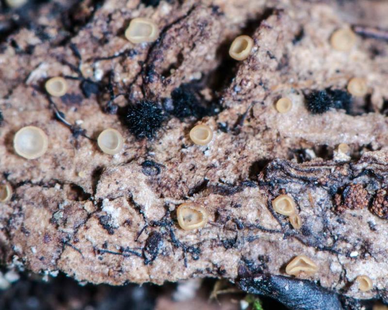
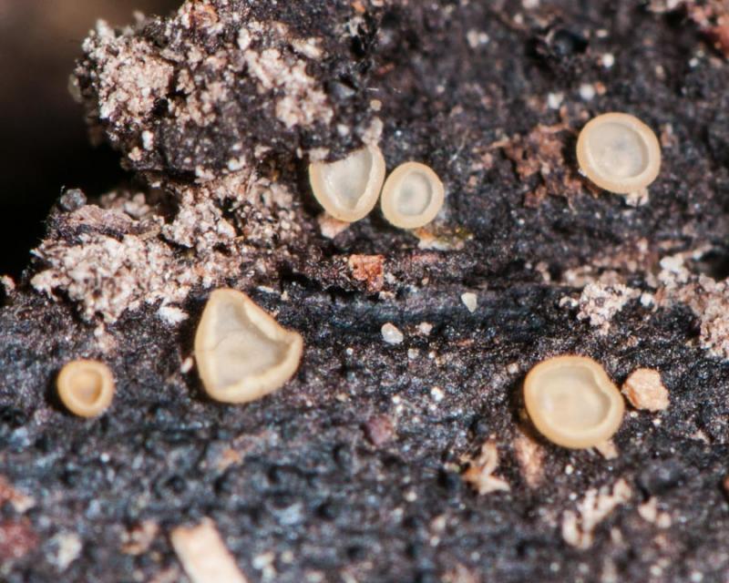
Castillo Joseba
Me mandan el material seco de Galicia,(EspaûÝa) re
Hyalorbilia inflatula 030514 176
Miguel ûngel Ribes,
06-08-2014 23:35
 Hi again
This cupulated yellow Hyalorbilia was relatively big (2 mm diam).
Spores cilyndrical ofô (5.9) 6.4 - 7.9 (8.7) x (0.9) 0.95 - 1.3 (1.6) ôçm;ô Q = (4.9) 5.3 - 7.7 (9.0) ; N = 42;ô Me = 7.1 x 1.1 ôçm ; Qe = 6.5
I couldn't see paraphysis, imposible to disgregate the sample.
Thank you.ô
Miguel û. Ribes


Hans-Otto Baral,
07-08-2014 09:01

Re : Hyalorbilia inflatula 030514 176
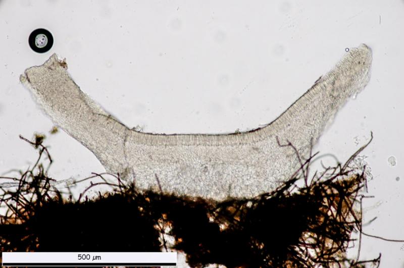
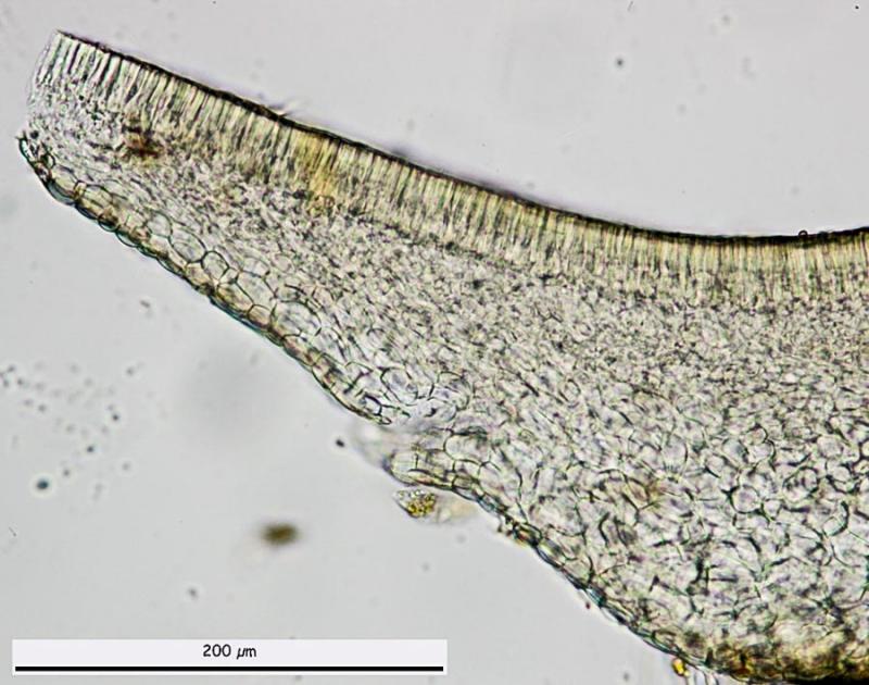
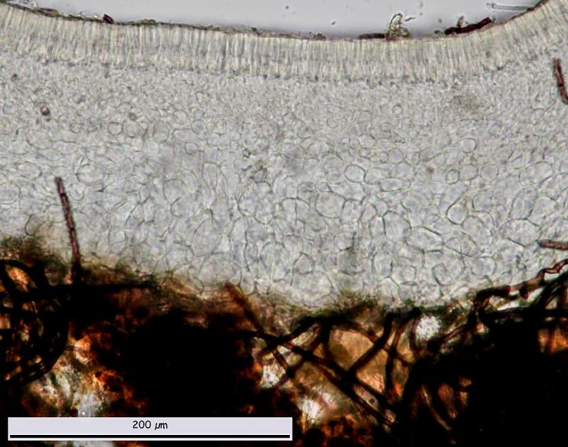
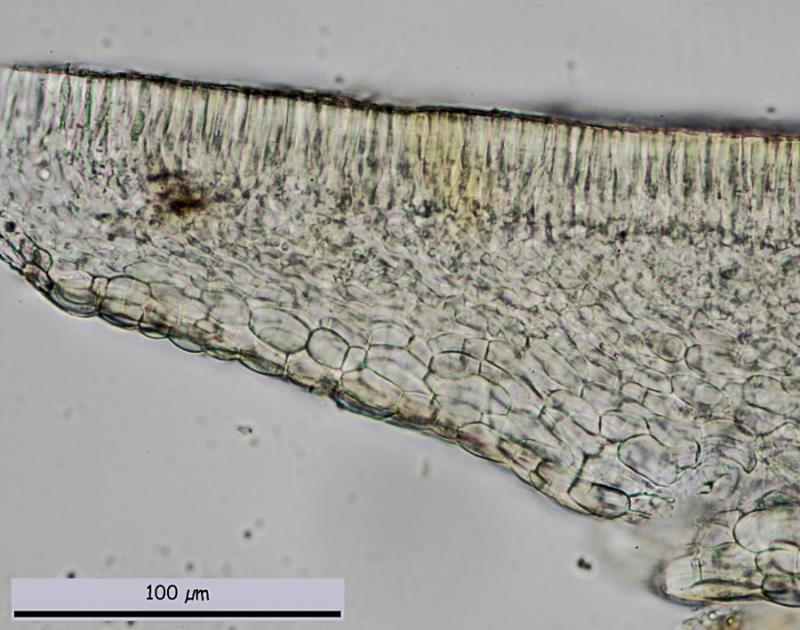
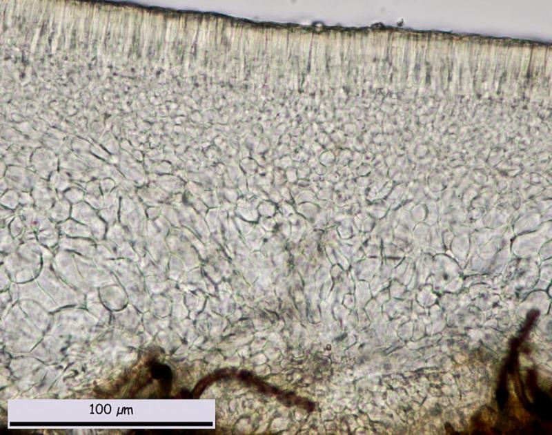
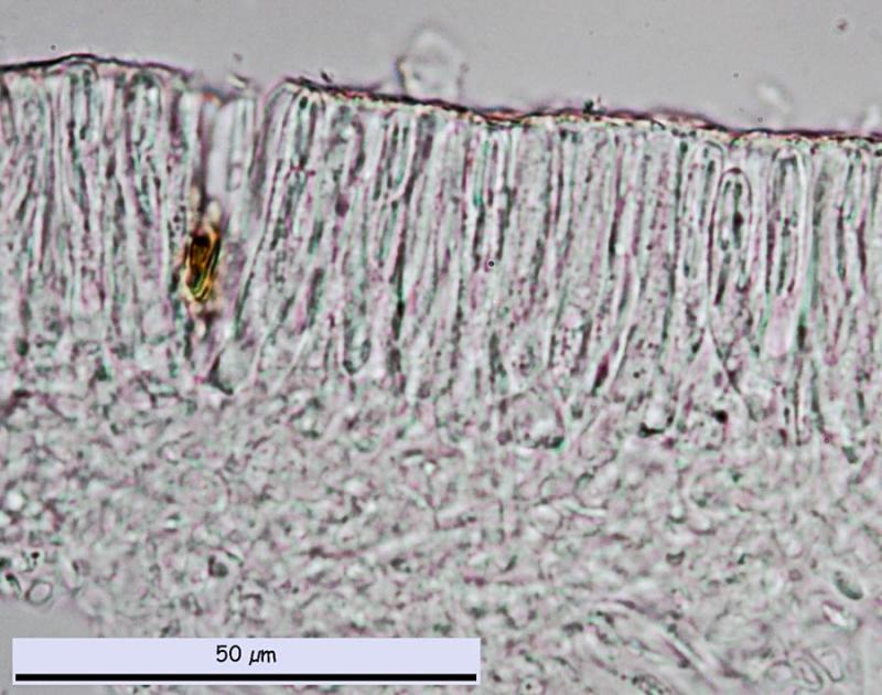
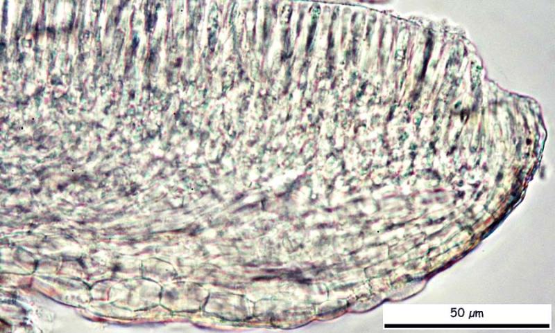
The chlorinaceous colour of the hymenium in overview section is due to the contents of the paraphyses. I think a closeup of such section would show them. Also do you have a closeup of the margin in section? I need to measure the width of the marginal cells.
Surely H. inflatula but that species is perhaps heterogeneous.ô
Is it from Tenerife or Spain? What substrate?
Spore width of 1.6 sounds strange. From your scale I arrive atô 5.5-7.7 x 0.8-1 ôçm
Zotto
Surely H. inflatula but that species is perhaps heterogeneous.ô
Is it from Tenerife or Spain? What substrate?
Spore width of 1.6 sounds strange. From your scale I arrive atô 5.5-7.7 x 0.8-1 ôçm
Zotto
Miguel ûngel Ribes,
07-08-2014 11:53
Re : Hyalorbilia inflatula 030514 176
It is from Pyrenees, Huesca, AûÝisclo. I am goingo to try to know the substrate as soon as possible.
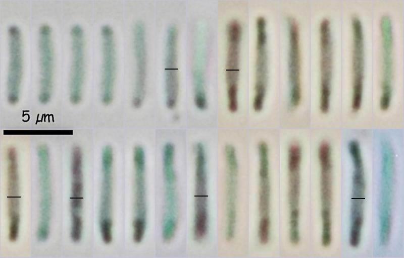
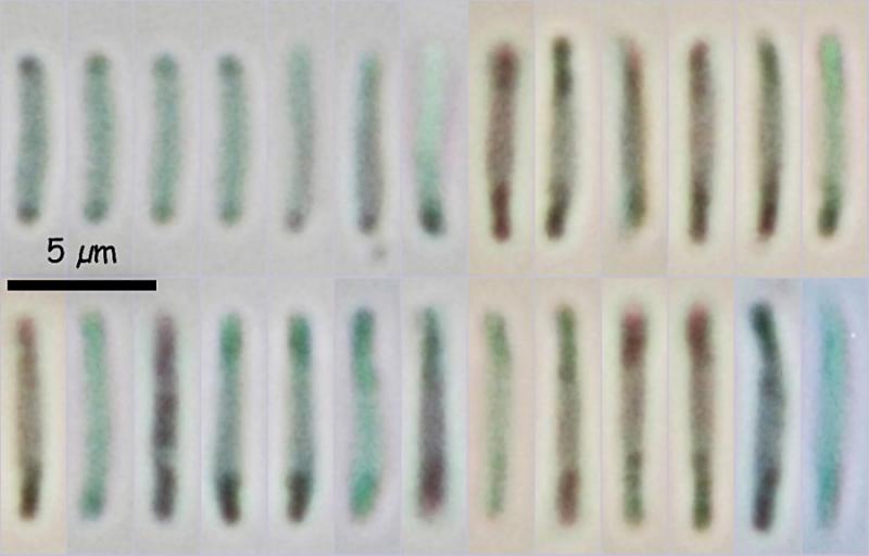
Here are the original measure of Piximetre. There is only one spore with 1.6 ôçm wide, but the statistical formula is so, anyway in my spore picture there are only 26 of the 42 spores measured and Me = 7.1 x 1.1 ôçm.
Attached 3 margin pictures. Thank you.
7.35 1.37
7.24 1.13
6.68 1.11
7.24 1.02
7.76 1.08
6.79 1.17
6.50 1.05
7.38 0.93
7.88 1.16
7.10 0.96
7.20 1.14
5.93 1.14
6.61 1.04
6.35 1.11
5.97 1.17
6.04 1.15
6.50 1.20
6.61 1.11
5.86 1.05
8.74 1.30
7.98 1.23
7.66 1.48
7.78 1.35
7.96 1.62
7.36 1.08
7.27 1.33
7.82 1.37
7.27 1.13
7.31 0.98
7.04 1.05
6.73 0.92
6.82 1.05
7.59 0.95
7.24 1.05
7.10 1.00
7.11 0.96
7.50 0.97
7.07 1.06
6.97 0.91
7.88 0.87
7.05 1.07
7.44 1.12


Hans-Otto Baral,
07-08-2014 11:59
Re : Hyalorbilia inflatula 030514 176
Thanks, this tells for the one with narrow marginal cells.
I do not trust Piximeter. Did you keep the pics where you measured them?
I do not trust Piximeter. Did you keep the pics where you measured them?
Miguel ûngel Ribes,
07-08-2014 13:05
Re : Hyalorbilia inflatula 030514 176
What measure method do you use? Could I ask you why you don't trust in Piximetre?
I could copy all the photos with the scale bar in a dropbox folder and sent it you.
Thank you.
I could copy all the photos with the scale bar in a dropbox folder and sent it you.
Thank you.
Hans-Otto Baral,
07-08-2014 13:27
Re : Hyalorbilia inflatula 030514 176
I simply take a centimeter and measure the scale bar. Then I measure spore with with the same centometer and calculate.
L:w ration of your spores looks like around 6-8, so a width of much over 1 ôçm is hardly possible.
It would be enough if you select a few of the wider spores with the piximeter bar and place it here (cut to have it small). Often the piximeter bar goes over the periphere of the spore.
L:w ration of your spores looks like around 6-8, so a width of much over 1 ôçm is hardly possible.
It would be enough if you select a few of the wider spores with the piximeter bar and place it here (cut to have it small). Often the piximeter bar goes over the periphere of the spore.
Hans-Otto Baral,
07-08-2014 16:54
Re : Hyalorbilia inflatula 030514 176
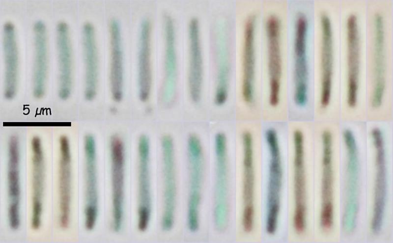
I made here a scale of 1 ôçm by reducing your 5 ôçm scale for 20%.ô
With your original photos with a 100 ôçm scale bar it is much more difficult.
With your original photos with a 100 ôçm scale bar it is much more difficult.
Hans-Otto Baral,
07-08-2014 17:18
Re : Hyalorbilia inflatula 030514 176
Sorry, I thought my text has disappeared

Miguel ûngel Ribes,
07-08-2014 19:51
Re : Hyalorbilia inflatula 030514 176
Hi Zotto
I think Piximetre is not the problem. The problems are mine, like this:
1.- The difficulty of stop the spores in water for the picture, so the result is "moved-spores"
2.- Moved-spores looks like bigger than real spores
3.- Small spores. The error measuring small spores is bigger than measuring big spores
4.- Moved + small spores = less defined outlines spores
5.- Fast measures and with low screen magnification = bigger measures
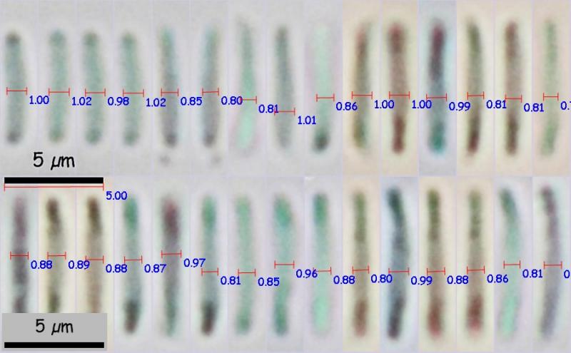
I have make a new measurement with the only 31 spores with better oultine and with great magnification in the screen, for greater precision in the measurements, and the results are significantly lower: (5.8) 6.0 - 7.8 (8.0) x (0.7) 0.8 - 0.96 (1.0) ôçm;ô Q = (6.1) 6.5 - 9.6 (10.5) ; N = 31;ô Me = 6.9 x 0.9 ôçm ; Qe = 8.0.
Attached are the picture with old measures, the picture with new measure and the same new picture manually recalibrated with the 5 ôçm scale bar and wide measures.
My question in the previous message was which program do you use to measurement?
Thanks again and best wishes.
I think Piximetre is not the problem. The problems are mine, like this:
1.- The difficulty of stop the spores in water for the picture, so the result is "moved-spores"
2.- Moved-spores looks like bigger than real spores
3.- Small spores. The error measuring small spores is bigger than measuring big spores
4.- Moved + small spores = less defined outlines spores
5.- Fast measures and with low screen magnification = bigger measures
I have make a new measurement with the only 31 spores with better oultine and with great magnification in the screen, for greater precision in the measurements, and the results are significantly lower: (5.8) 6.0 - 7.8 (8.0) x (0.7) 0.8 - 0.96 (1.0) ôçm;ô Q = (6.1) 6.5 - 9.6 (10.5) ; N = 31;ô Me = 6.9 x 0.9 ôçm ; Qe = 8.0.
Attached are the picture with old measures, the picture with new measure and the same new picture manually recalibrated with the 5 ôçm scale bar and wide measures.
My question in the previous message was which program do you use to measurement?
Thanks again and best wishes.
Hans-Otto Baral,
07-08-2014 21:15
Re : Hyalorbilia inflatula 030514 176
o.k., now I am happy :-)
I use a small slide rule. With the centimeter scale I measure the spores on the screen (though I usually measure directly at the mic), then I calculate the values in ôçm with the slide rule, sometimes also with my brain. Clear?
Movement of spores should not be a reason if you are able to have a shutter speed of 1/30 or shorter. But small-spored Orbilias are indeed not easy....
Could you please send me more detailed collection data? Maybe the substrate is still unclear.
Zotto
Miguel ûngel Ribes,
08-08-2014 00:47
Re : Hyalorbilia inflatula 030514 176
Ok, thank you, very clear. I am happy too.
Perhaps tomorrow I could sent you all collection data.
Best wishes.
Perhaps tomorrow I could sent you all collection data.
Best wishes.