

05-05-2026 22:40
Gernot FriebesHi,I believe this is a Plagiostoma growing on a Sa
06-05-2026 11:25
 Castillo Joseba
Me mandan el material seco de Galicia (España) re
06-05-2026 17:23
Thomas Læssøehttps://svampe.databasen.org/observations/10594257
28-04-2026 20:07
 Lothar Krieglsteiner
... on twig in the air at standing Ceratonia siliq
04-05-2026 18:13
 Stephen Martin Mifsud
ID request for what seems to be a true aquatic fun
04-05-2026 16:39
Stephen Martin Mifsud
ID request: This specimen was collected in Malta o
28-07-2011 18:31
 Alex Akulov
Dear FriendsToday I made the pdf file of Velenovsk
04-05-2026 09:50
Castillo Joseba
Me mandan el material seco de Galicia,(España) re
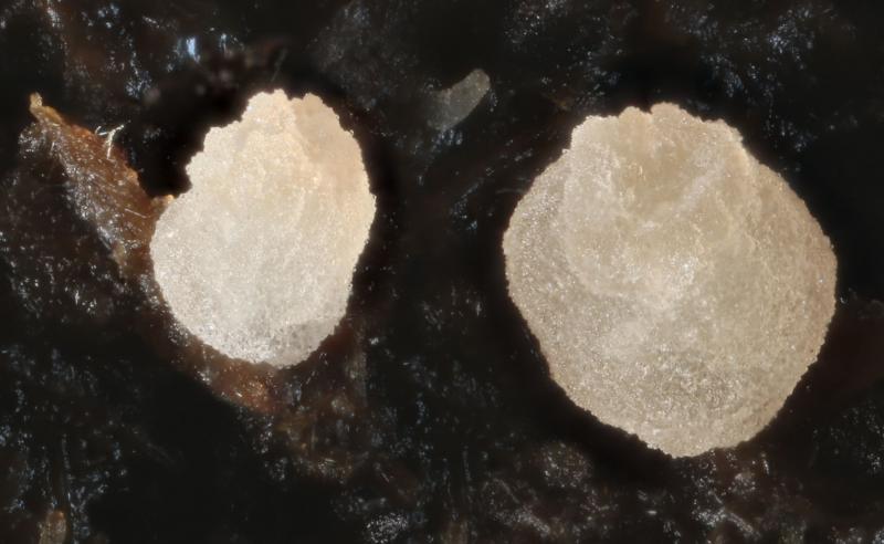
Thecotheus pelletieri - a close look
Chris Yeates,
10-07-2014 21:11
 This species has surprisingly few British records, I cannot believe it is actually rare however.
 Today I had a good chance to have a detailed look at this fungus, and it raised a question or two for me. I don't know how much of this is already in the literature, but it is certainly not mentioned in Doveri, but I dont have access to Aas' 1992 Monograph.
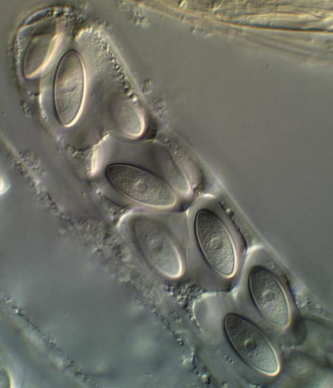
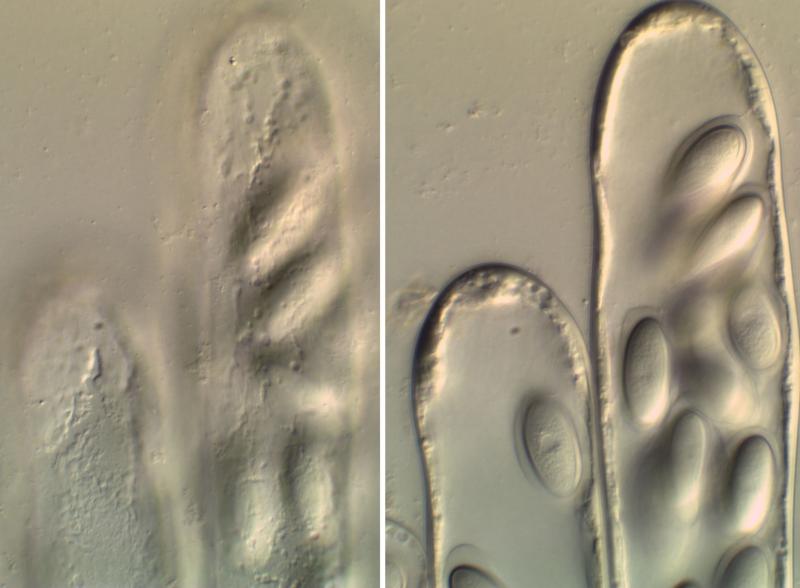
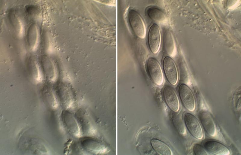
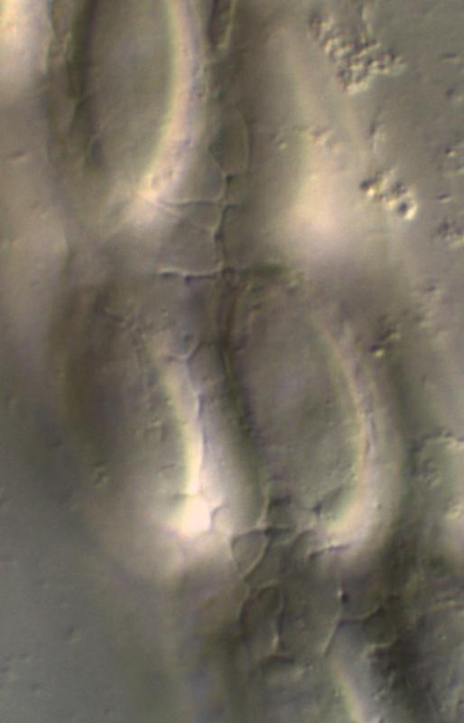
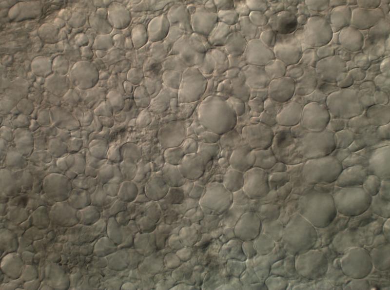
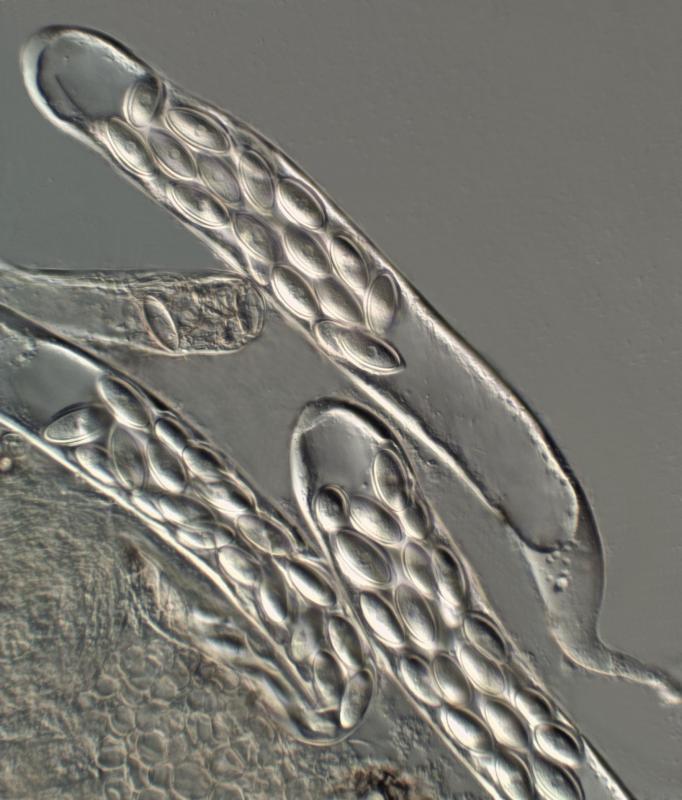
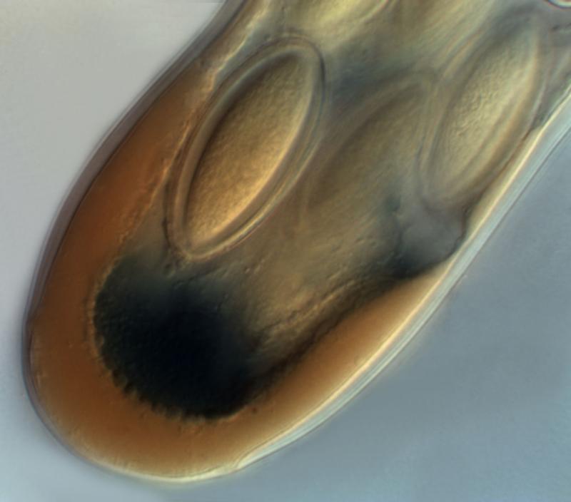
The spores are clearly not uniformly thick-walled, the walls being thickest at the equators and thinnest at the poles. But of more interest to me is the nature of the ascus wall. This seems quite complex (perhaps best seen in the accompanying image showing an ascus apex in Melzer's). Also the outer ascus wall seems to be irregular - with scattered pits and cracks (see the two images showing a combined surface view and optical section, and a close-up of one of these).
Perhaps these are just anomalies of this particular collection - I would be more than happy to hear any comments - operculate disco's are not one of my stronger areas.
Cordialement
Chris
Hans-Otto Baral,
10-07-2014 21:31

Re : Thecotheus pelletieri - a close look
Hi Chris
strange iodine reaction, did you compare in Lugol?
I wonder about the spore wall, isn't it the gel around? Your second pic shows the inflated gel, because the covering membrane lost its semipermeability and the ascus its turgor. On the third pic the ascus turgor is also lost but the membrane around each spore still intact, therefore the gel strongly dehydrated. You should easily test this by applying Melzer or the like to such "thick-walled" spores.
attached free spores with an equally medium thick wall (HB 7936c).
Zotto
Chris Yeates,
10-07-2014 22:09
Re : Thecotheus pelletieri - a close look
Hi Zotto
thanks for the comments - not tried Lugol; the reaction with Melzer is interesting and clearly depends on the stage of development of the asci. I have other photo's in which the whole wall of developing asci has turned blue, in other examples the blueing seems to affect different parts of the ascus (I didn't want to swamp AF with any more images!).
When you say "an equally medium thick wall" do you mean the effect is equal to that in my example or that the wall is of an equal thickiness? (English can be a very ambiguous language at times), because I actually see a similar difference in wall thicjness in your example too - particularly middle-right and bottom left spores.
best wishes
Chris
Hans-Otto Baral,
10-07-2014 22:30
Re : Thecotheus pelletieri - a close look
Yes, my photo shows the entire outer ascus wall light blue.
I wonder about the internal blue at the apex, it is not clearly the wall that is stained, or?
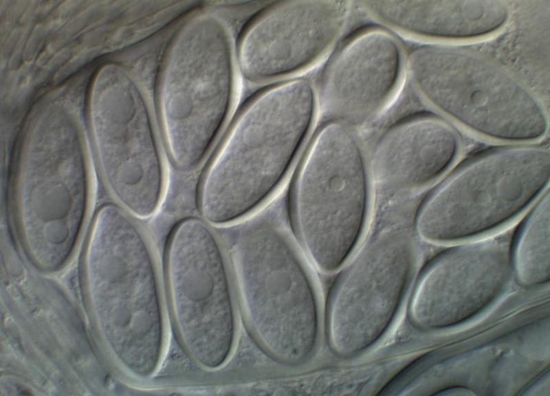
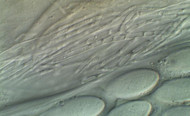
With equally thick I mean tha t in my photo the wall has the same thickness at the sides and the poles. But you are right, the bottom left spore is in focus and distinctly thicker-walled laterally. It seems I was irritated by the DIC-contrast which I am not used to see. The compressed gel is well seen on your photo of living asci (fourth), and it shows little refraction even there.
Zotto
I wonder about the internal blue at the apex, it is not clearly the wall that is stained, or?
With equally thick I mean tha t in my photo the wall has the same thickness at the sides and the poles. But you are right, the bottom left spore is in focus and distinctly thicker-walled laterally. It seems I was irritated by the DIC-contrast which I am not used to see. The compressed gel is well seen on your photo of living asci (fourth), and it shows little refraction even there.
Zotto
René Dougoud,
11-07-2014 08:07
Re : Thecotheus pelletieri - a close look
Chers Collègues,
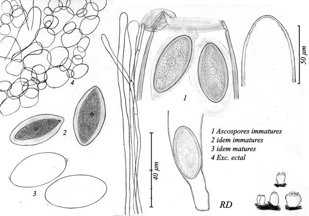
Les éléments microscopiques présentés sont manifestement issus d'exemplaires immatures. Ceci se traduit d'une part par la forme fusoïdes des ascospores et par l'épaisseur de la paroi sporale. En effet, lorsque matures les ascospores deviennent ellipsoïdales et la paroi mince. Ceci vaut (pour la paroi sporal) pour de plusieurs genres et espèces coprophiles. Par ailleurs, le muscilage qui entoure les ascospores disparait également à maturité et peu simuler des apicules (voir planche annexée).
S'agissant de la coloration par les réactifs iodés, celle-ci peut varier selon l'âge.
PS: lire sur la planche " 2 immature dans le melzer" Vous aurez relevé la présence du noyau, y compris pour les ascospores dessiné dans l'eau et encore dans l'asque.
Cordialement
René
Hans-Otto Baral,
11-07-2014 09:17
Re : Thecotheus pelletieri - a close look
Cher René
I suppose from your photo that the spore wall gets slightly thicker in the dead state due to turgor loss of the spore interior. How can you be sure that the spores in2 are more immature than those in 1?
Also I am not sure whether the gel disappeared before spore discharge. I could imagine, and we have often such a case in ascomycetes with gel sheaths, that the gel remains present until the spores are liberated. Then it strongly swells due to the loss of external turgor, and maybe remnants remain at the poles.
Sorry, I mistook your legend. So you consider 1 and 2 as immature. But this is probably a matter of definition. Maybe you consider spores as mature which I would term overmature. Because you did not draw the spore contenst in the "mature" spores in 3, it is impossible for me to estimate their maturity.
Zotto
Chris Yeates,
11-07-2014 13:43
Re : Thecotheus pelletieri - a close look
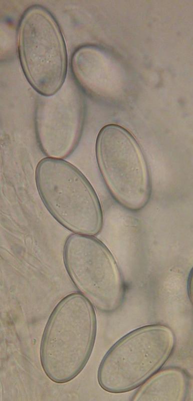
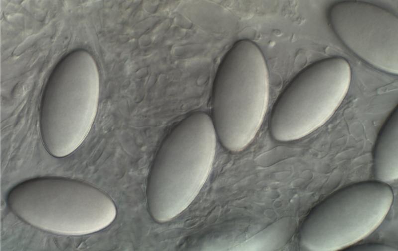
Un grand merci pour la clarification, René - j'apprends tout le temps! Voici ce que je pense sont matures, éjectés, spores (en l'eau). Est-ce correct? Ce sont le type de spores qui j'ai mesuré pour l'enregistrement.
Amitiés
Chris
Amitiés
Chris
René Dougoud,
11-07-2014 14:02
Re : Thecotheus pelletieri - a close look
Oui, en 1 - 2 les ascospores sont immatures, en 2 dans le MLZ, ont y relève la contraction d'éléments. En 3 les ascospores sont matures. ll reste sur une des ascospores matures, un peu de mucus sur les extrémités d'une ascospores. Non, les ascospores matures ne sont pas surmatures ! J'insiste sur le fait que les ascospores immatures ont des parois épaisses et qu'elle deviennent étroite à maturité, c'est ce qu'il faut surtout retenir. Je ne me souviens pas si, lorsque mature, le sporaplsme était visible. je pense que non, faute de quoi je l'aurais probablement aussi dessiné !
A noter que que AAs, dans sa monographie (in matériel et méthode), indique que la paroi de certaines ascospores immatures d'espèces peuvent mesurer jusq'à 4 µm d'épaisseur. Il indique que la paroi d'ascospores immatures ne se colore pas dans le bleu coton, mais qu'un fois mature elle se colore.
Cordialement
René
René Dougoud,
11-07-2014 14:17
Re : Thecotheus pelletieri - a close look
Oui Chris !
Amicalement
René
Amicalement
René
Hans-Otto Baral,
11-07-2014 17:08
Re : Thecotheus pelletieri - a close look
Thanks René! This is interesting that the spore wall stains in CB only at a late stage.
Mabe Chris finds fully mature asci and can observe spore discharge. Then we might be more sure. A swellable refractive spore wall I do not know in other Pezizales, at least I cannot remember.
Zotto
Mabe Chris finds fully mature asci and can observe spore discharge. Then we might be more sure. A swellable refractive spore wall I do not know in other Pezizales, at least I cannot remember.
Zotto