

05-05-2026 22:40
Gernot FriebesHi,I believe this is a Plagiostoma growing on a Sa
06-05-2026 11:25
 Castillo Joseba
Me mandan el material seco de Galicia (EspaÃąa) re
06-05-2026 17:23
Thomas LÃĶssÃļehttps://svampe.databasen.org/observations/10594257
28-04-2026 20:07
 Lothar Krieglsteiner
... on twig in the air at standing Ceratonia siliq
04-05-2026 18:13
 Stephen Martin Mifsud
ID request for what seems to be a true aquatic fun
04-05-2026 16:39
Stephen Martin Mifsud
ID request: This specimen was collected in Malta o
28-07-2011 18:31
 Alex Akulov
Dear FriendsToday I made the pdf file of Velenovsk
04-05-2026 09:50
Castillo Joseba
Me mandan el material seco de Galicia,(EspaÃąa) re
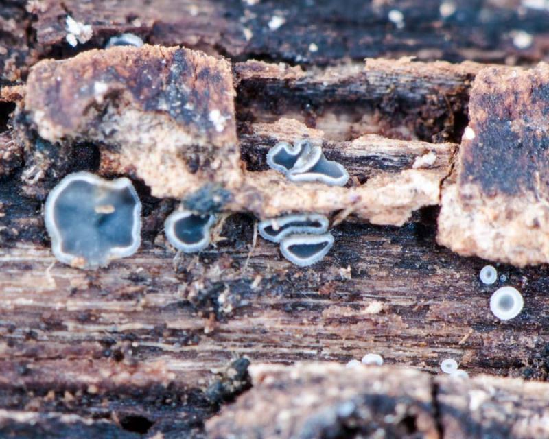
 Good night
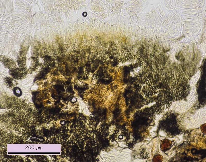
I have a new small Mollisia, not in conifer wood, perhaps Corylus or Fagus at 1.400 m. First macro picture are very young apos, second are more mature and the third one, very mature.
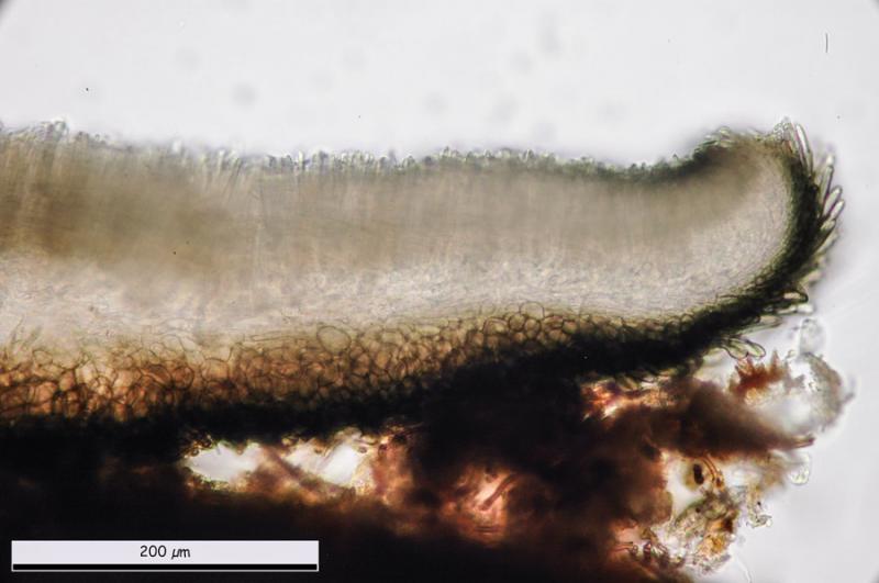
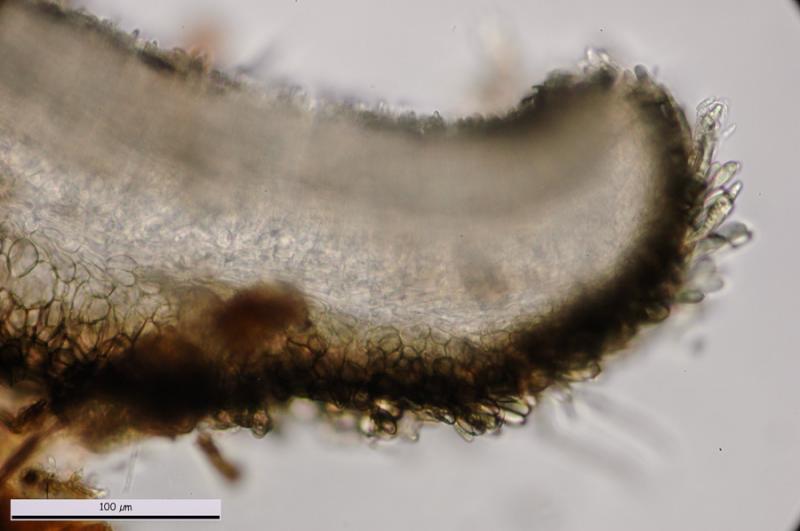
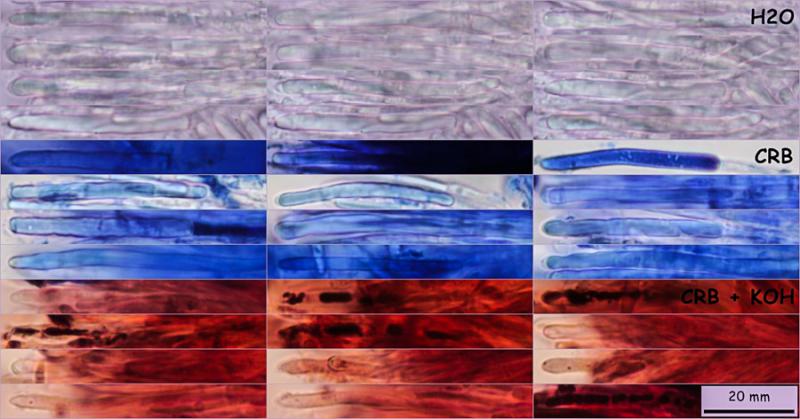
Ectal excipulum brown with subglobosum to angular cells, (11.5) 14.0 - 21.9 (25.7) Âĩm; Me = 17.9 Âĩm, more cilindrycal at margin and inside. Slightly yellow to green with KOH. Medullar excipulum subcylindrical to intricata?.
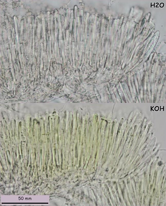
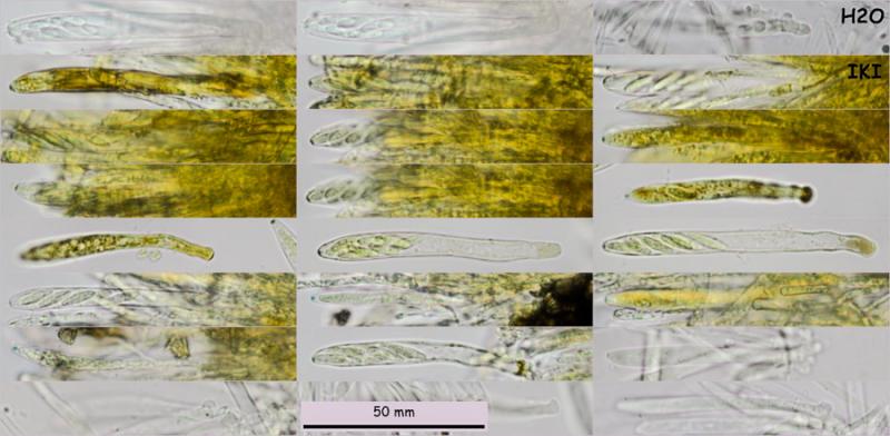
Marginal hairs brown, slightly claviform, more width at apo base and less at margin, (47.4) 49.3 - 65.5 (68.4) x (5.1) 5.6 - 7.5 (10.6) Âĩm; N = 28; Me = 57.2 x 6.7 Âĩm.
Asci bb with croziers, (61.1) 68.6 - 78.1 (82.9) x (5.6) 6.4 - 7.7 (7.9) Âĩm; N = 17; Me = 73.3 x 6.9Âĩm.
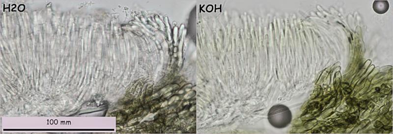
Paraphysis cylindrical with only a big VB, slightly yellow in KOH.
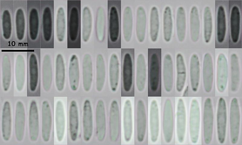
Spores with 0 oil LBs, only one septum in 1-2 spores observed, (9.1) 10.2 - 12.6 (14.2) x (2.4) 2.5 - 3.1 (3.5)Âĩm; Q = (3.2) 3.5 - 4.6 (5.0) ; N = 100; Me = 11.4 x 2.8 Âĩm; Qe = 4.0
Is it possible M. perelegans?
Thank you.


could be, and the older name for it would be M. olivascens.
Did you observe the yellow reaction at the moment when KOH comes in contact? Because it should be strongly yellow.
How large the apothecia?
The alternative M. elegantior has wider and also longer spores and IKI red apical rings, also it is KOH-
Zotto

To add to what Zotto said - I also think your collection could be, but I suspect it is more likely to be something else at the moment based on your images.
If it is Mollisia olivascens, the apothecia should be exceptionally large for Mollisia species (up to 4-5 mm or larger); the KOH reaction should be distinctly yellow (visible by naked eye in my collection), and the marginal hairs should be ~3.5-5 um wide (thickening at the base) and composed on multiple (~3-7) brown-pigmented cells. Â I think the ascospores may also have a tendency to be fairly variable in length, and perhaps also to be more pointed at the poles.
I haven't read Nannfeldt's protologue of Haglundia perelegans so I can't really comment on this synonymy, but I'd be very surprised if these species are not the same. Zotto's illustration of a paratype (HB7331) and Le Gal and Mangenot's recollection (HB6034) do seem to be the same species.
For comparison, I've attached are some images of my collection of Mollisia cf. olivascens (which I think also matches well with Zotto's illustrated collections on his DVD), and also Le Gal and Mangenot's illustrations of their recollection.
I hope that's of some use!
Cheers,
Brian
 Mollisia-olivascens-Felt.-Le-Gal-amp-Mang-0001.pdf
It is an old paper that I started to do on this species and Haglundia elegantior, and it is blocked as so many others by the monstrous and eternal monograph.Â
Zotto
Ah yes, I suspected you had something in the works for Mollisia olivascens! Nice - a Luxemberg epitype and several other collections is much better than my single Welsh specimen as an epitype!
Did you ever get an ITS sequence for any of your collections? Mine came out with a nice identical match to a named anamorph (see pages 190, 196, 202, 291, and 304 of my thesis). I believe Andreas Gminder also has a sequence from his phylogeny, but I've not seen the raw sequence yet. Â It would be nice to pin this species down properly both with morphological and molecular data!
Cheers,
Brian
No, I never worked with DNA or cultures in Mollisia. The only exception was this large undescribed species on Carex etc. ("sparganii") which is not a Mollisia but a Dennisiodiscus as I believe, with a nice anamorph with phialides like a brush. Maybe  Andreas has sequence.
Zotto
This collection has no more than 1-1,5 mm diameter, the yellow reaction of VBs is only a little more dark-strong than pictures, but no more, hairs have no more than 3-4 cells and are wider at apex [(5.1) 5.6 - 7.5 (10.6)]Â than base [(3.5) 3.9 - 5.7 (5.9)], ectal excipulum is subglobose-angularis, not prismatical like in Le Gal paper of M. olivascens, and spores are not so pointed. So I wonder mine is not M. olivascens.
Is not posible another similar species?
Best wishes.
Do you think could I ask directly to Aebi? Any mail?
Best wishes.
I do not have it as pdf:
Aebi, B. 1972. Untersuchungen uber Discomyceten aus der Gruppe. Tapesia - Trichobelonium. Nova Hedwigia 23: 49-112
Does anybody have?
Zotto
Aebi, B. 1972. Untersuchungen uber Discomyceten aus der Gruppe. Tapesia - Trichobelonium. Nova Hedwigia 23: 49-112
Thank you.
Her thesis abstract: http://e-collection.library.ethz.ch/eserv/eth:32343/eth-32343-01.pdf
MycoBank (ignore entries 6-12, they're wrong):Â http://www.mycobank.org/BioloMICS.aspx?Link=T&TableKey=14682616000000061&Rec=3362&Fields=All
You could compare your collection to the description of Mollisia villosa from Andreas Gminder's key, and also the similar species (but with broader ascospores) collected and examined by Ingo Wagner:Â http://asco-sonneberg.de/pages/gallery/mollisia-villosa-cf-130323-mcol-01jj35211.php?group_id=35131&position=82 . Â
I think it does look like a plausible match (or at least one to consider), although this is no guarantee that it is the same species. Â But having the original description would help a lot if anyone has it.
Cheers,
Brian


Hello,
Â
I have two sequences of Mollisia olivascens, but only ITS4 and one of Mollisia elegantior.
>Mollisia-elegantior_Jena_13_7_ITS4
CTTTAGAGTTGGGGGGTTCTGGCGAGCCACCGGGAGAACCCTGAAGCGAGAAGTTTTACTACGCTTAGAGCCAACCGGCACCGCCACTGGATTTGAGGGCCGCGGAACCGCGTGCCCCAACACCACGCCAGGCGTGAGTGGTCATAATGACGCTCGAACAGGCATGCCCCGSGGAATACCACGGGGCGCAATGTGCGTTCAAAGATTCGATGATTCACTGAATTCTGCAATTCACATTACTTATCGCATTTCGCTGCGTTCTTCATCGATGCCAGAACCAAGAGATCCGTTGTTGAAAGTTTTAACTATTATATAGTACTCAGACATCACTAATATTCAGAGTTTGGTCCTCTGGCGGGTACGTACGAGCAGAGCCCGCAGTGGAGGACCACAGCCCGCCAAAGCAACAAGAGTATGTAGACACGGGTGGGATACTCGGGAGAGCGCCCTTTCGGACGGTGCACCAAGAATCTTGTAATGATCCTTCCGCA
>Mollisia-olivascens_Jena_13_28_ITS4
TATGGCARGCGACCGAGGGGAACTCTATAGCGAGGAGATTTACTACSCTTARAGCCCACCGGCACCGCCWTTGATTTTAGGGGCCGCGAGACCGCRAACTCCAATACCAAGCTATGCTTGAGTGGCTATTATGACGCTCGAACAGGCATGCCCGGCGRAATACCACCAGGCGCAATGTGCGTTCAAAGATTCRATGATTCACTGAATTCTGCAATTCACATTACTTATCGCATTTCSCTGCGTTCTTCATCGATGCCARAACCAAGARATCCGTTGTTGAAAGTTTTAACTATTATATAKTACTCGGACATCACTAATATTCARAGTTTGGTCCTCTGGCAGGCACACSCGRATARAGTCCACGGKGGARACCACRCCCTGCCAAASCAACAARAGTARATAAACMCGGGKGGGGYCYMCCGGTTYCCCRGWAYCCTTTTTTAATGATCTTTCCGCA
>Mollisia_olivascens_Mo081
GGGTATCCCTACCTGATCCGAGGTCAACCTGTTAAAAAAAAATGGGGGGTTATGGCAAGCGACCGAGGGGAACTCTATAGCGAGGAGATTTACTACGCTTAGAGCCCACCGGCACCGCCATTGATTTTAGGGGCCGCGAGACCGCGAACTCCAATACCAAGCTATGCTTGAGTGGCTATTATGACGCTCGAACAGGCATGCCCGGCGGAATACCACCAGGCGCAATGTGCGTTCAAAGATTCGATGATTCACTGAATTCTGCAATTCACATTACTTATCGCATTTCGCTGCGTTCTTCATCGATGCCAGAACCAAGAGATCCGTTGTTGAAAGTTTTAACTATTATATAGTACTCGGACATCACTAATATTCAGAGTTTGGTCCTCTGGCAGGCACACGCGGATAGAGTCCACGGTGGAGACCACGACCTGCCAAAGCAACAAGAGTAGATAAACACGGGTGGGGTCTACCGGTTTCCCAGTATCCTTTTTTAATGAT
But I don't know how good they are. I have not succeeded in getting a useful tree when I tried to include the (appr. 40) new sequences to the hitherto sequenced ones.
@Brian: But the Mollisia atlantica you sent me the sequence is the same as the future type collection as well as several other collections - including collections from grasses!
Â
best regards,
Andreas
Hello,
Â
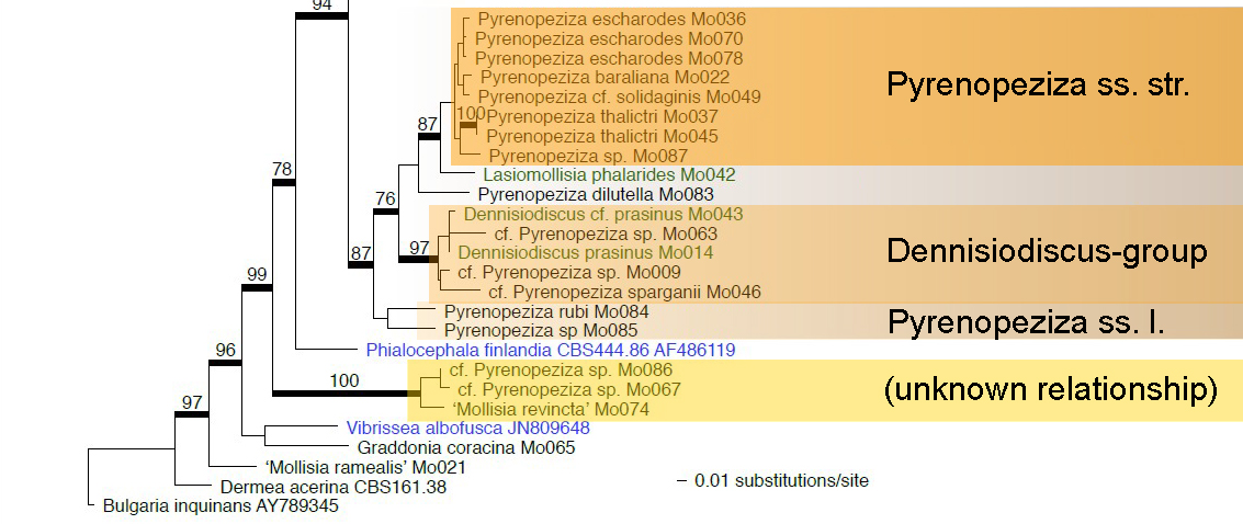
I have a sequence of this species and it is very close to what Zotto calls "Mollisia" "Gottmadingen" and to Dennisiodiscus. These species are one group within the Pyrenopeziza clade. Wether they can be kept separate on gerneric level or not will show up when more Pyrenopeziza species are included.
>cf_Pyrenopeziza_sparganii_Mo046
GGGTATCCCTACCTGATCCGAGGTCAACCTGTAAAAATAGGGGGTTGCTGGCAAGCAACCTACCGGACCCAATCGCGAGGAGTATTACTACGCGTAGAGCCGACAGGCACCGCCACTGATTTTAAGGGCCGCGGAACCGCGATCCCCAAGACCACGCGAGAGCGTGAGTGGTTATAATGACGCTCGAACAGGCATGCCCTACGGAATACCGAAGGGCGCAATGTGCGTTCAAAGATTCGATGATTCACTGAATTCTGCAATTCACATTACTTATCGCATTTCGCTGCGTTCTTCATCGATGCCAGAACCAAGAGATCCGTTGTTGAAAGTTTTAACTATTATATAGTACTCAGACTTCACTGAAAACAAGAGTTGGGGTCCTCTGGCGGGCGCGCAGCAGCCGGAGCCGCTGGCCTTGCGGCGGCCCGCCAAAGCAACAAAGGTAGTTTATTCAAGGGTGACTAGTGAGCTTACATCGTGGACCGGTTGCCCGACCACGAACGACTAGTTGTGTAATGAT
By the way, the Dennisiodiscus collections in the tree are as well from the Asco-seminaire in Melle (Marais de Potevin) as the "Mollisia" "sparganii". We had a collection of Dennisiodiscus prasinus there without the characteristic hairs on the excipulum - this is the collection "Dennisiodiscus cf. prasinus". It is the same, so D. prasinus can grow without hairs too and is characteristic nevertheless by this unique greenish colour of the hymenium.
I join the tree dated in 2012 here of this group.
Pyrenopeziza spec. Mo009 is "Mollisia" "Gottmadingen".
Pyrenopeziza spec. Mo063 is a very strange collection found on burnt branches of Ulex, looking macroscopically very much like any Mollisia, but having no vacuoles in the paraphyses (leg. Hiraud & Capoun).
Â
best regards,
Andreas
 Pyrenopeziza s.l. - Sequences
Hello,
I think that Tapesia villosa is a pretty good idea, I have little doubt about it.
I examined the type recently and I was also allowed to take some material for sequencing, but unfortunately the sequence did not work. I also have several collections from the alpin mountain and in three days I will be there again and collect hopefully also villosa then. All sequences hitherto except one didn't work, I don't know why.
A feature that I found in all my collections is, that the yellow KOH reaction is restricted to only some patches of paraphyses in the hymenium and that the yellow colour sticks inside the paraphyses and doesn't dilute into the medium. First I thought that this is due to old herbarium material, but it is also in fresh material. So the KOH-reaction is somewhat dubious in T. villosa, what would fit to the picture you show us.
I'm preparing a small publication on that species, which has not yet been combined into Mollisia yet. I wanted to wait on the molecular data of the type, but as the sequence didn't work, I think I will finish the paper without it.
best regards,
Andreas
Thank you Nicolas! Much appreciated!
@Miguel
Based on Aebi's description and illustrations, I also think Mollisia villosa looks like a good candidate identity in the absence of anything better. The ascospores are a little bit broader in your specimen, but I imagine Aebi's collection was examined when dry.
@Andreas
Your Mollisia olivascens ITS sequences are a 100% match to mine, except for sequencing errors in Mollisia olivascens Jena_13_28. So Mollisia olivascens does look like a good distinct morphological and phylogenetic species concept.
Your sequences are full-length ITS1-5.8s-ITS2 sequences, so they should align easily. Maybe you're having difficulty aligning them because they're backwards and reverse-complemented due to being sequenced using the ITS4 primer? You can easily tell when you blast-search the sequence since your query sequence has nucleotides counting up (e.g. 1-500), while your match is counting down (e.g. 500-1). You can reverse complement the sequence in MEGA (http://www.megasoftware.net/) or here: http://www.bioinformatics.org/sms/rev_comp.html. I find reverse sequencing using the ITS4 primer (or similar primers) usually works better due to introns just before the ITS1 region, but there are more reliable primers to use than ITS1F and ITS4 (I can send you the sequences and PCR protocol I used if you need them). Apologies if you already know all this!
I would be surprised if Mollisia atlantica also grows on grasses, but can easily believe that very close sibling species do - there are many clades in Mollisia where species seem to have host-jumped between grasses and lignicolous substrates/tree hosts. Were the sequences and morphology exactly the same?
The ITS2 region of species close to Mollisia atlantica are very strange and have high rates of mutation, so creating a reliable tree is quite tricky. Â There is a chance that species are more closely or distantly related than you would expect from the sequences. Did you receive the link to my thesis that I sent you? I can send you the relevant sections again if you prefer.
cf_Pyrenopeziza_sparganii_Mo046 fits in well with my current phylogenetic delineation of Pyrenopeziza, although it looks like it's in one of the basal clades. If you send me your other Pyrenopeziza sequences I'll be very happy to put them into my big phylogeny of Pyrenopeziza and related taxa and see where they come out.
Pyrenopeziza spec. Mo063 looks like it could be somewhere near Mollisia dextrinospora (actually a Pyrenopeziza) - I suspect "Mollisia" aquosa, Pyrenopeziza "ulicis", and maybe Pyrenopeziza laricina could also fall in this area. Did your species look something like these?
"Phialocephala finlandica" (actually currently in Cadophora, although it shouldn't be) is the phialidic anamorph of a member of Hyaloscypha - Pyrenopeziza and Hyaloscypha seem to be very closely related sibling genera. I would expect your Pyrenopeziza collections close to Mollisia revincta to actually be Mollisia species, but would need the sequences to properly look at that.
Cheers,
Brian
due to 4 days absence I could read your interesting conversation only now. I also reversed your sequences, Andreas  :-)  Better to work with them.
I have one question to Miguel-Angel: Where and at what altitude did you collect this? I ask because M. villosa (a good idea!) was described on Alnus viridis. The host genus Alnus is rather easily distinguishable by wood anatomy from Corylus (not to speak of Fagus).
I further wonder what about my collection H.B. 8251 on A. viridis at 1500 m, with spores *14-15.5 x 2.8-3 Âĩm and lacking KOH-reaction. Nicolas had something similar (IX.2009), with spores 14-20 x (2.2) 2.5-3 (4) Âĩm, VBs also KOH-.
Zotto
I must read all your mails slowly, are difficult to me, but answering Zotto, this collection is from Ordesa - San NicolÃĄs de Bujaruelo, 30TYN369309, at 1.400 m. I do not remember any Alnus there, but I am going to ask other persons who were there with me.
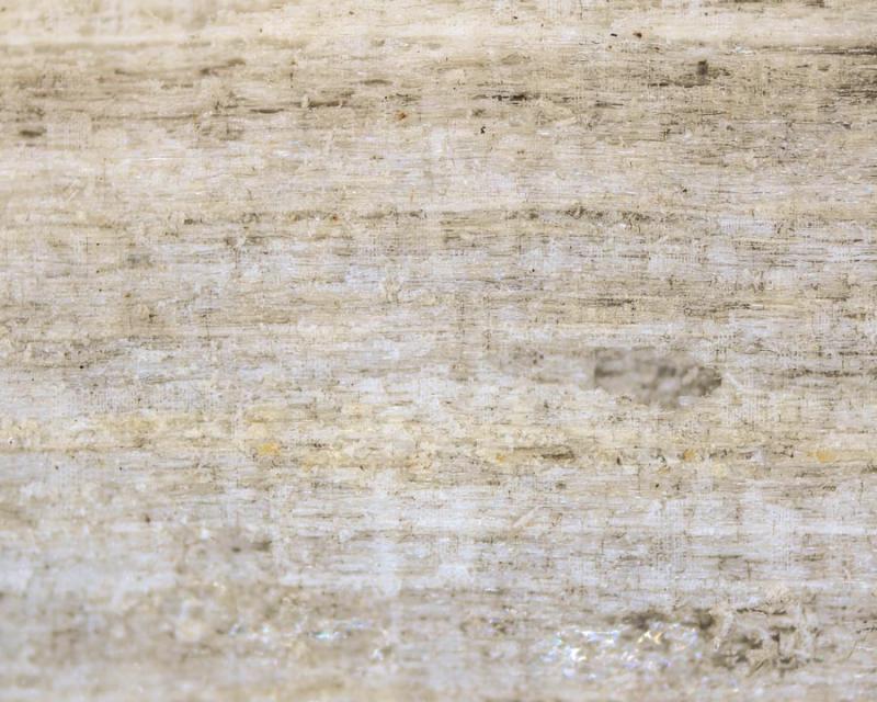
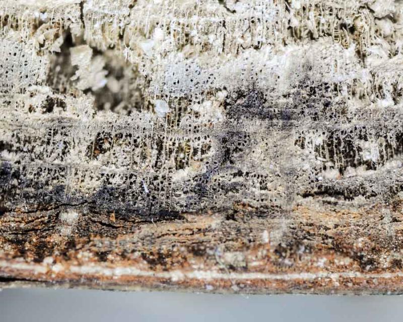
Perhaps latter I could make a wood transversal cut picture.
Thank you.
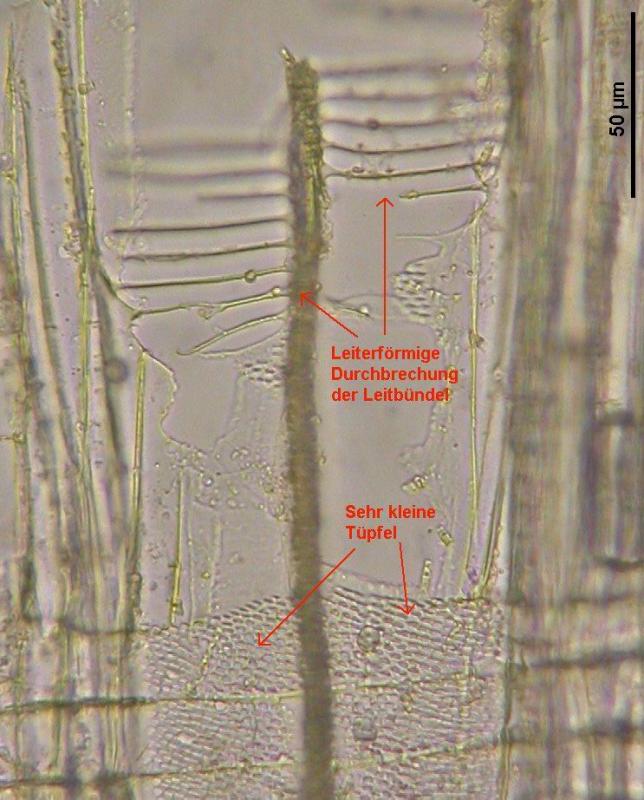
To identify Alnus you need to make a radial section for the ladder-like perforations of the pores. Fagus you can recognize by a hand lens (broad radial rays).Â
Zotto
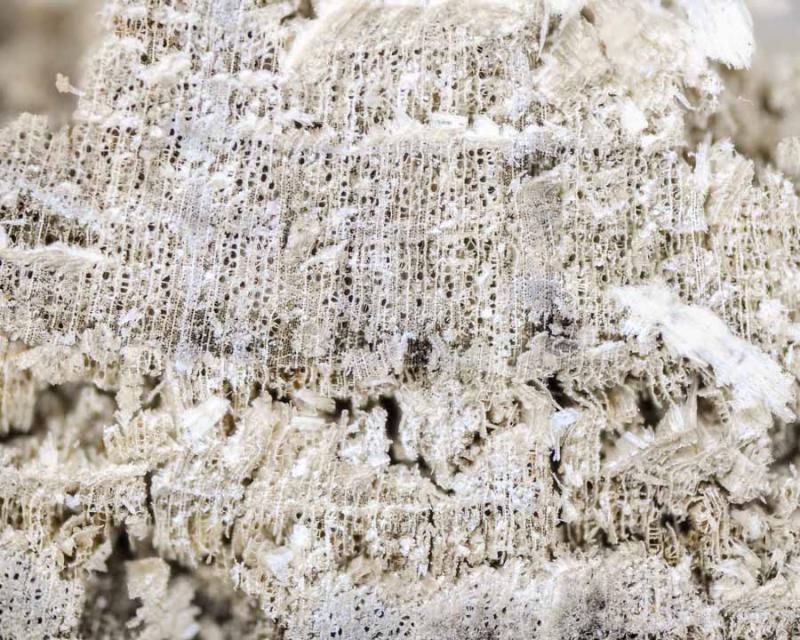
1.- Exterior
2.- Radial section
3.- Transversal section
4.- Transversal section
I think this is not Alnus, isn't it?
Thank you.

I suspect Alnus or Corylus. If you make a radial section and look at 100-400x at the pores it will be quite easy.
Example in attach
I have made the pictures at micro with 4x objective. Tomorrow I am flying to Tenerife until next Sunday, so next week I will try it 10x and 40x
Thank you.
Zotto
I have been reading slow all your coments and papers and I do not find another posibility than Tapesia villosa, as you said: spores, clavate hairs, slight yellow reaction inside paraphysis ...
Otherwise, I have been taken pictures of the wood in radial section, as Zotto said, at 100x and 200x, in order to determine wether it is Alnus or not, but I can't decide it, sorry. Here are the pictures.
Thank you again.


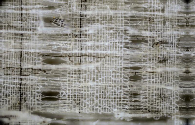
I think I see the ladder-like perforations on your two last photos, especially onm the last in the lower right region.
You have no possibility for a closeup to these?
Zotto
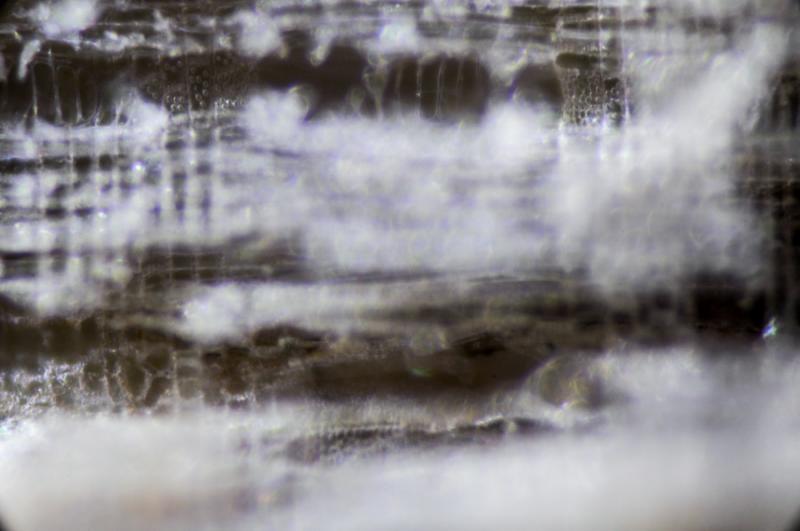
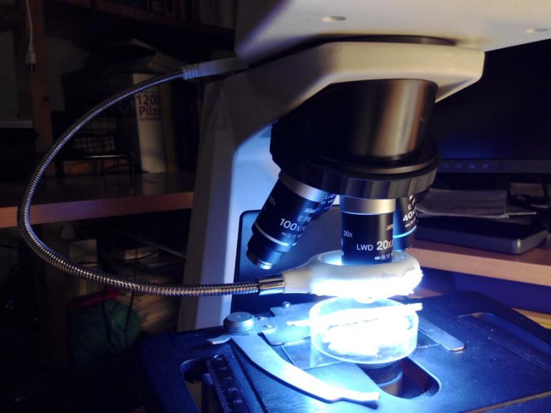
No, my pictures are not made with dark field, are made with 20x and 40x normal objectives and with a ring of leds (20x) and small individual 1 led (40x).
First photo is with 20x, second and thrid with 40x.
Isn't it?
Thank you.


Yes, this must be Alnus (oer Betula), no other native tree with such many spokes in the ladder.
Seems I am overlooking ascofrance mails,....
Here you have two links:
http://www.dx.com/es/p/usb-powered-flexible-neck-18-led-super-bright-white-light-lamp-50080#.U-KWAfl_vEc
http://www.dx.com/es/p/usb-powered-flexible-neck-1-led-white-light-lamp-black-166728#.U-KWQPl_vEc
Thank you again for your help.
Best wishes.


And did you photograph a thin section or simply a thick piece of wood by this reflected light method?
In order to fix the sample propertly for a stack (usually a wood, branch, ...) and to make a correct management for a good frame I use, bottom to top:
* A porta. This allows you to move the sample with the microscope controls as usual.
* A small Petri dish. Sometimes I fill the dish with water to avoid desiccation.
* A piece of plasticine (plastilina in spanish) inside the Petri dish to fix the branch.
* The sample fixed to the plasticine at the desired position.
Good photos.