
28-04-2026 22:51
 Bernard CLESSE
Bonsoir à toutes et tous,Pourriez-vous m'aider à

28-04-2026 21:50
 Pablo Sandoval
Hola a todos,Espero se encuentren bien. Hace mucho
27-04-2026 18:05
 Lothar Krieglsteiner
... still attached at standing tree. The green con
28-04-2026 20:33
Vitus SchäfftleinHello, I found Trochila ilicina on Ilex aquifoliu
28-04-2026 20:07
Lothar Krieglsteiner
... on twig in the air at standing Ceratonia siliq
27-04-2026 20:52
Lothar Krieglsteiner
Found on hanging tiwg of Olea europaea in dried-ou
27-04-2026 18:48
Tony MoverleyCollected 23rd April 2026, Norfolk, EnglandSwarms
27-04-2026 17:41
Lothar Krieglsteiner
.. Algarve, same leaf than the last post. The con
27-04-2026 17:16
Lothar Krieglsteiner
.. Algarve, moist lying.The conidiomata look like
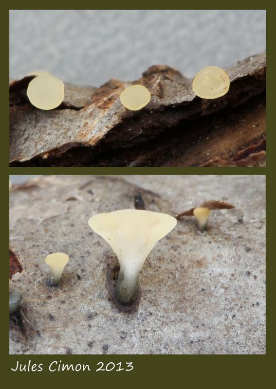
Bonjour !
Voici un disco stipité et inconnu de nous.
Merci de nous aider !
Amitiés ! Roland
Données :
Date de récolte : 20 / 06 / 2013
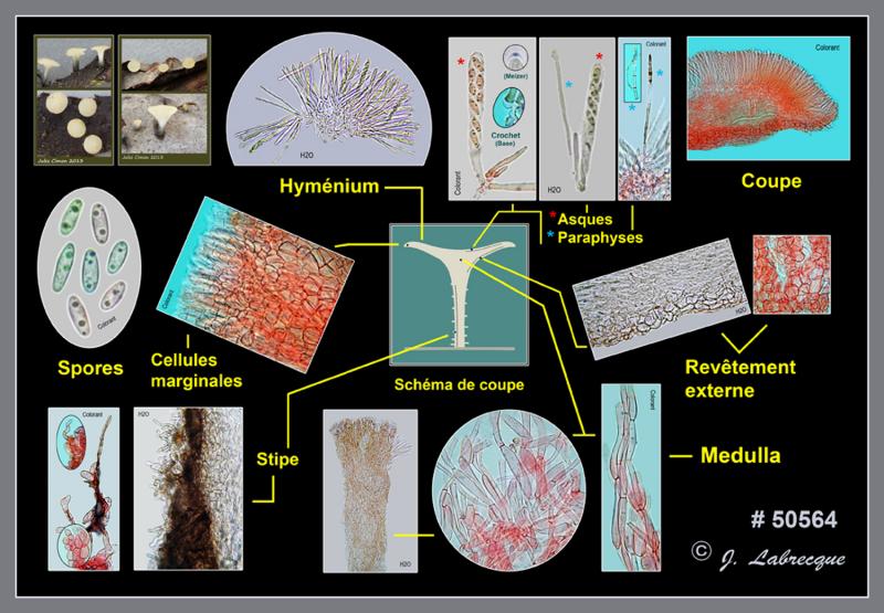
Substrat : débris ligneux à demi submergés (feuillu?) d'une forêt mixte Ascome 7 mm de hauteur totale Apothécie 1 mm de hauteur, 2 mm de diam.
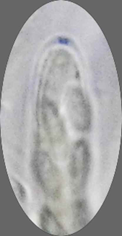
Asques à 8 spores partiellement bisériées, avec crochetés à la base et appareil apical amyloïde, jusqu'à 76 x 9 µm
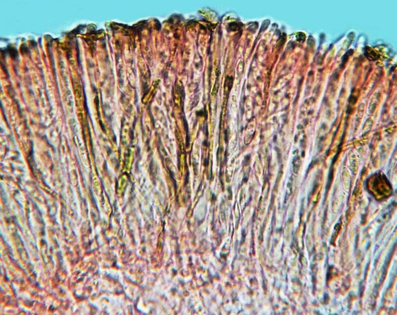
Paraphyses cylindriques, septées, parfois à une petite ramification cylindrique au tiers inférieur, légèrement élargies et parfois couvertes d'une substance hyaline (gélifiée?) à l'apex, pigmentées de brun ochracé olivâtre à ocre doré, à contenu jaunâtre > 80%, jusqu'à 75 x 3,5 µm, dépassant les asques de 3-6 µm
Réaction négative dans le KOH
Spores ellipsoïdes, lisses, non septées, avec 2 petites ou moyennes guttules, hyalines à jaunâtres, 7-12 x 2,5-3,3 µm, 8,4 x 2,9 µm en moyenne, Q = 2,89
Medulla en textura epidermoidea / intricata, ± ascendant, à cellules hyphoïdes polymorphes, hyalines, de taille variable, 25-90 x 5-30 µm
Excipulum ectal en textura globulosa / globulosa-angularis, à cellules à paroi légèrement épaissie, 15-34 x 13-32 µm
Revêtement externe formé de cellules globuleuses à ellipsoïdes en chaîne (?), à pigment brun à olivâtre, 16-18 x 12-15 um
Cellules marginales cylindriques à l'apex et à segments progressivement plus gros vers la base
Revêtement caulinaire brun olivâtre à brun foncé, avec cellules terminales sinueuses, cylindriques à clavées

important woud be here to see living paraphyses with their contents. Moellerodiscus I want to exclude. The apical ring looks Calycina-like, but I am not sure, the resolution is too low. Min. 200% would be necessary for this plate to better work with it. Could you perhaps send me by mail?
Zotto
Yes, I will, Zotto !
Roland
On the photo of paraphyses I can see that they contained VBs, though this is far away from the aspect of the living state.
Now, this species fits, in my opinion Hymenosc. vernus, with one exception: the asci clearly arise from croziers while H. vernus is consistently without. Typical for H. vernus is the blackish base of the stipe. H. vernus is frequent on Alnus but may occur also on Fagus and Salix. Did you ever record H. vernus in your area?
There is also a Hym. foliicola Abdullah et al. from England which grows on twigs (with a characteristic anamorph recorded on leaves), which is similar to H. vernus, but the ascus base was not studied by the authors.
Zotto
Zotto, yes we have record H. vernus., but without croziers.
see : http://www.flickr.com/photos/19369983@N06/4425309001/in/photolist-7K3Sc4-koZLwm
Here, we have croziers.
It may be not H. vernus.
I will seek on H. foliicola.
Thank you ! Roland
On this plate you can see, as an example, an artifact of dead cells: the below pic (base du pied) shows strongly wrinkled hyphae, but they are smooth when alive.
Zotto