
19-04-2026 21:23
Steve ClementsBonjour, I found this anamorphic fungus on old pl
19-04-2026 20:46
Steve Clements1 mm diameter approx spherical conidiophores on pl
12-04-2026 17:56
 Hardware Tony
Found on dead stems in February earlier this year
17-04-2026 19:16
 Enrique Rubio
Hi to everybodyI would appreciate any assistance r
14-04-2026 05:32
Ethan CrensonHi all, A few weeks back a friend pointed out som
17-04-2026 15:14
 Bruno Coué
Bonjour.Récoltes du 16/04/2026, sur feuilles mort
12-04-2026 15:52
Gernot FriebesHi,I'm looking for help with this anamorph collect
14-04-2026 21:52
Gernot FriebesHi,found on dead leaves of Carex elata. Conidia: 4
16-04-2026 22:09
Buckwheat PeteHello, I'd like to ask about this older specimen:
15-04-2026 19:33
Fátima Durán ManzanequeHi!! I need help, I found this Ascomycete but I d
 Hi again,
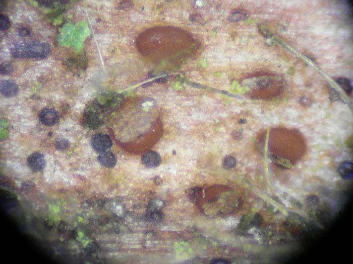
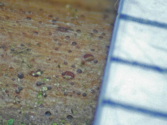
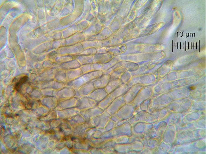
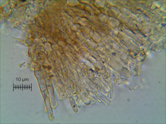
on that rubus-twig I found a second fungus of the Naevioideae. They are very difficult, I think.
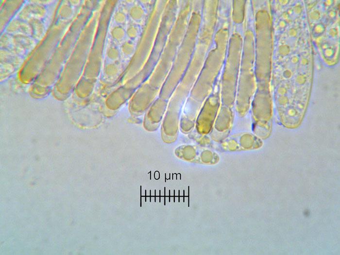
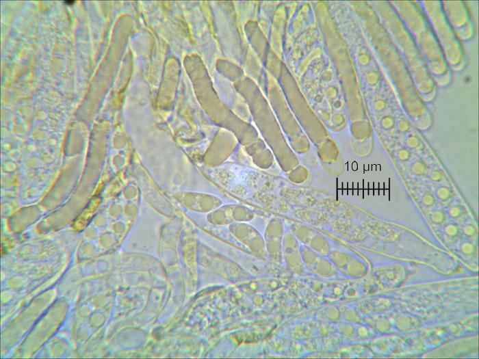
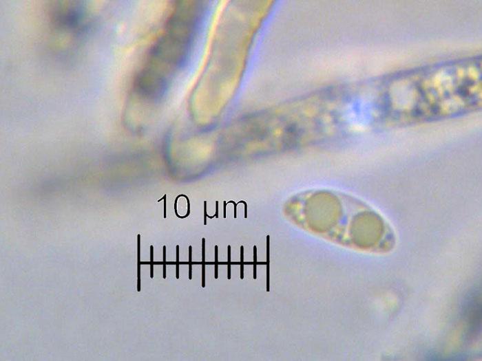
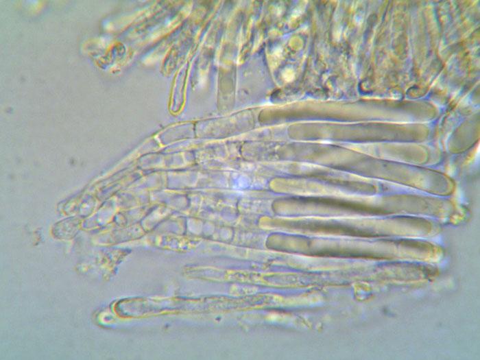
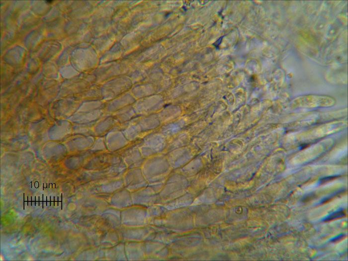
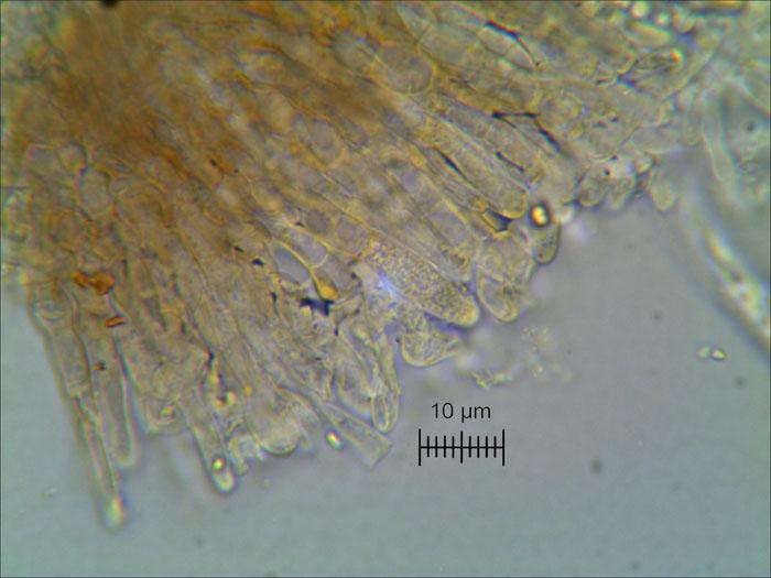
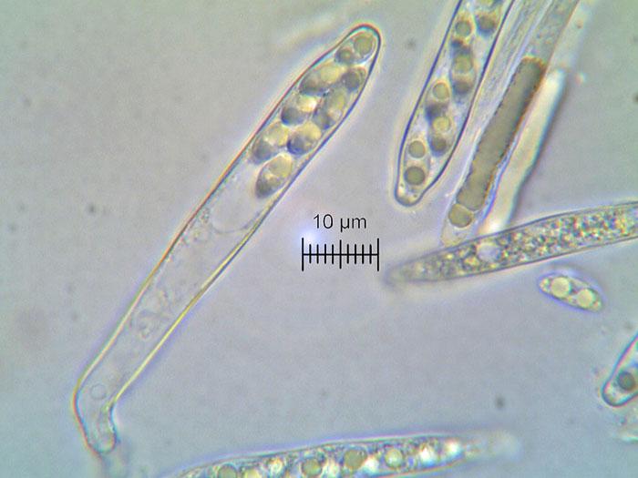
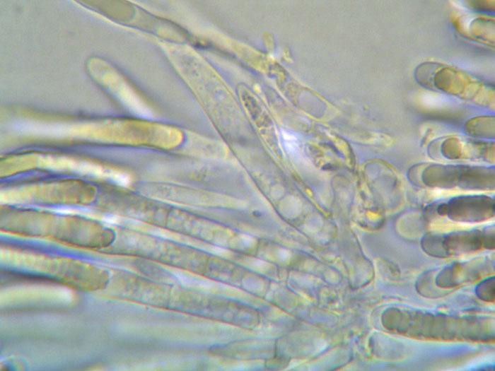
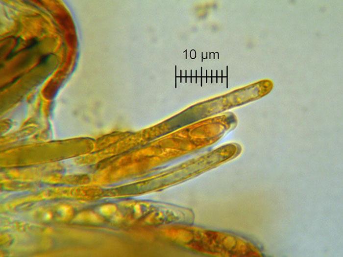
This one opens with a lid like in Trochila, apothecia up to 0,2 mm, spores ellipsoid, hyalin with two large guttules and some smaller ones, (9-10) 9,63x3,94 (3,5-4) µm. Asci cylindric to clavate, 50-69 x 7-8 µm,porus-reaction Ikl positive, dull-violett to blue, biseriat with croziers. Paraphyses with strongly refractive content, cylindrical, 4 µm wide. Marginally hairs? up to 45 x 4 µm, 5 cells, 4 are light brown, the cell at the end is clavate, hyalin.
Thank you for your help.
Regards Maren
?
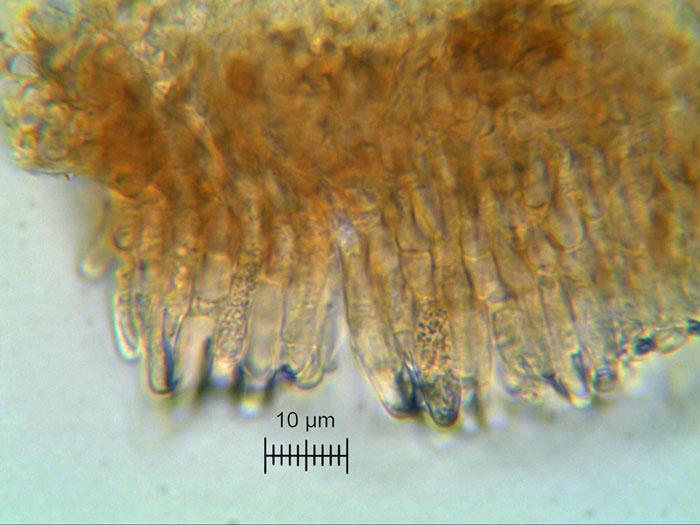


I overlooked this, sorry. It is not Naevioideae but a relative of Trochila as you compared. Hysterostegiella would be an option, but the paraphyses are there always lanceolate. H. dumeti would be on Rubus but has much smaller spores with a low oil content (as also all the other Hysterostegiella species treated by Hein 1983).
I know a similar fungus, in which I never saw a lid like here, though it is erumpent and pushes the epidermis aside, see HB 3802. I used to identify this at first as Duebenia cf. blyttiana, but only until I studied the type of it. Now I have it as "Duebenia-like" in Hysterostegiella, though it could better fit in Trochila. In my HB 5801 the paraphyses are actually slightly lanceolate. Contrary to yours the asci are alway inamyloid there.
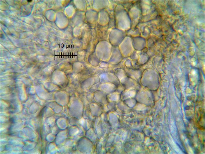
The excipulum is covered by crystals. Is this also in yours? I think one of your middle pics show crystals.
Zotto