
02-05-2026 12:42
Alain BRISSARDBonjour à tousJeuidi 30 avril dernier on m'a remi
02-05-2026 13:06
Pauline. PennaBonjour Please can someone help me with this id
01-05-2026 22:45
 Thierry Blondelle
Bonjour à tous, Une récolte sur bouse séchée d
28-04-2026 20:07
 Lothar Krieglsteiner
... on twig in the air at standing Ceratonia siliq
14-04-2026 05:32
Ethan CrensonHi all, A few weeks back a friend pointed out som
28-04-2026 20:33
Vitus SchäfftleinHello, I found Trochila ilicina on Ilex aquifoliu

30-04-2026 10:28
Rot BojanHello, by appearance I would say that I am dealing
27-04-2026 18:48
Tony MoverleyCollected 23rd April 2026, Norfolk, EnglandSwarms
27-04-2026 20:52
Lothar Krieglsteiner
Found on hanging tiwg of Olea europaea in dried-ou
Coloration des cloisons
Michel Hairaud,
07-01-2005 17:49
 Bonjour, Je pense avoir déterminé Arachnopeziza obtusipila , très conforme à la description donnée par Zotto Baral (CD ROM no 2) mais j'ai des difficultés à mettre en évidence les cloisons des spores. Que pouvez vous me conseiller ? (ai essayé Bleu coton et Rouge congo)
 Merci. Michel hairaud.
NC NC,
07-01-2005 18:30
Re:Coloration des cloisons
It is almost impossible to color the septa in ascospores, but one can see them more clearly if one uses a cytoplasmic stain. One I used for many years was to mount in a 1% solution of ERYTHROSIN B, and then to move to a drop of 50% slightly acidified aqueous glycerine (a drop of concentrated HCl added to 50 mil of diluted glycerine). This stains the cytoplasm pink, and the unstained septa are then clearly visible. There are other stains that work. Probably Baral covers this topic, or will discusss it with you if you e-mail him at <zotto@t-online.de>.
The Arachnoperzizeae was the subject of my Ph.D. thesis in 1950, and has been of continued interest to me ever since!
Dick
REPLY TO: RPK1@cornell.edu
Richard P. Korf, Emeritus Professor of Mycology, Cornell University
May-Oct phone (607) 273-0508, fax (607) 273-4357
Nov-Apr phone (727) 322-0112, fax (727) 321-1460
VISIT THE MYCOTAXON WEBSITE at <www.mycotaxon.com>
.
NC NC,
07-01-2005 19:37
Re:Coloration des cloisons
J'ai trouvé cette espèce il y a quelques années tous près de chez moi (Eastern Flanders, Belgium), sur branche épaisse de Pinus sylvestris. L'espèce ne semble que former jusqu'à 3 septes très tardivement et les psores perdent en même temps leur contenu lipidique (index 4 avant formation de cloisons). Je fais toujours mes observations dans de l'eau potable (ajoutant de l'IKI à la fin, après description complète, pour contrôler l'amyloidité).
Conseil: Garder la collection dans une boite en atmosphère humide, et contrôler l'évolution des spores chaque deuxième jour.
Bernard Declercq
Conseil: Garder la collection dans une boite en atmosphère humide, et contrôler l'évolution des spores chaque deuxième jour.
Bernard Declercq
NC NC,
08-01-2005 00:42
Re:Coloration des cloisons
Bonsoir Michel,
J'ai 2 récoltes de Arachnopeziza obtusipila et les spores même mures n'étaient pas cloisonnées. Certaines (overmature ou mortes ?) en avaient 3 ou 4 . J'avais même noté "fausses cloisons ?". Je pense que le conseil de Bernard est judicieux.
Si ça peut conforter ta détermination voici une description rapide des 2 récoltes :
24/03/1999 men 3010 B 08 Chehery Discos blancs poilus 0,2- 0,3 mm sur cône Larix
spor en massue 28-35 (37) X3,2-3,8 µm Poils droits septés parois épaisses 6-9 cloisons 125 X 4-4,5 µm coniques mais à bouts arrondis Hyphes du subiculum cloisonnées 2,5 µm
Paraphyses multifourchues 1,5 µm. Asq 80-95 X 10-14 µm J+
28/04/2004 Ile de Ré Salières Porte en Ré ? Sur cones Pïn maritime 0,15 mm 0,3 de haut;
spores remplies de petites guttules sauf au centre lipid 5 25-31 X 3,5-4 µm
Poils 100-125 X 3,5 -5 (7) µm : 5 (2 à 8) cloisons bouts arrondis quelques concretions hyalines
Asques 60-75 X 10-15 µm à crochet IKI bb apex conique
Amitiés
Robert
Michel Hairaud,
08-01-2005 01:09
Re:Coloration des cloisons
I am very grateful to you, Dick, for this reply.
I will send macro and micro pictures of this collection and may be you will confirm my conclusion. Amazingly enough, the species was first created by Grelet who once lived at about 5 kilometers from my place.
As far as Grelet is concerned, I take the opportunity to ask if there is a contemporary synonym for Arachnopeziza zonulata Roll. Cf. in Grelet) ? Moreover, would it be possible to get a modern key for this Genera ? For the difference in spores measures found in litterature is quite astonishing.
Merci aussi à toi, Bernard. C'est bien dans l'eau que j'ai ''deviné'' les cloisons, ce qui m'a fait en effet penser que les cloisons n'apparaissent que tardivement. Bien sûr, l'utilisation du Melzer fait nettement apparaître des ''cloisons'' mais je m'en méfie car il s'agit, me semble t'il, surtout de séparations entre masses lipidiques.
Michel Hairaud,
08-01-2005 01:20
Re:Coloration des cloisons
Merci Robert pour ta réponse.
Les dimensions que tu donnent correspondent en effet à celles de ma récolte.
Il y a un caractère très frappant, c'est la coloration progressive du subiculum en jaune vif , ainsi que des poils (en vue macro). On retrouve des amas de ''mucus '' jaune vif sur les poils.
Je suis un peu rassuré sur le fait que tu aies aussi eu des difficultés à voir les cloisons. Mais quand on voit les dessins de Zotto, on se demande si on sait bien utiliser un microscope !!!
Michel Hairaud,
10-01-2005 22:04
Re:Coloration des cloisons
Bonsoir,
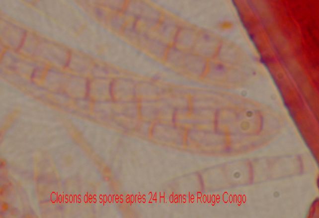
Dans le prolongement de notre discussion, je me suis aperçu qu'en laissant la préparation dans le rouge congo 24 heures, on apercevait alors nettement les cloisons, comme le montre le fragment de photo joint.
Je propose d'autres photos de cette espèce dans la base.
Amitiés
Michel
Dans le prolongement de notre discussion, je me suis aperçu qu'en laissant la préparation dans le rouge congo 24 heures, on apercevait alors nettement les cloisons, comme le montre le fragment de photo joint.
Je propose d'autres photos de cette espèce dans la base.
Amitiés
Michel
NC NC,
10-01-2005 23:52
Re:Coloration des cloisons
I am not sure that congo red stains cell walls. You may be seeing cytoplasm between oil drops. If you use a cytoplasmic stain the walls are colorless and the cytoplasm stains. I am not very familiar with congo red, so I could be mistaken. Does the outer wall of the spore also stain red? I cannot see that clearly in your photomicrograph. I do know it is easy to confuse cytoplasm between oil drops with septa. There are relatively few stains that stain septa, but many that stain cytoplasm.
Dick
Dick
NC NC,
10-01-2005 23:54
Re:Coloration des cloisons
When using a cytoplasmic stain (eosin, erythrosin) the cytoplasm stains and the septa are colorless.
Dick
Dick
Michel Hairaud,
11-01-2005 00:16
Re:Coloration des cloisons
I agree with you, Dick, the microphoto is rather poor.
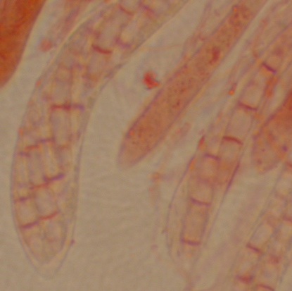
Here is another one, with the same Red congo then KOH, not much better I confess, but it shows that spores have regular transversal stains. Would Cytoplasm stain so regularly between oil drops ?
I'll try eosin or so. Did you have a look at the other pictures in the Database ?
Michel
Here is another one, with the same Red congo then KOH, not much better I confess, but it shows that spores have regular transversal stains. Would Cytoplasm stain so regularly between oil drops ?
I'll try eosin or so. Did you have a look at the other pictures in the Database ?
Michel
NC NC,
11-01-2005 00:24
Re:Coloration des cloisons
I did not see the database, nor understand how to access it. If it is cytoplasm, it would surely stain between oil drops and could be mistaken for septa. The reason I asked about the cell wall of the rest of the spore is that if the septra stain in congo refd thjen the rest of the cell wall should stain equally red.
Dick
Dick
Christian Lechat,
11-01-2005 06:40

Re:Coloration des cloisons
Hello ,
I have just put on-line Michel Hairaud's index card concerning Arachnopeziza obtusipila
best regards,
I have just put on-line Michel Hairaud's index card concerning Arachnopeziza obtusipila
best regards,
NC NC,
11-01-2005 16:38
Re:Coloration des cloisons
Dear Christian,
I seem uninformed. How would I find the on-line index card? ;(
I seem uninformed. How would I find the on-line index card? ;(