
28-04-2026 20:07
 Lothar Krieglsteiner
... on twig in the air at standing Ceratonia siliq
04-05-2026 18:13
 Stephen Martin Mifsud
ID request for what seems to be a true aquatic fun
04-05-2026 16:39
Stephen Martin Mifsud
ID request: This specimen was collected in Malta o
04-05-2026 09:50
 Castillo Joseba
Me mandan el material seco de Galicia,(EspaûÝa) re
02-05-2026 12:42
Alain BRISSARDBonjour û tousJeuidi 30 avril dernier on m'a remi
02-05-2026 13:06
Pauline. PennaBonjourô Please can someone help me with this id
01-05-2026 22:45
 Thierry Blondelle
Bonjour û tous, Une rûˋcolte sur bouse sûˋchûˋe d
14-04-2026 05:32
Ethan CrensonHi all, A few weeks back a friend pointed out som
28-04-2026 20:33
Vitus SchûÊfftleinHello, I found Trochila ilicina on Ilex aquifoliu
Hymenobolus agaves anamorph
Miguel ûngel Ribes,
12-03-2013 00:39
 Good night
Perhaps someone remember this Hymenobolus agaves: http://www.ascofrance.fr/search_forum/10909
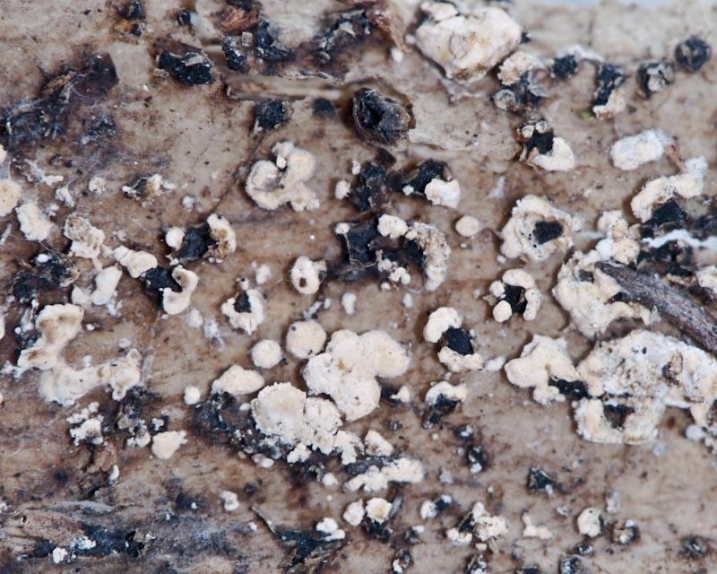
Rubûˋn has foung more collections in another Canary Island, La Gomera. In some collections, between H. agaves apothecium, are growing too a white-orange anamorph, 2-5 mm broad, relatively hard (it is posible to cut it).
Is it posible the anamorph of H. agaves? How to study this anamorph?
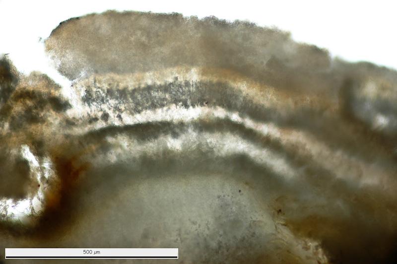
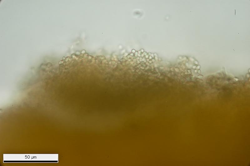
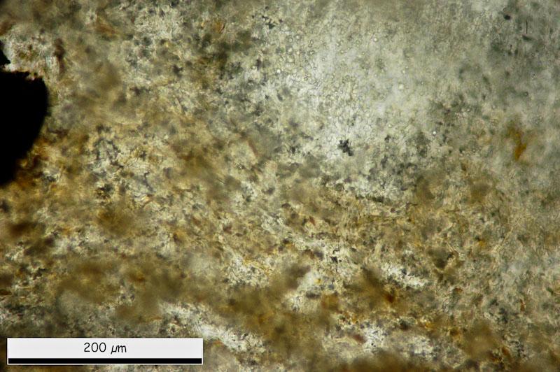
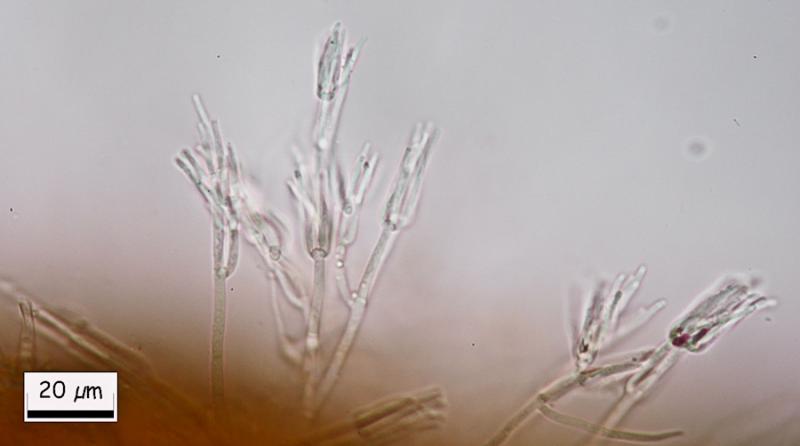
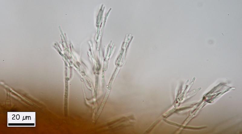
This are general views.
Thank you.
Miguel ûngel Ribes,
12-03-2013 00:44
Re : Hymenobolus agaves anamorph
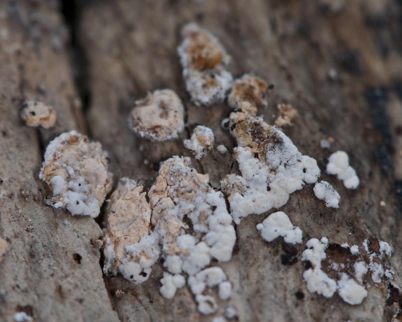
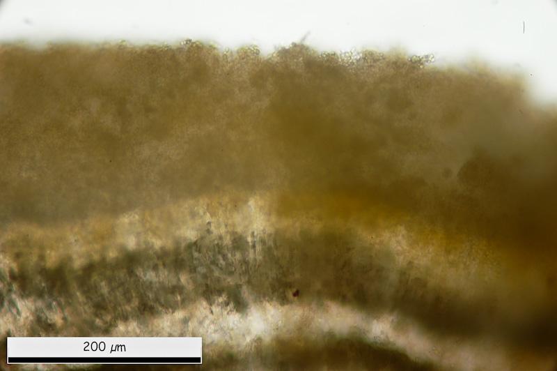
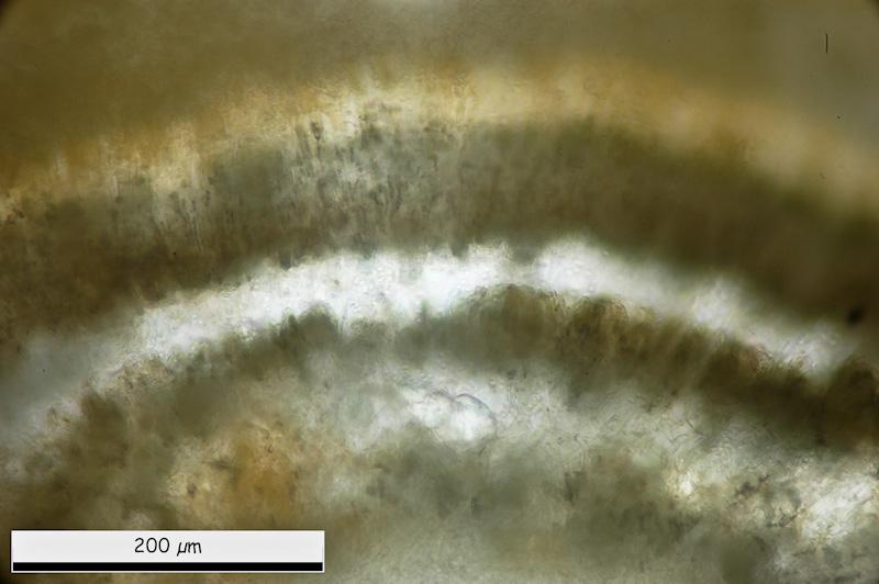
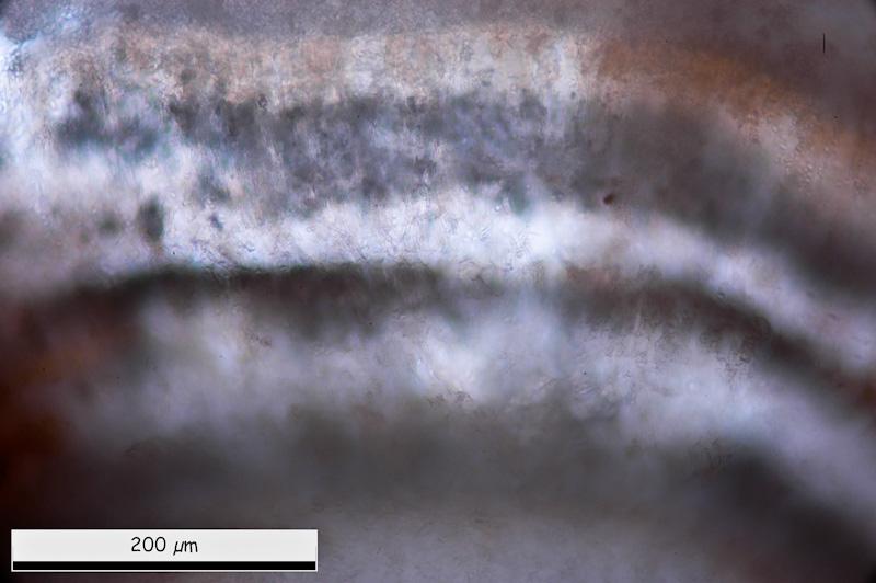
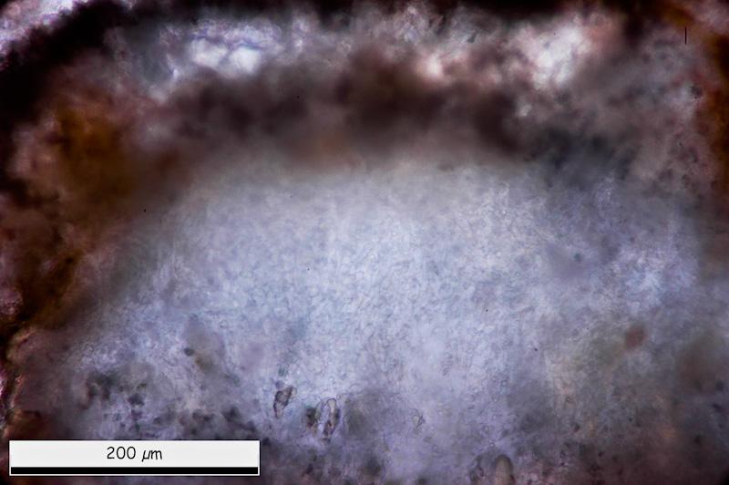
General micro views. It is posible to see some layers. External one with more-less rounded cells.
Miguel ûngel Ribes,
12-03-2013 00:50
Re : Hymenobolus agaves anamorph
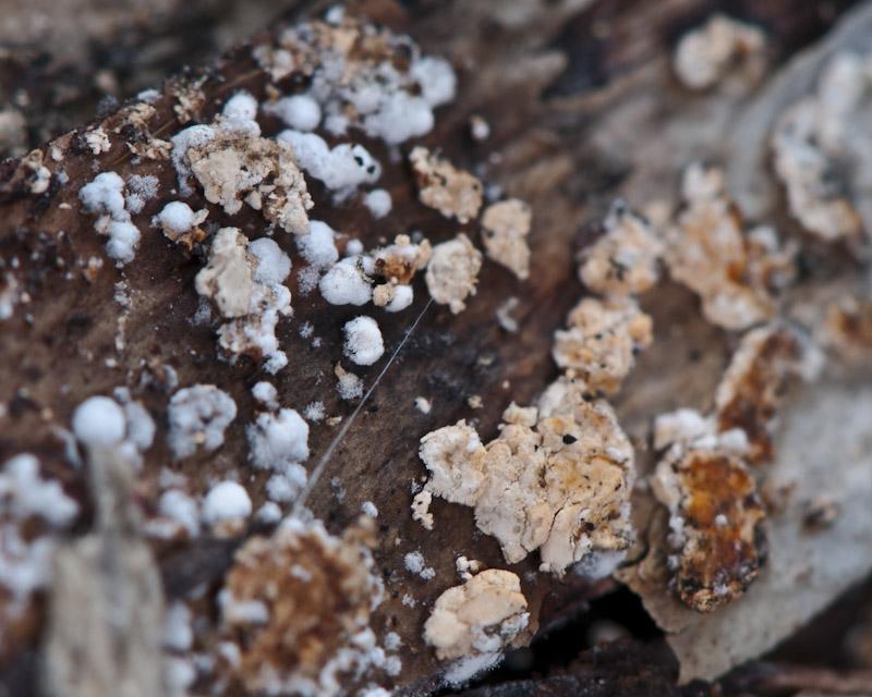
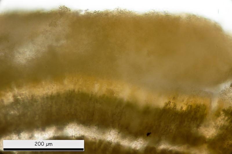
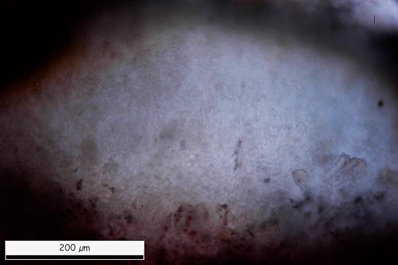
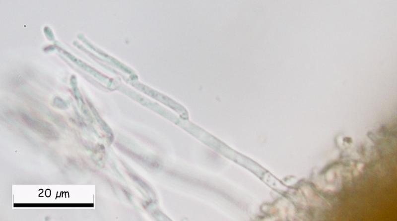
Inside, tow black lines. And in medular area a white area with globose-angular structure mixed with cilyndrical cells.
Miguel ûngel Ribes,
12-03-2013 00:53
Re : Hymenobolus agaves anamorph
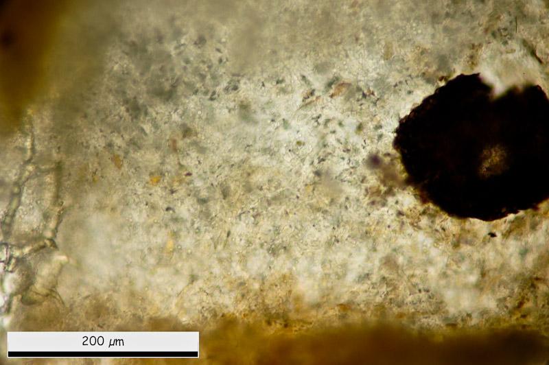
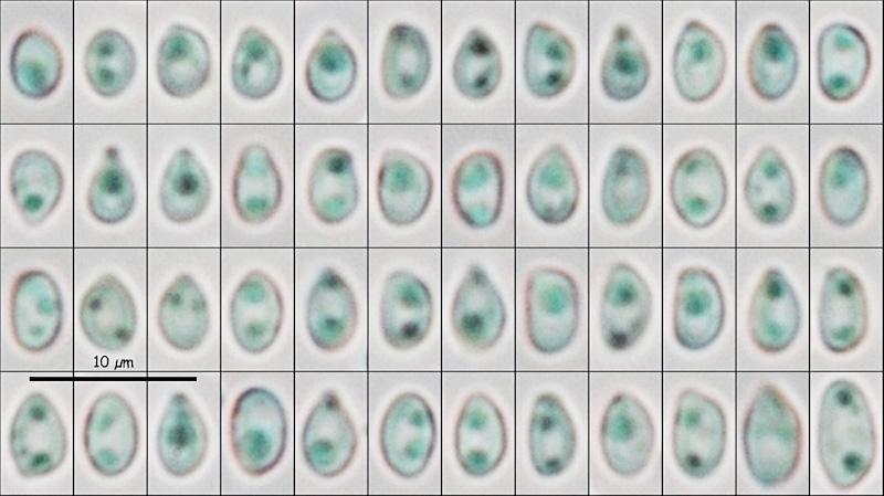
Eliptical conidiospores in water:
(4.06) 4.49 - 5.21 (6.63) x (2.87) 2.94 - 3.35 (3.57) ôçm
Q = (1.28) 1.40 - 1.70 (1.86) ; N = 52
Me = 4.88 x 3.14 ôçm ; Qe = 1.56
(4.06) 4.49 - 5.21 (6.63) x (2.87) 2.94 - 3.35 (3.57) ôçm
Q = (1.28) 1.40 - 1.70 (1.86) ; N = 52
Me = 4.88 x 3.14 ôçm ; Qe = 1.56
Miguel ûngel Ribes,
12-03-2013 00:55
Re : Hymenobolus agaves anamorph
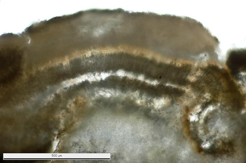
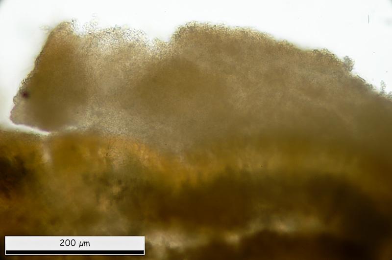
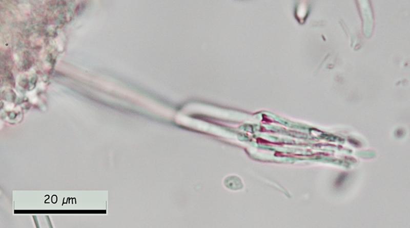
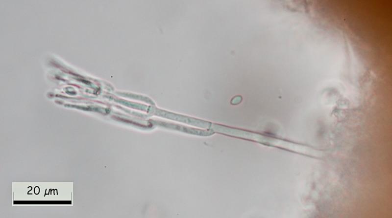
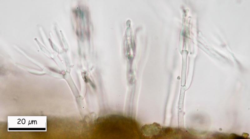
And finally, at the margin, this conidial structure.
Than you in advance.
Miguel û. Ribes
Than you in advance.
Miguel û. Ribes
Hans-Otto Baral,
12-03-2013 08:06

Re : Hymenobolus agaves anamorph
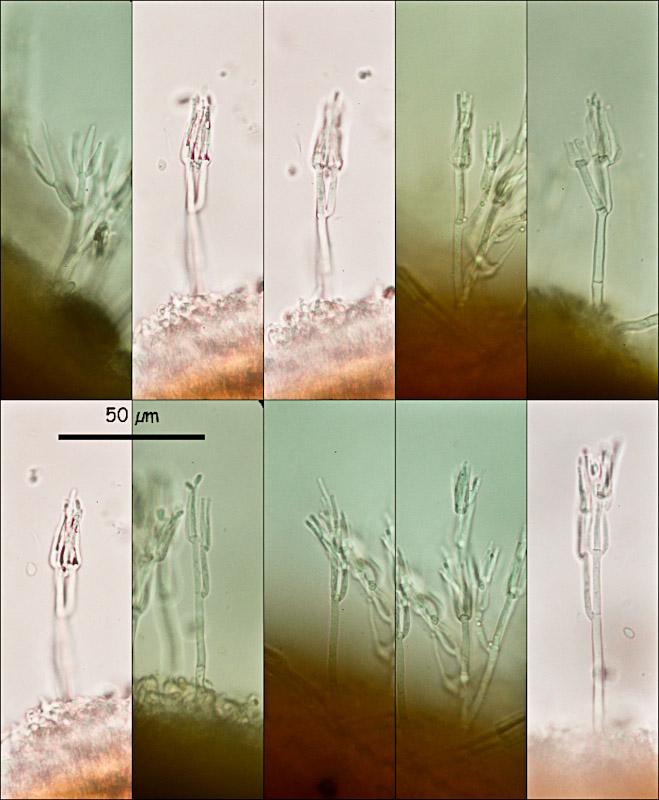
Great, Miguel! Could you please show us a closeup of the conidiogenous cells, were the conidia emerge? I assume they are phialidic. Then we can search in Genera of Hyphomycetes, or someone has an idea.
I have given the previous Hymenobolus specimen for sequencing, I am curious where it could belong.
Zotto
I have given the previous Hymenobolus specimen for sequencing, I am curious where it could belong.
Zotto
Miguel ûngel Ribes,
12-03-2013 11:27
Re : Hymenobolus agaves anamorph
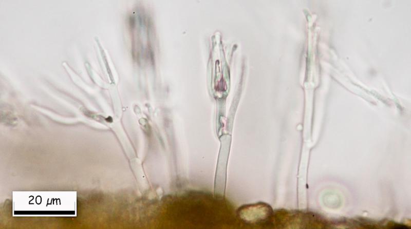
Here there are.
Hans-Otto Baral,
12-03-2013 22:56
Re : Hymenobolus agaves anamorph
Hi Miguel
Walter Gams answered me that this is ô clearly aô Clonostachys, probably Clonostachysô solani (Harting) Schroers & W. Gams, which is quite common, often fungicolous, and the anamorph of a Bionectria. So certainly not belonging to Hymenobolus.
Zotto
Walter Gams answered me that this is ô clearly aô Clonostachys, probably Clonostachysô solani (Harting) Schroers & W. Gams, which is quite common, often fungicolous, and the anamorph of a Bionectria. So certainly not belonging to Hymenobolus.
Zotto
Miguel ûngel Ribes,
13-03-2013 00:15
Re : Hymenobolus agaves anamorph
Hi Zotto, Superb.
Thank you again to resolve this puzzle.
See you.
Thank you again to resolve this puzzle.
See you.