

22-04-2026 20:54
 Enrique Rubio
Hi to everybody.This Pyrenopeziza grew in moist le
24-04-2026 03:16
 David Chapados
Found while looking at something else from wood in
22-04-2026 01:06
 Richard VALERI
Bonjour à tous.Je vous présente cette Nectria s.
22-04-2026 20:17
 Marian Jagers
Is anyone familiar with the Hyphomycetes genus Pse


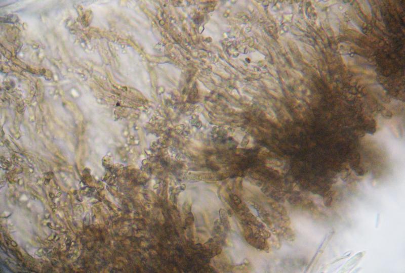
Hello, dear friends!
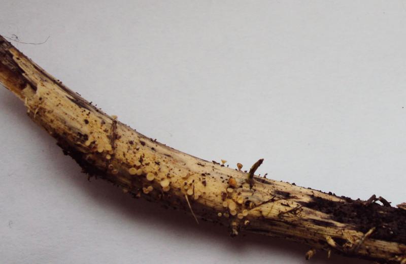
These 2 specimens some time ago i identidied as C. cyathoidea. Now I see some differences in spore morphology, and I wonder whether one of them could be C. pallida.
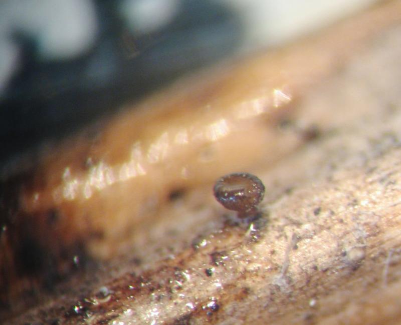
The 1st was examined in fresh condition, the 2d in exciccated state.
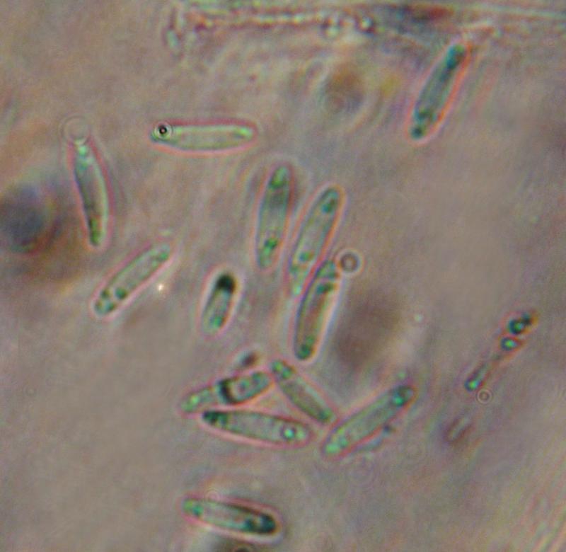
So, the 1st specimen was collected in oak forest, on Urtica dioica rotten stem.
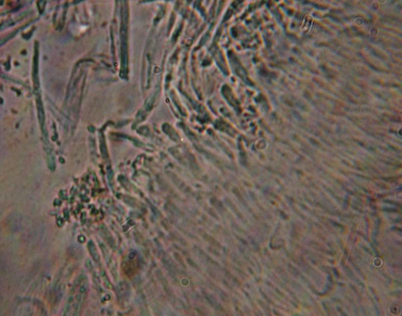
Spores 7,3-12,6*2,2-3,6 um, with 1-3 small oil drops on each end.
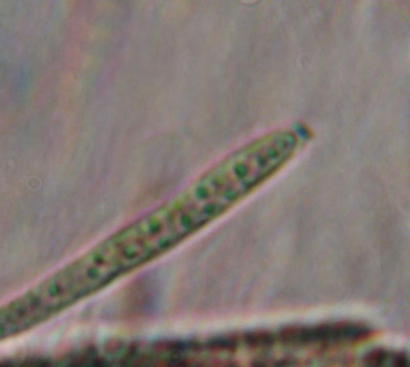
Asci IKI B, with croziers, 49-68*3,6-5,5 um
Cheers,
Irina


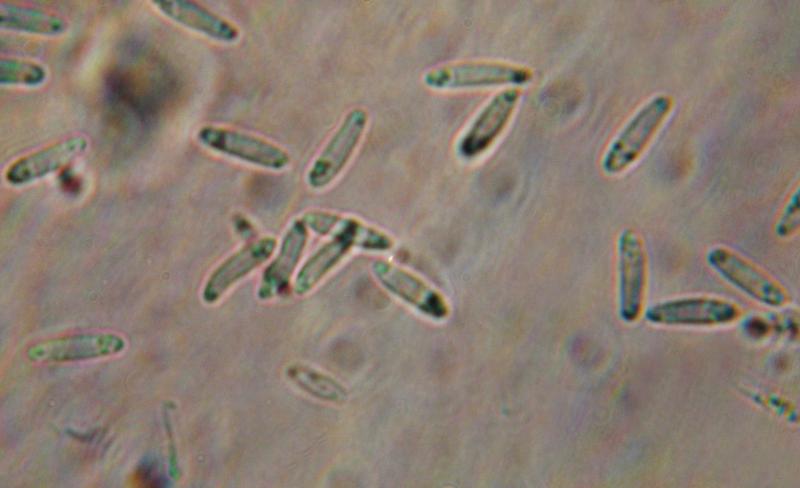
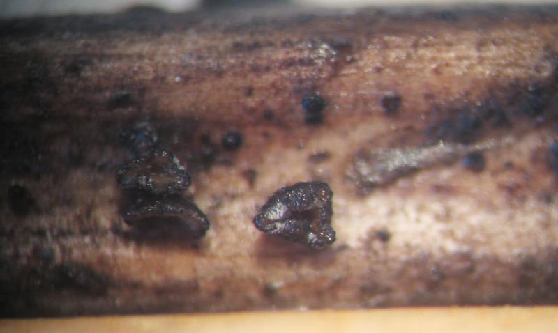
The 2d one was collected on a meadow, on rotten herbaceous stem.
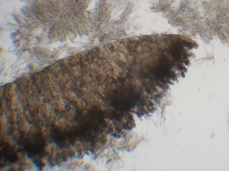
Spores 9,1-12,7*1,8-2,7 um, fusiform, sometimes slightly S-shaped
Asci IKI B, with croziers, 44-60*3,8-5,5 um


in order to confuse you a bit :-)
C. pallida is a species with marginal teeth, at least as I understand it. It was treated by Breitenbach & Kränzlin under the wrong name C. dolosella. The marginal teeth are not shown on their photo, but they are mentioned, and I reexamined their material:
your whitish specimen could well be C. cyathoidea, quite a variable species. Are the spores actually up to 3.6µm? Regrettably, only the spores are alive in your preparations. Maybe you press too strong. The apical ring photo seems to exclude hymenoscyphus repandus.
The brown one reminds me of C. cacaliae.
Zotto

Hello, Zotto!
And thank you for answer.
Yes, I know about marginal teeth in C. pallida, but in my opinion they probably could be poorly visible/destructed, so on. The 2d one was collected in dry condition, so I cannot say surely whether it was brown in living state or not.
With best regards,
Irina