
23-05-2026 11:44
 Charles Grapinet
Hello, I am having trouble identifying this copro
23-05-2026 18:57
Sylvie Le GoffBonjour à tousRécolté sur une branchette de Sal
22-05-2026 14:44
 Lothar Krieglsteiner
in unripe condition citrine yellow, then soon fadi
22-05-2026 21:35
Steve ClementsBonjour, I expected this find on old wood on our
22-05-2026 18:12
Lothar Krieglsteiner
... in moist chamber from Portugal.As the fungus s
22-05-2026 20:08
Ethan CrensonHello all, Yesterday in NYC I was visiting an e
11-01-2022 16:36
 Jason Karakehian
Hi does anyone have a digital copy of Raitviir A (
20-05-2026 17:47
Margot en Geert VullingsWe found this Mollisia on dead Juncus stems mown l
22-05-2026 14:47
Gernot FriebesHi,superficial ascomata collected on bark of a liv
Will someone help me identify?
Thank you in advance.
Regards
Mirek


A very hard topic for such a novice in this topic as me.
I had to review everything that is available on the internet before I came to any conclusions.
Initially, I tried to compare my collection to T. fuckeliana. However, the features did not suit me at all, although on the asco-sonneberg website I found collections identical to mine, signed just as T. T. fuckeliana;
http://asco-sonneberg.de/pages/gallery/nectria-fuckeliana-100325-mcol-0123451.php?group_id=7071&position=16
However, I measured the spores visible in the pictures themselves and their size is rather very similar to mine and not as stated in the description so I gave up this option.
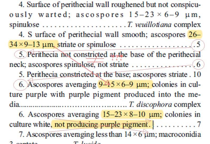
Then I used the work "The genus Thelonectria (Nectriaceae, Hypocreales, Ascomycota) and closely related species with cylindrocarpon-like asexual states - 2016". I may be wrong but it seems to me that it is written with errors. There are large inaccuracies in the key (see scan No. 01).
Yesterday I came to the earlier work of the same authors and according to her my collection is the closest to Thelonectria discophora;
"Phylogeny and taxonomic revision of Thelonectria discophora
(Ascomycota, Hypocreales, Nectriaceae) species complex - 2013 ".
(See scan 02)
Today I have measured a greater number of spores and their dimensions are practically perfectly consistent with this description!
(11.4) 11.9 - 15.1 (16.2) × (4.6) 4.9 - 5.9 (6.2) µm
Q = (2.1) 2.2 - 2.8 (3); N = 34
Me = 13.4 × 5.5 µm; Qe = 2.5
Individual dimensions of the spores are given in the picture nr. 03
Christian, fruiting bodies are not overripe. I showed germs germinating but there were very few. In my opinion, the fruiting bodies are of the perfect age for microscopy. In my collection there are completely immature spores and free spores that are already germinating. However, the vast majority of spores are moderately mature, with ornamentation already formed. This time I have measured just such.
I compared other species but in their case the size of the spores is not compatible with mine!
Thank you for the hint!
You'll agree with me?
Regards
Mirek


There is no experience with this type.
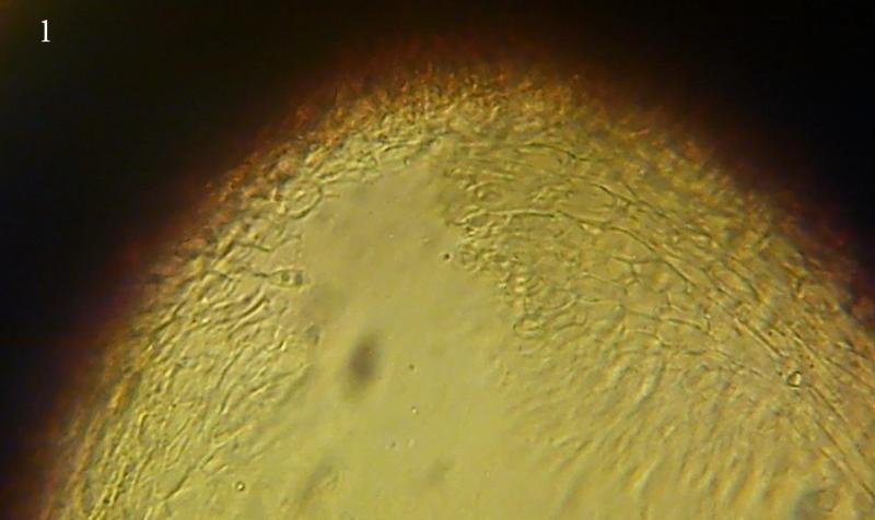
I prepared some photos. Maybe you can read the necessary qualities from them?
Mirek


http://www.centrodeestudiosmicologicosasturianos.org/?p=15486
It is true that my photo is not as perfect as Enrique but you can see sufficient arrangement of the cells.
Is this how it was supposed to look like?
Regards
Mirek


Your help was priceless!
Mirek