
24-03-2026 21:37
Elisabeth StöckliBonsoir,Sur bois (tronc) très pourri de conifère
25-03-2026 10:35
Hulda Caroline HolteHello,I collected this species growing on a dead b
26-03-2026 15:31
 Åke Widgren
Hello,I found this one in October last year, on r


25-03-2026 22:23
 Marc Detollenaere
Dear Forum,On a debarked stem of Tilia, we found s
24-03-2026 15:44
Åge OterhalsI hope someone can confirm the name of this collec
25-03-2026 20:53
François BartholomeeusenDear forum members,On 23 March 2026, I found sever
23-03-2026 20:16
 Miguel Ángel Ribes
Good eveningI'm unable to identify this Coprotus o
25-03-2026 15:06
 Bernard CLESSE
Bonjour à toutes et tous,Pourriez-vous me confirm
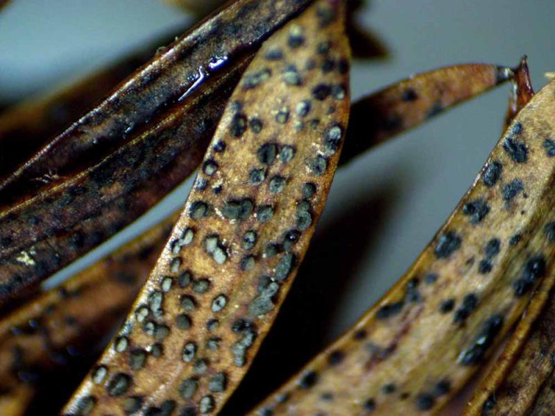
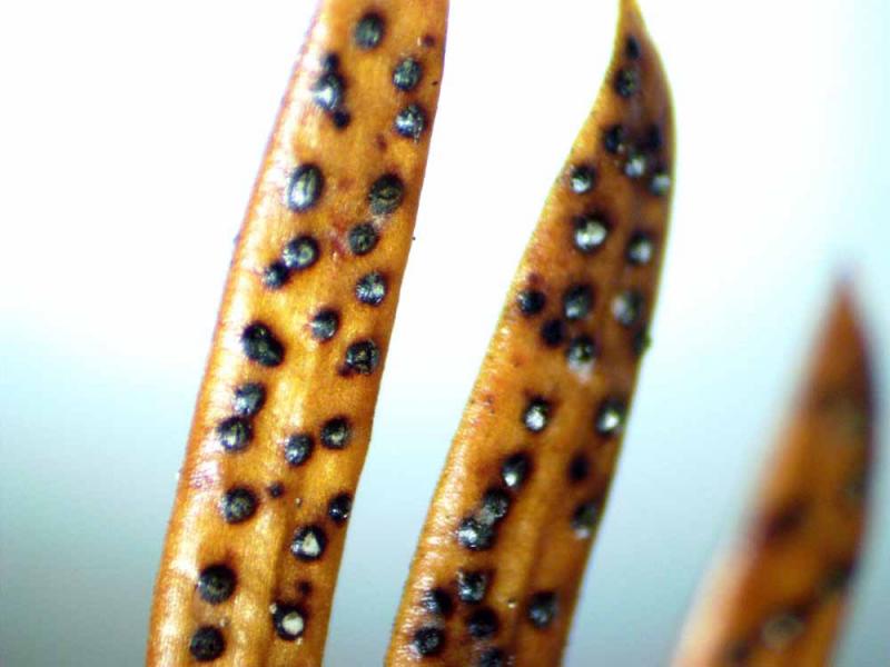
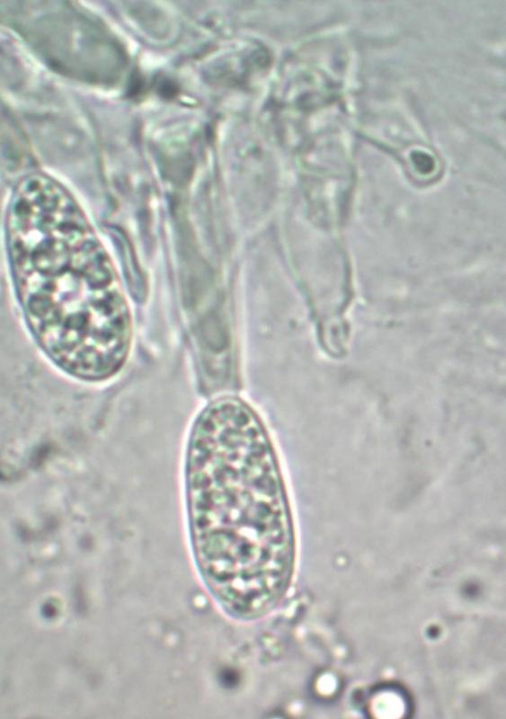
 Hola, tengo esta muestra desde hace un mes en casa, en camaraa humeda y acaban de abrir , pero para mi sorpresa solo veo lo que pueden ser conidios, he realizado varias preparaciones, de la masa esporal,de muestras cerradas , de muestras abiertas y solo he logrado ver estos posibles conidios ya que Ascas no he visto,
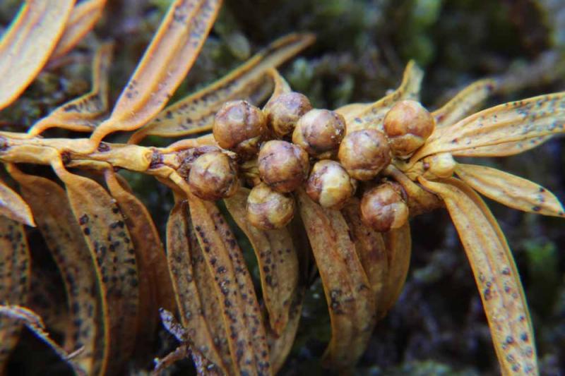
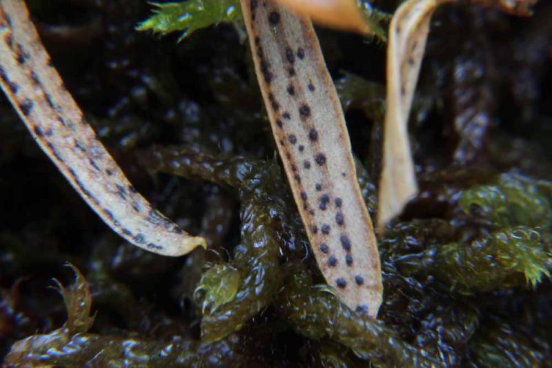
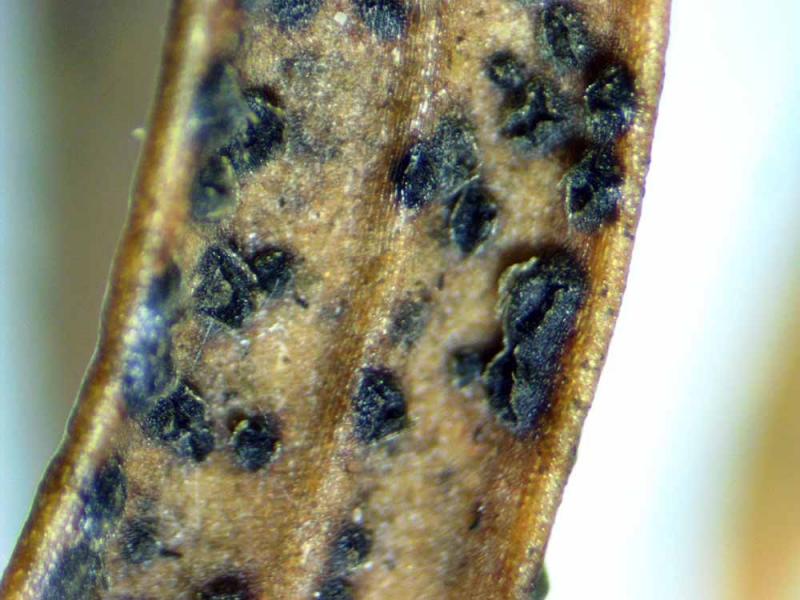
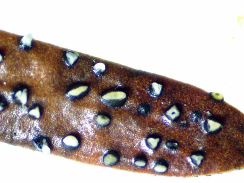
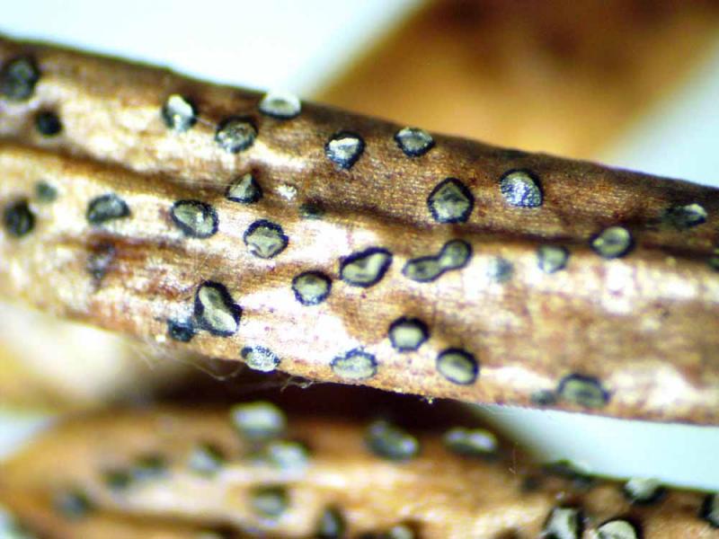
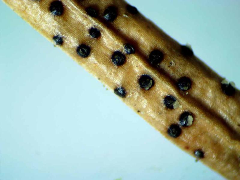
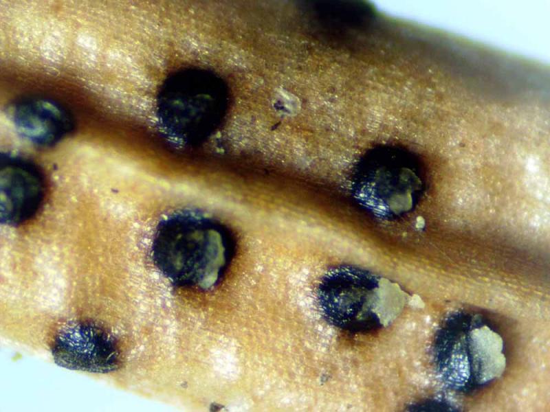
no se si alguno de Ustedes esta familiarizado con los hongos de Aciculas de Taxus baccata. pudiera ser algun Lophodermiun o Colpoma,
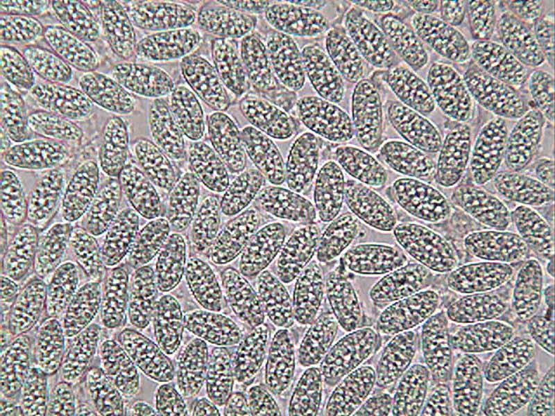
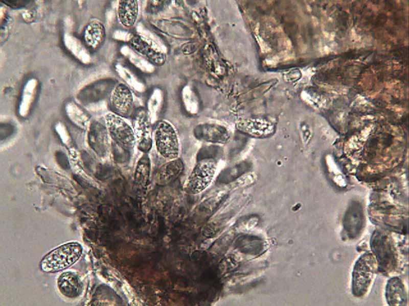
La fructificacion mide entre 0,17--0,29 largo x ancho0,15--0,18.
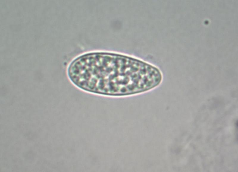
Conidios o Esporas 15--18 x 7--8.
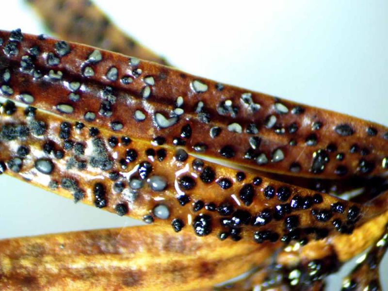
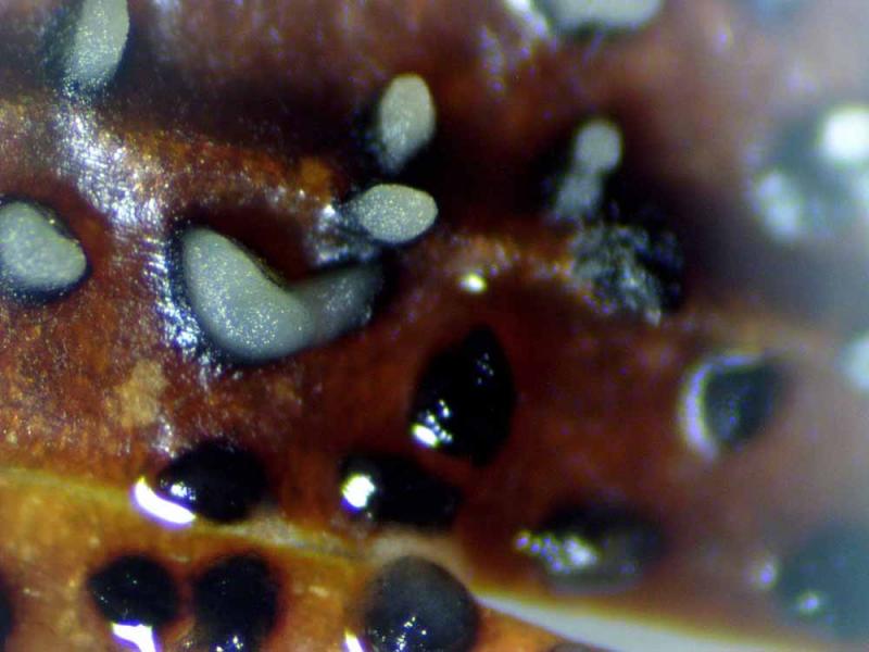
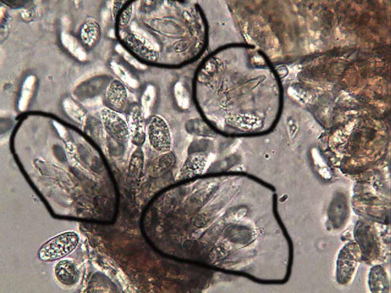
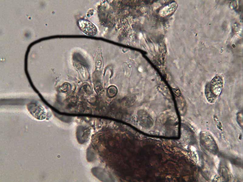
En fotos 17--21--29--31, pongo unos elementos que no se si pueden ser Conidioforos.
Un saludo
Rafael


Despite the geographical disruption and distinct host, your fungus seems to be a Zelandiocoela species. However, as the genus is monotypic and the spore size of your specimen doesn't fit with the only species described, perhaps you have found a new species.
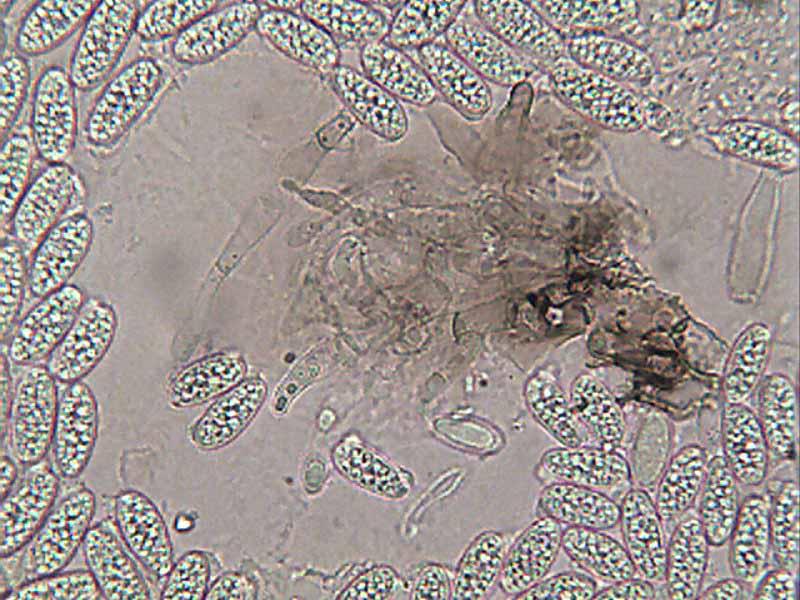
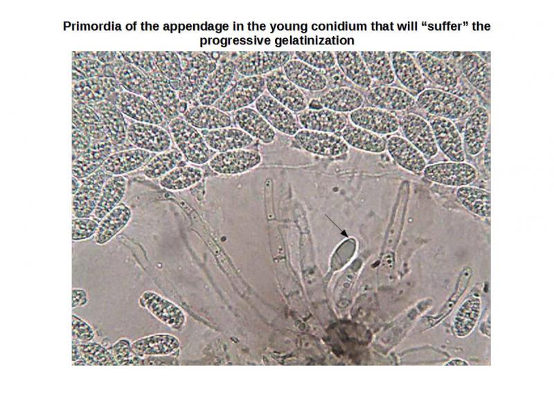
The appendage type of your fungus seems to fit with the H-type [extracellular appendages arising from structural changes (progressive gelatinization) in appendage primordia (wall areas) delimited on the developing condium], according Nag Raj.
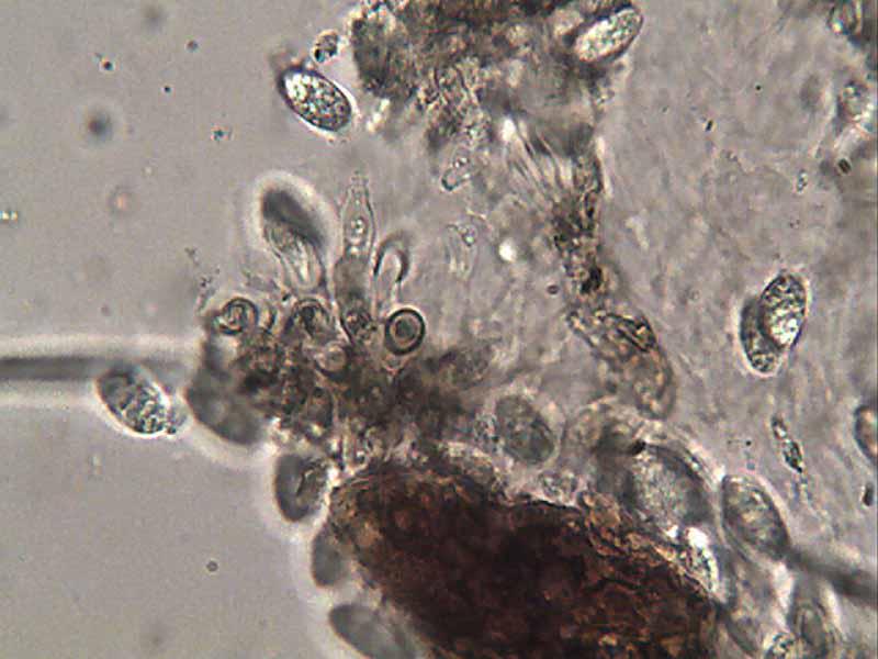
In your image 9, you can see a primordia of the appendage in the young conidium attached to the conidiogenous cell.
There are other genera similar with Zelandiocoela, but they can be distinguished by the type of appendage, spore septation, and conidioma type.
A good transversal section of your specimen will help to clarify this.
Attached follow the description of the genus and a illustration extracted from Nag Raj's monograph of Coelomycetes Anamorphs with Appendage-bearing Conidia
 Zelandiocoela-0001.pdf
parece que el Hongo como Usted dice va bien con la descripcion, pero falla en medidas de conidios, y tambien el habitat,
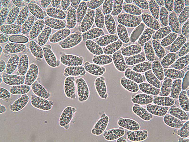
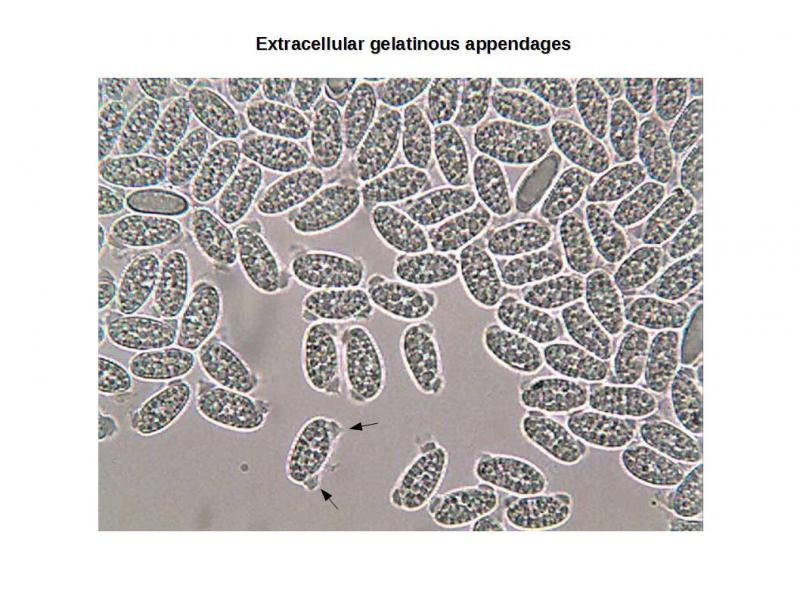
he intentado ver mas apendices conidiogenos y a sido imposible, solo he visto conidios, la mayoria de ellos, en los apendices con una masa gelatinosa excentricas.
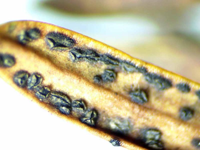
envio fotos macro por si le dan una idea a seguir. o si Usted sabe a quien puedo enviar, tengo mas material,
mañana intentare ver dichos apendices.
Rafael

Creo que no he sido claro, los apéndices son visibles en todos los conidios en tu fotos. Mire en las fotos abajo yo he indicado con flechas.
Lo que puedes hacer son secciones transversales para mirar se el conidioma es picnidial o no.
No conosco nadie que tienes trabajado con eso hongo, pero yo gustaria de recibir una muestra si usted puedes enviarme. No lo puedo garantir que voy conseguir identificarlo con seguridad, pero puedo intentarlo.
Saludos,
Dartanha

I think this is Diplodia taxi; not uncommon on dead Taxus leaves in Europe. !See edit below!
The conidium formation shown in your "photo 9", on a long conidiophore is typical of Diplodia; quite different from the Zelandiocoela which shows short conidiophores with collarettes.
Eventually the conidia turn brown and become 1-septate, but for a long time they remain colourless and full of tiny guttules, as here. The dimensions given in books are slightly larger than what I find - I suspect they are based on the mature, septate, conidia.
Compare with:
http://www.librifungorum.org/Image.asp?ItemID=19&ImageFileName=SyllogeFungorum3-102.jpg
and this image
un saludo

how about Cryptocline taxicola? Do these conidia also rise from collarettes? I am not sure by these pictures: http://www.invasive.org/browse/subthumb.cfm?sub=77798
Best
Martin
I must admit that when I saw what I interpreted as "bipolar gelatinous appendage", surely not present in Diplodia species, I forgot to pay attention to the conidiogenesis.
However, I still in doubt about this fungus, a transversal section of the conidioma will clarify if it is acervular or picnidial, thus will help to exclude some genera.
Rafael, if you dont mind I would like to receive a sample of your specimen.
Please let me know if you could send it to me, so I will give you my post address.
Dartanha
mandeme su direccion de correo para el envio
Rafael
su foto se parece mucho, pero no veo en las esporas ninguna con lo apendices gelatinosos como en la que yo mando,
Sabe Usted si esto no es un caracter constante???????
en el Pdf que le mando tampoco lo nombran,
Un saludo
Rafael

No hay pigmentacion oscura.
No hay septacion.
A pesar de que las fotos son malas se ven dos tipos de celulas conidiogenas, envio fotos por si sirven.
Rafael
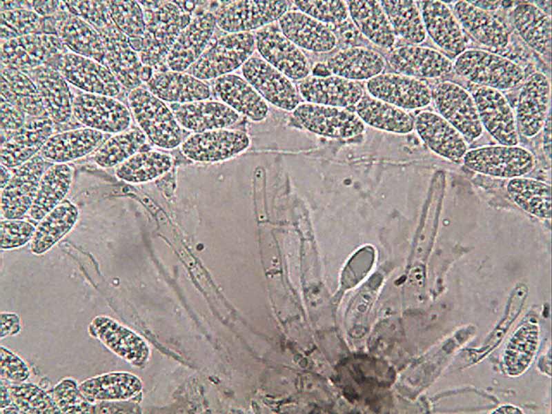

no ostiolo
Rafael
So probable Martin was right, it could be Cryptocline.
But there are others genera like, Rhodeosiopsis.
Saludos,
Dartanha
He leido la descripcion de Cryptocline taxicola, y dice que tiene esporas truncadas en la base y no es el caso.
Las fotos de Conidios tampoco tienen en los lados de los apices esa masa gelatinosa-mucosa, la tiene central, no se si este detalle cambiara de esporas inmaduras a maduras.
Podria ser una buena pista, pero a lo mejor otra especie.
Un saludo
Rafael
according to Sutton 1980 Cryptocline is a heterogenous genus with different kind of conidiogenesis and conidial morphology. I don't have a more modern work at hands. I append two things: first the protologue of this species under Gloeosporum taxicolum by Allescher. He describes the conidia as elliptical or oblong with obtuse ends, filled with a"cloudy" content or oildrops. Size 10-16x 5-8 µm.
Secondly I attach a paper by Safrankova that shows a microphoto of the conidia very similar to yours and gives a measurement for the conidia of 10–18 × 4,5–8,4 µm.
Both would be well in the range what you measured in your find.
Best regards
Martin

Although there was no obvious evidence that it was parasitic (as one would expect with Cryptocline, it was on leaves still attached to the tree. In addition, although the conidia of Diplodia taxi are reported to remain colourless for quite some time I would expect them to be thicker-walled than these. Finally, the conidiomata do look acervular rather than pycnidial.
Attached is a useful monograph of Cryptocline, with good drawings of C. taxicola. Fortunately the site where I collected this is very close to my home - I shall re-investigate!
Cordialement
Chris
thank you for the Morgan-Jones paper, I searched for in vain. Would be interesting now to see the conidiogenous cells of Rafael's find. Clearly should have collarettes in case of Cryptocline.
Best
Martin
Chris is right that Diplodia species initially have hyaline 0-septate conidia but in my experience you can always find some mature brown 1-septate conidia.
Marcus
best wishes
Chris
and thanks Chris for the Morgan-Jones paper.
All the best to all.
Dartanha
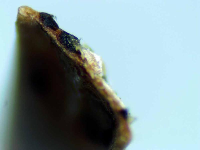
He realizado muchas preparaciones y he sido incapaz de ver mas conidioforos, solo los vi en la primera preparacion en ejemplares recien fisurados y asomando la masa conidial.
Un saludo
Rafael