08-11-2024 17:36
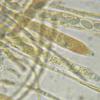
Juuso ÄikäsRecently I posted here my finding of small white a

04-11-2024 17:32
Yves AntoinetteBonjour, je pense qu'il peut s'agir de Trichoderma

23-10-2024 17:27
Hi againThese tiny apothecia (100-200 µm) were gr

06-11-2024 20:07
• Macro and habitat suggest Hymenoscyphus s.l.,

04-09-2024 21:02
 Stephen Mifsud
I have found an interesting Xylaria growing on fal

06-11-2024 11:59
 Stephen Mifsud
I am trying to identify a Cheilymenia sp. using ke

05-11-2024 18:00
Karen PoulsenHello, Can anyone help with this one? On twigs o
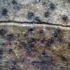
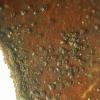
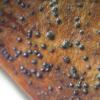
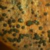
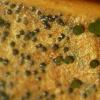
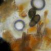
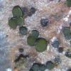
On the leaves of the Rhododendron. I found this black fruitbody's. I know there are a couple like
Trochila, Phacidium. But I don't hane any idea if there's a name for the ones on the Rhododendron. Maybe one of you has an ides about this?
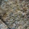
The second picture is the leave on the bachside,
Thanks
Hannie

Zotto

I still have the leaves and maybe tomorrow I'll try at again, Thanks a lot
Hannie
Hello,
It looks so enormeous like Trochila laurocerasi on Prunus laurocerasus to me.Only the crack in the leave has much from Rhododendron.
Best,
Marco
Hello,
Yes,and I had no doubts.I never seen any ascomycetes on leaves of Rhododendron ponticum.You can discriminate Prunus laurocerasus and Rhododendron by the fact that Prunus has saw-toothed (gezaagd ) edges of the leaves, Rhododendron is smooth.When alive you can tell the differences by other characters as well.
Hannie, I am not the person to give you critics,but one thing I must say.Please go out in the field next season with a plant-book.(Heukels of Heimans en Thijsse. )And please,keep searching for fungi.Fungi are sexy .Fungi are alllmost the ultimate fhantasy.(But I recently heard that there are other things competing with that. )
Groet,
Marco
Hello Marco, so I can say that this is Trochila laurocerasi? I'll never forget the difference between these leaves. And thank you so much, my poblem is solved.
Hopefully I have some time to go with a book for the names of the plants, I always search for fungi and during the summer I'm fond of insects and vlinders.
Thanks a lot, best wishes Hannie
Hello Hannie,
There are two species of Discomycetes (obsolete name, I know )known from leaves of Prunus laurocerasus, these are Trochila laurocerasi and Phacidium multivalve.But ,seen the habitus (gregarious ) and the spore you depicted,there is practically no doubt that your fungus is the Trochila.
Yes,I can get lyrical about fungi,that is not the case with flying things.
Best,
Marco

- inverted world -
Zotto

this finding looks to me as it is neither one of them but a immersed pyrenomycete, the species I held for Eupropolella brittannica is more irragular opening as Trochila laurocerasi and has a different apical ascus-ring, more Hymenoscyphus-like, the spores seems to be multiguttulate. T laurocerasi has a Calicina apical ring and spores with 2 big guttules and several small ones.
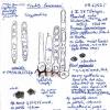
photo's from my interpretation of E. britannica.
Stip
Hello,
Yes,I am aware of the existence of the Eupropopella,and it could be that there are pyrenomycetal species known from Prunus laurocerasus,but this way the discussion goes getting a bit theoretical don't you think?There are also coelomycetes and hyphomycetes known from the Prunus.
But look at the photographs !Do you SEE an Eupropopella or a pyrenomycete,or are you having ultimate phantasy's?
Best,
Marco

did you indeed find E. britannica on Prunus? In NL?
Tell me please the spore size. The apical ring recalls that of Trochila craterium, see my drawing Trochila craterium, HB 6373.JPG. VBs lacking in paraphyses?
Now I see that also Enrique studied a find of E. britannica on Prunus, showing these apical rings. Here VBs are lacking.
A striking difference between Trochila and Eupropolella concerns the paraphyses which contain refractive VBs in Trochila but none in Euprop., in living material of course.
My guess is that Trochila belongs in the Hemiphacidiaceae because of its similarity to Sarcotrochila, which was sequenced and falls there. VBs are comon in that family. But for Eupropolella I am not sure, I still have it in the Naevioideae.
Zotto
Hello,
Reading all these reatrions, there is only one thing to do I think? The leaves on the picture are not too good. This afernoon I'll have a look if I can find new leaves and put it all again under the microscoop, I like to be sure if everything is ok.
Thanks,
Hannie
Hello,
As I am not mistaken,there are just a few ascomycetes known from leaves of Prunus laurocerasus.These are Aulographum hederae,a Diaporthe,a few Mycosphaerella 's ,A Phacidium, and Eupropolella britannica.All species with septate spores !
On the,by the photographs from Hannie,depicting by the way clear leaves,we see a hyaline amerospore,and the only ascomycete on P.l leaves which has that is Trochila laurocerasi.Hence my statement that the fungus on the photographs is that species.
I am not a philosopher,otherwise I would say that only reality is the ultimate phantasy !
Best,
Marco

But thanks for drawing my attention to spore septation. Actually, E. britannica has finally 1-2-septate spores, as I can see also on Enrique's plate, but at first they are amerospor, and I strongly guess that they are ejected that way (fresh material with living asci would solve this question).
It is possible that Trochila laurocerasi spores remain non-septate even when overmature, I cannot remember to have seen such.
But spore size is different between the two.
Zotto
Hello Zotto,
Oh yes,it is so easy !It is easy as it is.Do you really see another species on the pictures?
Best,
Marco

as I said I do not see one of them in this collection, as the matter is, it has too less featues for a determination. I showed the pictures of Eupropolella for an example of the species.
Hannie shows spores that look brown to me and without any oil content. The apothecia of Trochila lauroceraci are more uplifting above the leaf-surface. and spores with a high oil percentage. I did grow both species to study the apothecial development.
I attach photos o f development of T. lauroceraci in 4 days

I have no idea so far, the spores are from which fruitbody, photo 1, 2 or 3 becouse they look as 3 different species. The one with the shining fruitbodies could become Trochila laurocerasi if you keep the leaves moist for a few days. When no fruitbody is left I think it is almost impossible to say what it is becouse you need a fruitbody to get to a genus.
groeten,
Stip
Hi Stip,
That s good idea, to keep these leaves wet for a couple of days. I will try it again after a few days and see what it can be. Thanks for helping,
groeten
Hannie
Hello,
I think that the shiny fruitbody 's are shiny because of the epidermis of the leaves lying over the fruitbody 's of Trochila laurocerasi,with its odd Dutch name Laurierkersputdekseltje or something like that ,and that there are possible some contaminatng spores in the slide. I still see just one species.
Best,
Marco

cheers
Stip

cordialement
Chris
Hello Chris,
a little difficult for me to inderstand, but I will look for what you wrote these days. Thank you
best wishes Hannie

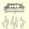
Here is the page of Grove 1935
http://www.cybertruffle.org.uk/cyberliber/01209/0291.htm
Do you know on which pages the figures can be found? For Ceuthospora hederae (p. 289) Grove cites Fig. 19b.
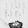
Here my sketch of it.
Zotto

here is the plate by Grove. Just the contours of the spores but maybe matching picture 17.
Regards
Martin
Update: Sutton 1980:473 shows fan-shaped appendices at the ?basal end of the conidia of C. lauri as they are depicted in Zotto's drawing as well. It would be helpfull to produce a sharper image of the conidia of Hannie's collection.

Desmazières Phacidium laurocerasi Desm. (1846) is actually younger than C. lauri Grev. (1827).
So it would be great if someone goes there and takes a sequence of both ana- and teleomorph of these Trochilas to ascertain their connection.
The appendages of the conidia are probably at the base (proximal), but I am not sure
Zotto

this explains a lot of unsatisfying collections.
cheers,
Stip
Hello,
Sorry, I was not able to sit behind teh pc yesterday, so I fount all the massages this morning.
I can't complain about the help you give to me. I'll try to explain for myself the things all of you wrote, because the technical words are somtimes difficult for me to understand.
I think it is an imperfect sort of Ceuthospora hederae or of the Ceuthospora lauri?
Just this morning I put the leaves into some water as Stip suggested. Did I understand it well that someone had to take a look at the spores?
What is the best I can do at this moment to be sure?
thank all of you again,
Hannie

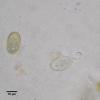
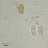
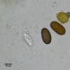
I think I can solve the mystery of the third fungus with the brown ovoid conidia. Yesterday I collected some dead leaves of P. laurocerasus at the parking lot of a supermarket and kept them over night in a moist box.
Besides the Trochila I found tiny black spots that turned out to have an ostiolum and produced the same kind of conidia as in Hannie's mount.
This third fungus shoud be Diplodia laurocerasi Westend.
Best regards
Martin
Thank you Martin, there come some light in the darkness. I had this leaves once before under the bino and found the same cells as yours in the first picture of you. I thougtI did somerthong wrong.
Thanks again, Hannie

Grove 1937: 52 took D. laurocerasi as a synonym for D. tecta. But this is not adopted by IF or MB.
Does someone have it?
Update: Superfluous question. I did not realize that the figures on the plate are not in numerical order, all is complete....
Best regards
Martin
Hello all,
I did my homework again today with beteer results (I think)
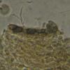
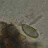
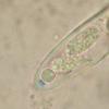
Here are some new picrures. I made the leaves wet and put them yesterday in a little box. This afternoon it didn't work in the beginning and I wanted to give it up. Just one leave, I thougth and I'm happy I did so.
Thanks everybode for your help.
Best wishes Hannie

Zotto
Hello Zotto, I'm glad that this got better. You'r right, in the last two pictures I used a drop of Melzer so Im coud see if the tops of the asci got blue. Thanks
Hannie

http://www.gbif-mycology.de/HostedSites/Baral/IodineReaction.htm
Zotto
Zotto, that's right the spores are also in Melzer.
I've heard about Lugol. Last summer I was visiting a lot of farmers in this area. Someone told me that I could get it at a farmer who used it with his cows. No farmer had heard about Lugol and afterwards I forgot is.
I will read the site you linked. In a quick look I think that when used KOH and afterwards with Lugol the ascitop colors red? Does it has to be KOH 5 or 10 %? Thanks again.
Hannie

No, not with but without KOH you can get a red reaction, depending on the fungus. After KOH you will have only blue and thus you loose a valuable character.
Melzer suppresses the red reaction, that is why hemiamyloidity and especially the red reaction was overlooked for so long.
Melzer also masks the spore contents as you see in your example. Oil drops in spores are very important characteristics.
Zotto

This spring I have been asked to lead a workshop on "Ascomycetes for Beginners" for the British Mycological Society - trust me I will make sure everyone knows the value of Vital Taxonomy ;-)
Chris
Zotto and Chris,
I always start with the preparation with water, mostly afterwards I use melzer. It's difficult for me in what order I have to use other stuff. It should be nice we als had a course "Ascomycetes for Beginners". We (I ) could learn a lt of it.
Groeten
Hannie


