02-05-2026 12:42
Alain BRISSARDBonjour à tousJeuidi 30 avril dernier on m'a remi
02-05-2026 13:06
Pauline. PennaBonjour Please can someone help me with this id
01-05-2026 22:45
 Thierry Blondelle
Bonjour à tous, Une récolte sur bouse séchée d
28-04-2026 20:07
 Lothar Krieglsteiner
... on twig in the air at standing Ceratonia siliq
14-04-2026 05:32
Ethan CrensonHi all, A few weeks back a friend pointed out som
28-04-2026 20:33
Vitus SchäfftleinHello, I found Trochila ilicina on Ilex aquifoliu

30-04-2026 10:28
Rot BojanHello, by appearance I would say that I am dealing
27-04-2026 18:48
Tony MoverleyCollected 23rd April 2026, Norfolk, EnglandSwarms
27-04-2026 20:52
Lothar Krieglsteiner
Found on hanging tiwg of Olea europaea in dried-ou
Thanks in advance!
Bharati

>"this is a very common error, apparently impossible to erase. Not less mature but alive!
>You have plenty of ejected living spores, which are fully mature, as I defined the term mature (1992).
>"It seems to me that the mature spores are only 1-septate and get 3-septate only when overmature. But >this you can find out when testing the numerous ejected spores with MLZ."
Will do.
>"I am not sure why your first micro image shows dead elements only, although being in water. I suspect pressure or drying and rewetting´as reason?
>Your ascus measurements sound like referring to dead asci, considering the low width. I see no living ascus in your pics."
Pressure is what I suspect. I will review my current collection of photos (I have more and of different mounts) and better yet make a new set of slides after I read the 1992 paper.
>"The spores are quite large, so L. lonicerae is impossible."
Noted. Based on the Raitviir key I get from 1-->2-->3-->9 which would rule out L. lonicerae – am I missing something again?
Bharati
Doi 10.5943/mycosphere/15/1/25) and copied an account by Peter Johnston which I found interesting:
I did not forget my homework assignment on this collection. :-) Life sometimes interferes with mycological pursuits.
I updated my inaturalist observation to include: an image of the dead spores showing septation (see photo 6; and one showing living asci with those guttulate spores (see photo 7); and relabeled photo 5 to reflect the fact that they are living spores.
Thanks again,
Bharati
All this is very illuminating for me – what structures within spores are what and what is visible in what media - , so some more questions in my ongoing tutorial: :-)
1) What do you make of the septation in the version with KOH?
2) In your Google Drive collection for L. subflavidum
a) How would you interpret the MLZ image embedded in 100x_spo_koh+rc_24038_apomad0420.jpg?
b) In the image titled Lasiobelonium subflavidum cf., 23.VI.94-2a.jpg, (JM Castro 23-3-94), the note below the spore drawing says that 1 septum is clear, 2 (others) are less clear. The next drawing from this series, labeled 2b shows a variation in septa.
Thanks
Bharati
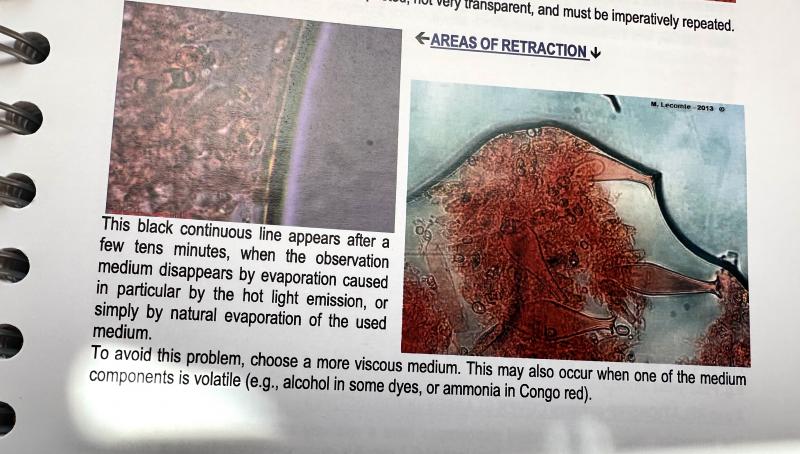
I am attaching a photo of that paragraph from p. 77, in case others don't have a copy.
On pp. 41-42, he includes some options for viscous media including glycerinated water.