29-11-2024 21:47
Yanick BOULANGERBonjourJ'avais un deuxième échantillon moins mat

27-02-2026 17:51
 Michel Hairaud
Bonjour, Quelqu'un peut il me donner un conseil p

27-02-2026 16:17
 Mathias Hass
Hi, Found this on Betula, rather fresh fallen twi

27-02-2026 12:56
Åge OterhalsFound on fallen cones of Pinus sylvestris in midle

27-02-2026 11:21
 Yannick Mourgues
Hi to all. Here is a specie that can may be relat

26-02-2026 15:00
Me mandan el material seco de Galicia, recolectada

24-02-2026 11:01
Gernot FriebesHi,found on a branch of Tilia, with conidia measur

23-02-2026 11:22
Thomas Læssøehttps://svampe.databasen.org/observations/10584971
• The lack of condiomata with so many apothecia seems distinct to many other examples fruiting on smaller and less decayed pieces of wood at the moment.
• Spores, structure of the medullary, abundant druses, and less inflated paraphysis apices support section cylichnium.
• Consulting Baral's key (2000), it must be A. cylichnium var. cyclichnium, based on the croziers (and almost uninflated and longer paraphysis apical cells, habitat, and maybe the conidial LBs).
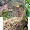
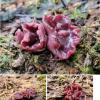
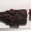
Habitat: On the top and sides of a large angiosperm trunk, possibly Fraxinus, sodden and well decayed, lying on the ground, damp and shady area near several streams, flooding in very wet weather, mixed deciduous woodland, Low Weald, early October, after lots of rain, close to flooding.
Associates: A variety of fungi (espc. basidios) have been seen fruiting from the trunk throughout the year, currently Galerina marginata noticeable.
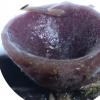
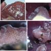
Apothecia: < 1 x 2 cm, usually 3-10+ caespitose,10+ groups on the trunk, purple, mostly cupulate to more discoid, appearing sessile or with a short and thick stipe, superficial, opaque, shape and margin appear variable when larger and more mature, possibly linked to exposure, margin round to several distinct lobes to undulating and densely scalloped, usually narrowing to a darker edge, receptacle paler, more pinkish, with an uneven-fleshy appearance, seen as many whitish lumps on the surface under low magnification (possibly druses), disc darker, vinaceous, waxy-gelatinous appearance, +/- uneven, sometimes with wart-like islands, initially convex, becoming plano-convex, sometimes eventually concave and umbilical, a light tomentum visible all over under low magnification, tough-gelatinous texture when sliced, internally lower part appears more gelatinised with a wet appearance.
Asci: Cylindrical-clavate, croziers, sometimes with a downward basal protrusion, apex bluing, rings bb or rarely rb, form looks typical for Ascocoryne, 2-2.5 seriate when turgid, apex rounded when turgid, becoming more conical-subtruncate when flaccid, acute-rounded when immature and sometimes rings not present or inamyloid.
Spores: Ellipsoid-fusoid, ends rounded-acute, often slightly heteropolar with base more elongated, inequilateral to very slightly curved in profile view, rarely slightly s-shaped or alternately curved, lower spores seem longer, filled with many smallish LBs (no large LBs seen), OCI 4-5 when fresh and mature, apparently uninucleate, some mature spores in a dead ascus appear to have 3-5 septa, one free spore seen germinating after some hours in water, with an ellipsoid outgrowth from each pole.
Spores free in water or in mature vital asci: (21.2) 21.8-26.8 (27.7) × 5.0-6.1 (6.3) µm, Q = (3.9) 3.92 - 5.1 (5.2), n = 13, mean = 24.4 × 5.5 µm, Q mean = 4.5.
Paraphyses: Narrow filiform, not to very slightly inflated at apex, with many smallish VBs in upper part of apical cell, coalescing somewhat over time, lower part of hymenium looks pink and more vinaceous en masse, sometimes branching from the central septa.
Ectal: Text. primatica-globosa, gradual transition in shape from medullary, hyaline globosa at surface, underneath a layer of dense pigment, purplish, continues to the margin, chains of globose-obpyriform cells at the margin, several cells protruding, shortish hyaline hyphae extending from the surface, branching occasionally, some druses and many crystals on the surface.
Medullary: Text. intricata, hyaline but more pigmented toward subhymenium, low part with more narrow and gelatinised hyphae, in the upper part and close to subhymenium with broader and more dense hyphae, many druses (clusters) and crystals of varying sizes, individually approximately octahedral, some cells with a relatively large globose refractive body inside (SCB?).
Subhymenium: Pigmented, dense with paraphysis bases.
Excudate: Some hyphae appear pigmented but pigment also exuded, clumping in the excipulum and hymenium, strongly gelatinised hymenium (epithecium), pigment seems to dissolve somewhat in KOH and more slowly in water.
Anamorph: Seem to be related, conidiophores on the surface of the excipulum, like the protruding hyphae, (botryoid?) conidia growing in small bunches at septa or apices, on individual short thin stalks, conidia 2.3-2.8 µm diameter, hyaline, globose, with several small or one larger LB.

There is no evidence that these anamorphic structures are related, but I could also see no overt evidence of parasitism and the short hairs are present on all of the apothecia. I looked at a water mount that I left wet, and I found some more conidia growing in the same way but most of these are more elongated, cylindrical-obovoid.
 Sections-0009.jpeg