
05-06-2026 12:10
 François Freléchoux
Capitotricha sp. sur Lonicea caerulea Caractères
05-06-2026 11:02
Thomas Læssøehttps://svampe.databasen.org/observations/10596691

04-06-2026 11:36
Gernot FriebesHi,found on Vaccinium myrtillus.Asci: IKI –, 8-s
19-05-2026 10:27
Patrice TANCHAUDBonjour, récolte récente sur terre retournée i
04-06-2026 18:39
Gernot FriebesHi,I collected this species in two different locat
22-05-2026 13:29
Gernot FriebesHi,I am curious to hear your opinion on this mater
04-06-2026 10:50
François Freléchoux
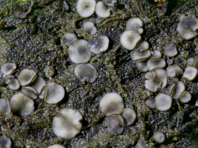
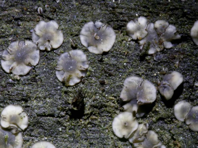
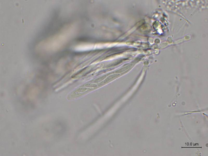
Bonjour, J'ai trouvé hier un petit asco observé
Mollisia lividofusca ?
Thierry Blondelle,
11-02-2024 17:40
 Hi
HiI would like to have your opinion on this harvest of Mollisia on fir branch peeled in a humid environment
Floriform apothecia up to 3 mm, gray in color but whitish discolored.
Subiculum++
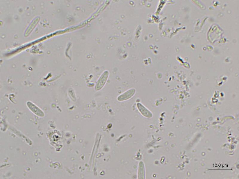
Spores 8-12 x 2.5-3
Asci 44-51
IKI bb
KOH-
OIC 0 to 1
Clavicated marginal cells
Subhymenium rather dark in color
I turned to M. lividofusca.
What do you think ?
Ingo Wagner,
11-02-2024 18:33

Re : Mollisia lividofusca ?
Hello Thierry!
You found it on dead wood in airspace, is this right?
Your thought with M. lividofusca is very likely.
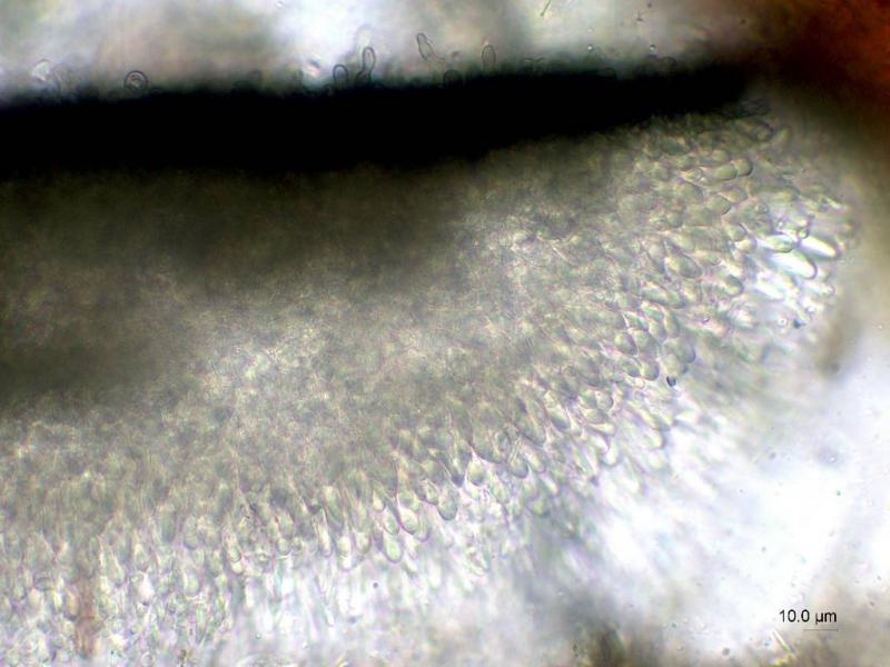
Macroscopically, the brown exterior of young apos up to the edge (margo) fits well, and also the brownish color of the fruit layer when it dries.
In the lower part of the ascus you can also see the longer, curved spores.
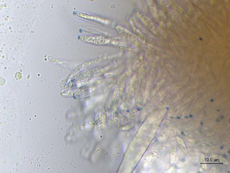
However, viewed microscopically, the find is in bad condition.
You found it on dead wood in airspace, is this right?
Your thought with M. lividofusca is very likely.
Macroscopically, the brown exterior of young apos up to the edge (margo) fits well, and also the brownish color of the fruit layer when it dries.
In the lower part of the ascus you can also see the longer, curved spores.
However, viewed microscopically, the find is in bad condition.
You can try a cross section, this species sometimes has a brown subhymenium in this state.
https://asco-sonneberg.de/pages/gallery/subhym-dunkel-bei-lividofusca39765.php
Greetings
Ingo W
Thierry Blondelle,
12-02-2024 17:55
Re : Mollisia lividofusca ?
Thanks a lot Ingo for your appreciation
it was found on the side of the branch facing the ground
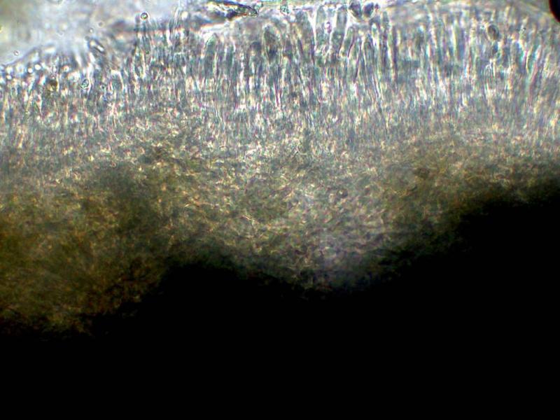
My cross section is not really ideal for observation.
I'll try a better one.
Are the longer and curved spores at the base of the ascus specific to lividofusca?
Thierry
it was found on the side of the branch facing the ground
My cross section is not really ideal for observation.
I'll try a better one.
Are the longer and curved spores at the base of the ascus specific to lividofusca?
Thierry
Ingo Wagner,
12-02-2024 20:23
Re : Mollisia lividofusca ?
Hello Thierry!
"It was found on the side of the branch facing the ground."
And the branch touched the ground, where the apos grew?
"Are the longer and curved spores at the base of the ascus specific to lividofusca?"
I missed them in your spore picture AND there are species whose spores are more uniformly straight.
"My cross section is not really ideal for observation"
You can pick out a piece of the fruit layer. Normally the subhymenium is attached to it. Best seen at about 100x magnification.
If you don't see this, it doesn't matter. I often don't recognize the darkness of subhymenium in Mollisia lividofusca.
Greetings
Ingo