
15-05-2026 13:33
Sylvie Le GoffBonjour à tousJe serais très reconnaissante enve
16-03-2011 14:31
roman vargas albertoHi. I would like some opinion about this Peziza
14-05-2026 05:36
Ethan CrensonHi all, I haven't paid much attention to Lachnu
10-05-2026 23:17
 Andreas Gminder
Hello,today we found in a moist steep decidous for
11-05-2026 12:32
 Bernard CLESSE
Pourriez-vous m'aider à identifier cette héloti
13-05-2026 15:26
 François Freléchoux
Bonjour,Voici une récolte faite il y a quelques j
12-05-2026 15:41
 Nicolas VAN VOOREN
Dear Ascolovers, especially interested in Pezizale
13-05-2026 12:05
 Thierry Blondelle
Bonjour à tous,J'aimerais avoir confirmation de c
28-04-2026 20:07
 Lothar Krieglsteiner
... on twig in the air at standing Ceratonia siliq
27-04-2026 20:52
Lothar Krieglsteiner
Found on hanging tiwg of Olea europaea in dried-ou
Mollisia sp.
B Shelbourne,
16-01-2024 12:19
 I've had a go at identifying the Mollisia that I found the chytrid parasitising.
I think it could be Mollisia lividofusca (Fr.) Gillet, 1882. Any feedback appreciated.
This species seems to be 'incorrectly' named Tapesia lividofusca in Index Fungorum (later publication).
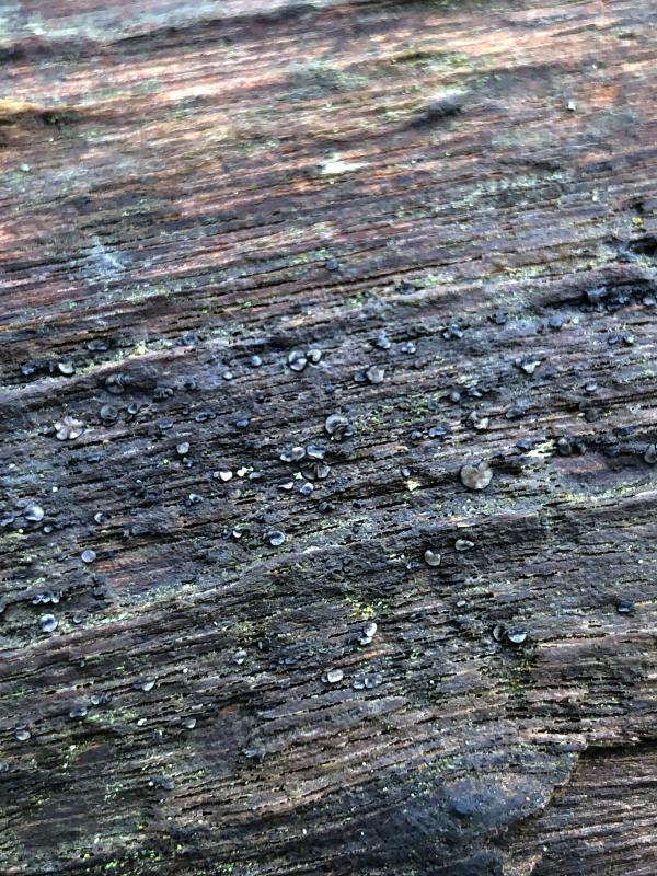
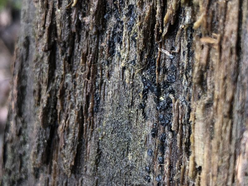
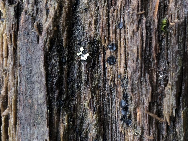
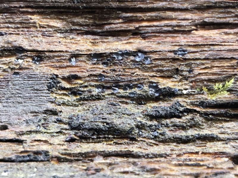
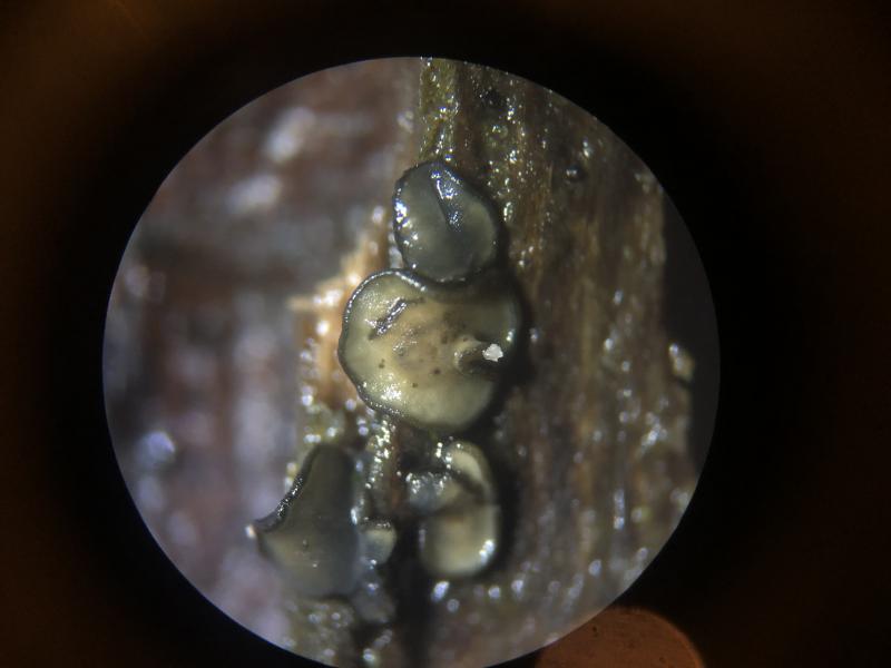
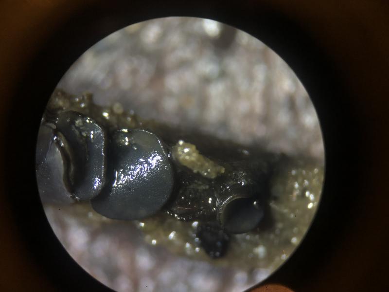
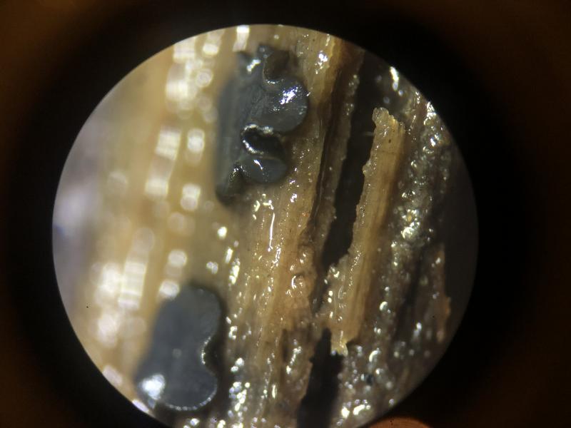
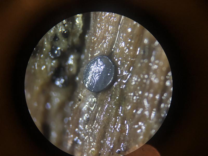
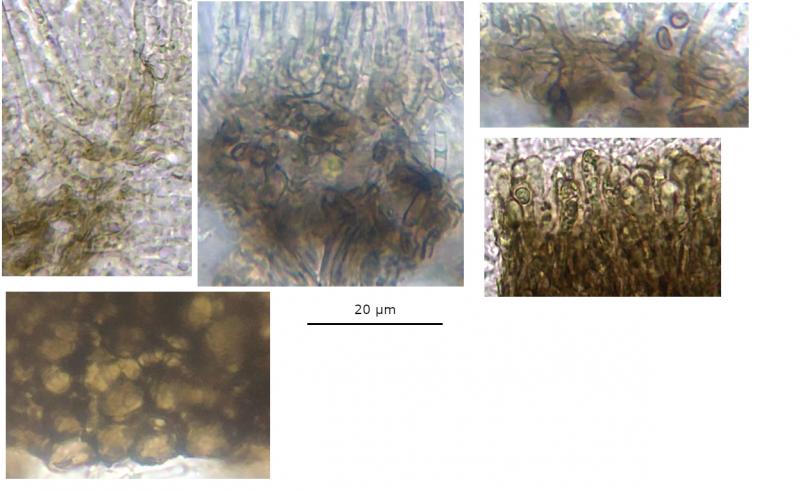
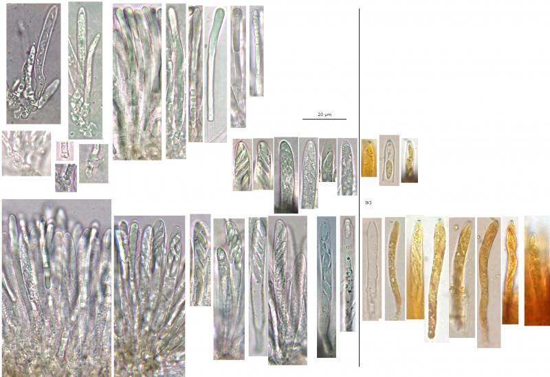
Ascomata: Apothecial, discoid, <= ~2 mm diameter, blackish-grey exterior, margin concolourous and uneven, disc pale whitish-grey when dry and darker bluish-grey when hydrated, margin in-rolled even in maturity, sessile, starting more urceolate, becoming acetabuliform, eventually +/- plane, often flexuous to irregularly lobate in maturity, glittering/micaceous especially exterior.
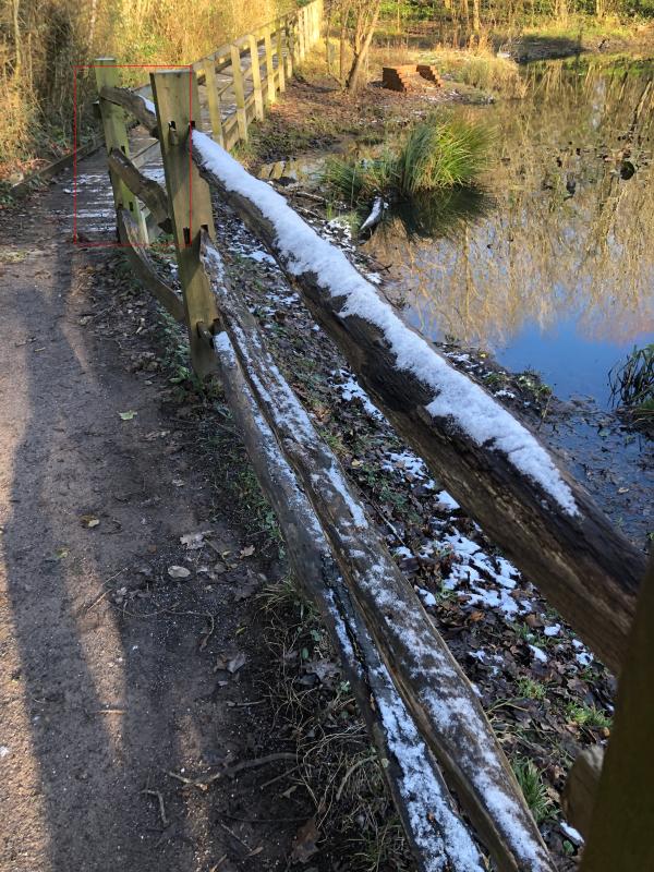
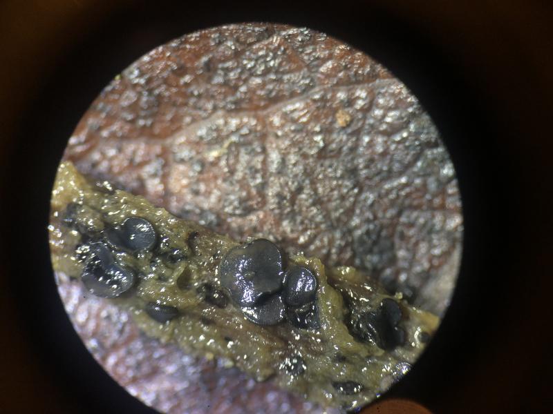
Habitat: Observed twice in January, gregarious to clustered, on decayed wooden fence rails, unidentified deciduous wood, top (more exposed) and middle rails of fence, ~0.5 – 1.25 m from the ground, presumably xeric, top and bottom sides of rails, decorticated parts, often in depressions and crevices, next to a pond in mixed deciduous woodland, southern England.
Some snow settling between observations, no noticeable effect on ascomata that were collected after snow but free from snow when collected.
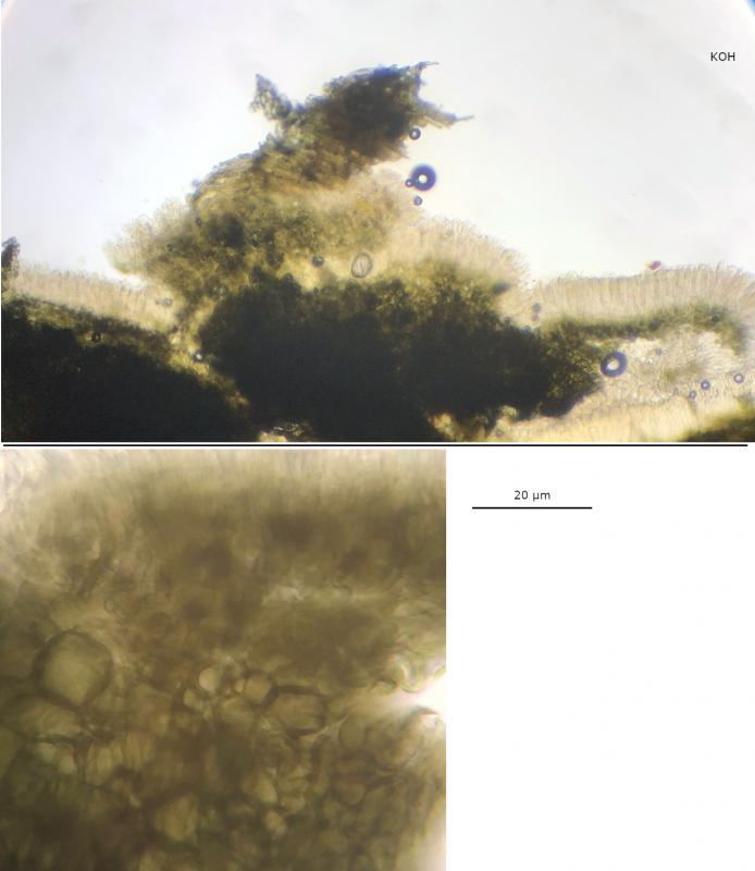
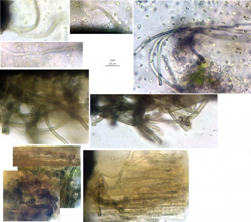
Associated: Some ascomata parasitised by intra-hymenial Chytridomycete sp., evidenced by many sporangia appearing like asci in the hymenium, significantly supressing the host asci, and some motile zoospores ejected in water mount (see http://www.ascofrance.com/forum/77885/some-very-swollen-asci-on-a-suspected-mollisia-sp).
Subiculum appears to be part of an algal and dematiaceous biofilm that is visible on substrate around many ascomata, small/young thalli of lichens and bryophytes close by, tiny orange ascomata from an Orbilia sp. (indistinct macromorphology) also on the middle rail, basidiomata of Stereum cf. hirsutum on other corticated rails.
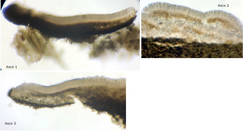
Preparation: Three ascomata from the middle rail examined microscopically, the first ascoma was stored for several days (in a damp container) prior to examination, but the following two were collected later and examined within hours of collection, water was used to mount unless otherwise stated.
Note that hymenial characters have been confirmed in the unparasitized ascoma (2), because clearly distinguishing the parasite 'rhizoids' has not been possible.
IKI: Apical ring bb, standard reactions elsewhere.
KOH: Ectal excipulum more brownish-olivaceous in thin sections, maybe a similar reaction in subhymenium, no reactions from hymenium.
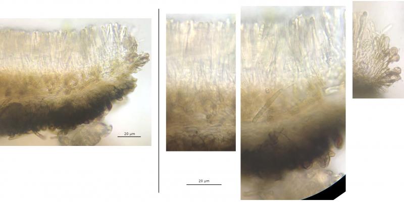
Hymenium: Ochraceous en masse, no epithecium/exudate or gelatinous matrix identified.
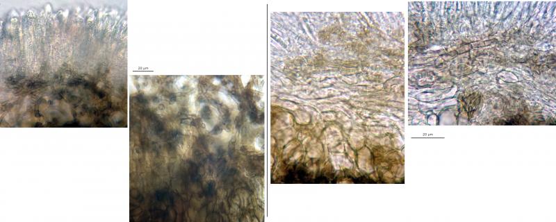
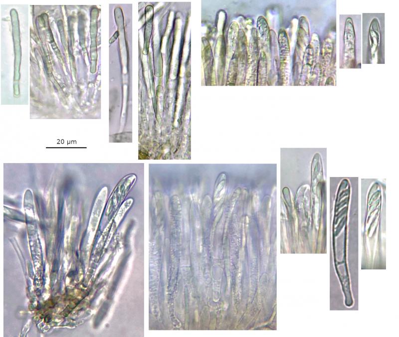
Paraphyses: Filiform, sometimes gradually widening toward apex, rarely slightly swollen at apex, apex rounded, almost-completely filled with large cylindrical yellowish-hyaline VBs, 3 - 4 septa towards base, terminal cell longer.
Asci: 8-spored, croziers +, apex rounded, maybe slightly more truncate apex in front view.
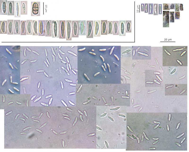
Vital – 2 - 3 seriate, single spore at apex, mildly clavate, pars sporifera ~30 - 40%,
63.2 - 77.7 × 6.2 - 7.5 µm, N = 8.
Dead – usually uniseriate, more cylindrical, pars sporifera ~70-90%, often opaquely full of tiny LBs,
45.6 - 66.3 × 4.1 - 5.6 µm, N = 8.
Spores: Cylindrical to more allantoid or fusiform, occasionally narrowly-clavate, occasionally narrow-end slightly curved, inequilateral in profile view, aseptate, nucleus often visible, occasionally a few tiny shadowy LBs towards poles, OCI 0 (- 1).
Vital spores measured in squash mount (shorter spores probably immature) –
(7.6) 8.7 - 12.9 (13.9) × (2.3) 2.4 - 2.9 (3.2) µm,
Q = (2.6) 3.2 - 4.9 (5.1), N = 32,
Me = 10.5 × 2.7 µm, Qe = 3.9.
Subhymenium: Loose brownish hyphae.
Medullary excipulum: Hyaline to brownish, textura porecta?
Ectal excipulum: Blackish, textura globosa-angularis, some thick-walled.
Marginal cells/hairs: Brownish, usually one cell protruding, apices pyriform with large globose VB, or more cylindrical-hyphal with several medium-size VBs.
Subiculum: Anchoring hyphae brownish to hyaline, hyphae thick-walled, septate, clear contents, lots of hyphae found in/on the substrate with algae.
Possible anamorph: Diplococcium-like, seems micronematous, conidia - brownish, clear contents, 1 - 2 septate, basal cell usually wider, slightly constricted at some septa, acropetal, catenate? (maybe chains of two found),
4.7 - 9.4 × 3.1 - 5.7 µm,
Q = 1.4 - 2.2, N = 7,
Me = 7.1 × 4.3 µm, Qe = 1.7.
Ingo Wagner,
16-01-2024 13:12

Re : Mollisia sp.
Hello!
I think you are right with M. lividofusca.
Greetings
Ingo
I think you are right with M. lividofusca.
Greetings
Ingo
B Shelbourne,
16-01-2024 14:16
Re : Mollisia sp.
Thank you very much for looking, Ingo. I don't think I would have got so close without your wonderful key!
Ingo Wagner,
16-01-2024 16:05
Re : Mollisia sp.
Unfortunately the key is still very buggy.
I will add pictures in the future, for example of the spores.
The genus Mollisia is quite difficult.
I will add pictures in the future, for example of the spores.
The genus Mollisia is quite difficult.