
28-04-2026 20:33
Vitus SchäfftleinHello, I found Trochila ilicina on Ilex aquifoliu

30-04-2026 10:28
Rot BojanHello, by appearance I would say that I am dealing
27-04-2026 18:48
Tony MoverleyCollected 23rd April 2026, Norfolk, EnglandSwarms
14-04-2026 05:32
Ethan CrensonHi all, A few weeks back a friend pointed out som
27-04-2026 20:52
 Lothar Krieglsteiner
Found on hanging tiwg of Olea europaea in dried-ou
28-04-2026 22:51
 Bernard CLESSE
Bonsoir à toutes et tous,Pourriez-vous m'aider à
29-04-2026 08:01
Lothar Krieglsteiner
... on twig attached to small tree of Citrus auran
29-04-2026 10:44
Lothar Krieglsteiner
growing at moist, drying-out soil at the side of a

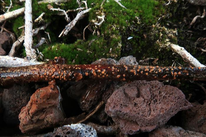
 Hello.
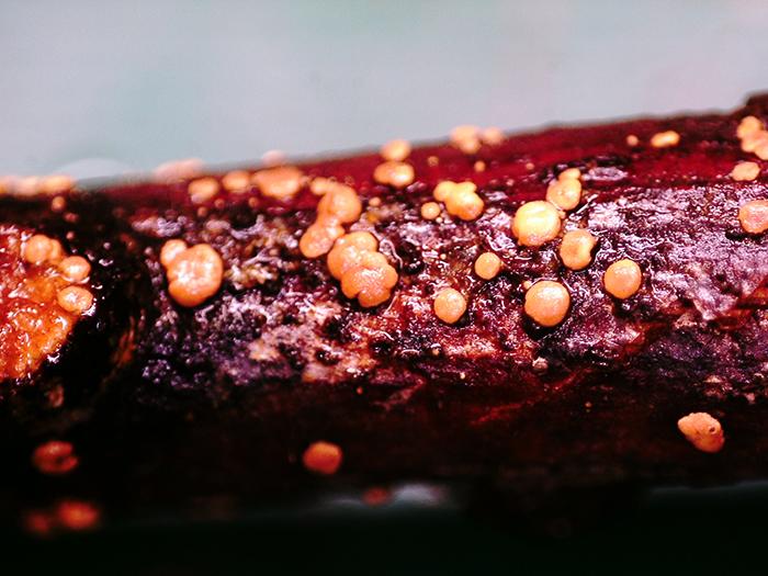
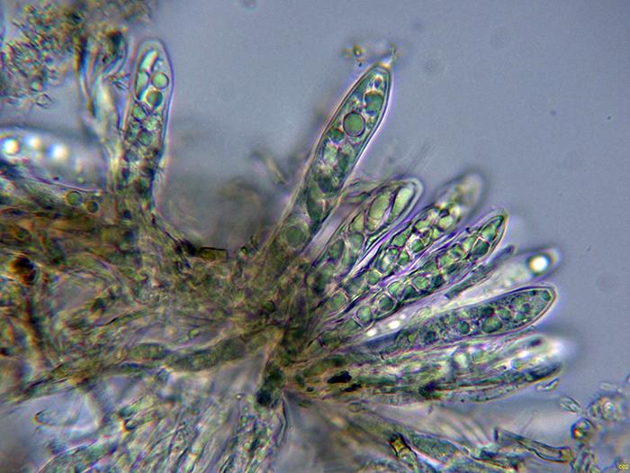
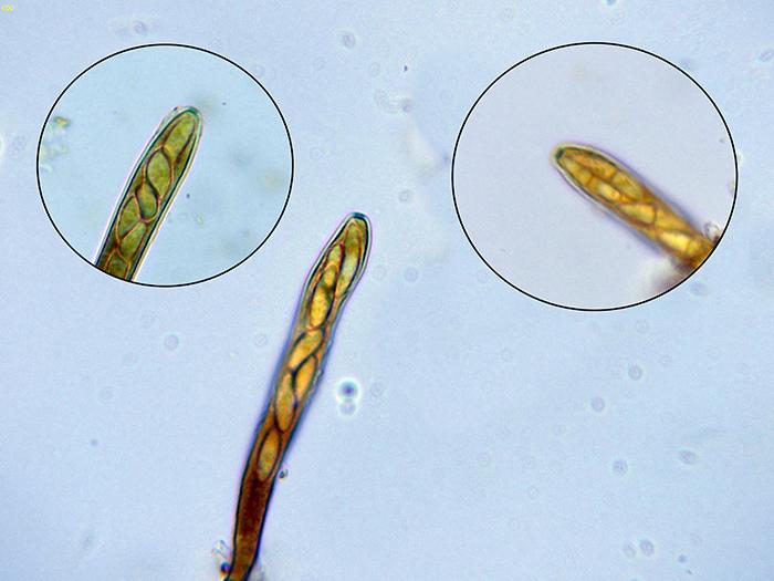
Some apothecia sprouting massively on a thin trunk of Laurus nobilis.
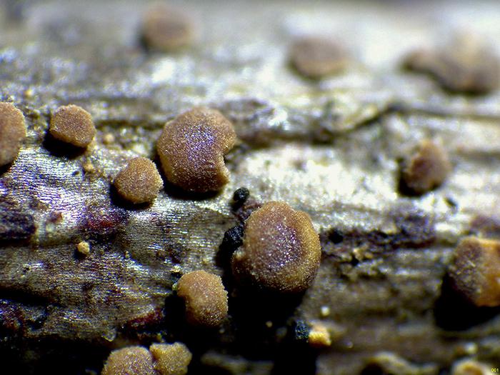
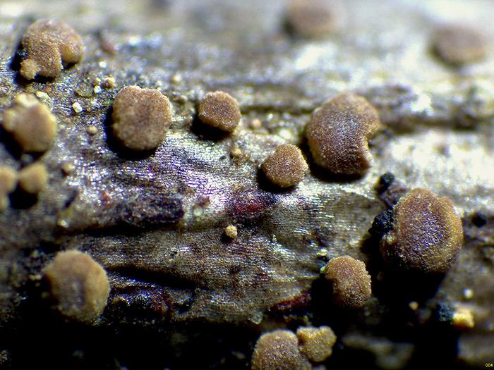
Spherical in shape, somewhat flattened, orange when wet and greenish brown when dry. with a diameter of between 0.2 to 0.7 mm.
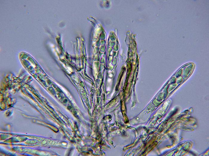
Octosporic asci, with uncinules at their base and amyloid reaction of their apical apparatus.
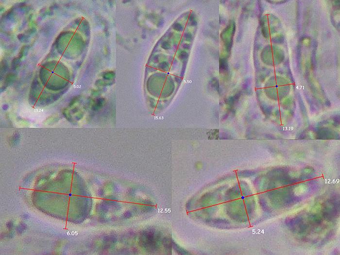
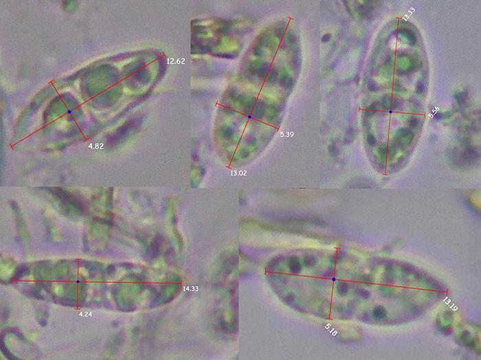
Moriform ascospores, poorly septate and with free spore measurements of (12.5) 12.6 - 14.3 (15.6) × (4.2) 4.7 - 5.6 (6) µm.
Based on the data obtained, everything seems to fit with what could be considered Claussenomyces prasinulus, but I already studied Clausennomyces prasinulus last year and the reaction of the apical apparatus to iodine was negative, and I can't find anything more similar than Claussenomyces or Vexillomyces.
Any opinion from you will be well received.
Thank you very much in advance.
Kind regards.
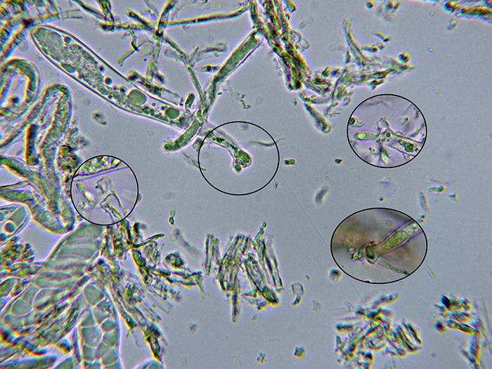
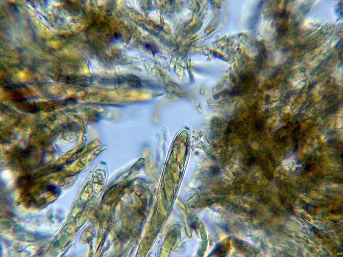
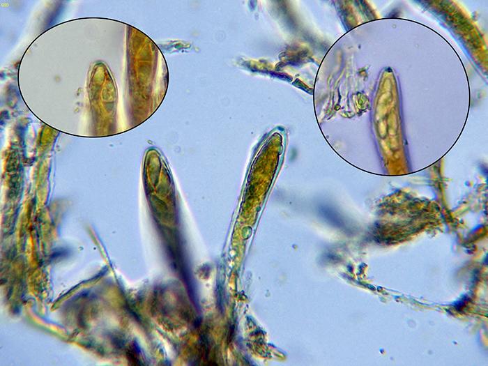

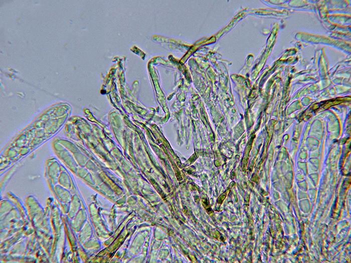
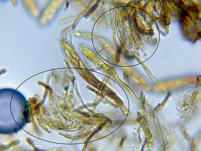
The apothecia were not at their best and unfortunately the ascos were dead.
I add a couple of images of the paraphyses, one in water and the other in Lugol, these filiform, septate paraphyses, with intracellular pigment, some branched and that do not protrude above the level of the asci.
Again thank you very much for your help.
Kind regards.
It fits very well with the little information I have been able to find on Rodwayella sessilis.
Kind regards.
The information in your folder is very interesting, including a very detailed description of microscopy.
Kind regards.