28-04-2026 20:07
 Lothar Krieglsteiner
... on twig in the air at standing Ceratonia siliq
04-05-2026 18:13
 Stephen Martin Mifsud
ID request for what seems to be a true aquatic fun
04-05-2026 16:39
Stephen Martin Mifsud
ID request: This specimen was collected in Malta o
04-05-2026 09:50
 Castillo Joseba
Me mandan el material seco de Galicia,(España) re
02-05-2026 12:42
Alain BRISSARDBonjour à tousJeuidi 30 avril dernier on m'a remi
02-05-2026 13:06
Pauline. PennaBonjour Please can someone help me with this id
01-05-2026 22:45
 Thierry Blondelle
Bonjour à tous, Une récolte sur bouse séchée d
14-04-2026 05:32
Ethan CrensonHi all, A few weeks back a friend pointed out som
28-04-2026 20:33
Vitus SchäfftleinHello, I found Trochila ilicina on Ilex aquifoliu
 I think this could be in the Mollisia olivaceocinerea group but I'm not sure how to separate the teleomorphs of the three species M. oblonga, M. nodosa, and M. mallochii. Also, the OCI seems quite variable or hard to interpret with no experience, and there are some similarities to M. cinerea group. Any feedback is appreciated.
The genus/family seems to be another tricky one. I have tried to use an unpublished key by A. Gminder (hard to get a good match), a (more recent one) by I. Wagner, and comparison with some of the examples in the 'Mollisia on wood & bark' folder in the drive of H.O. Baral.
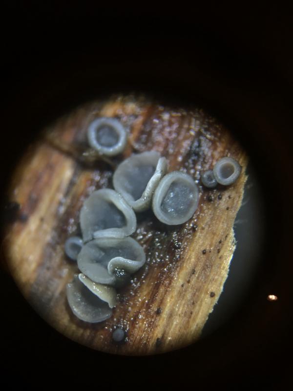
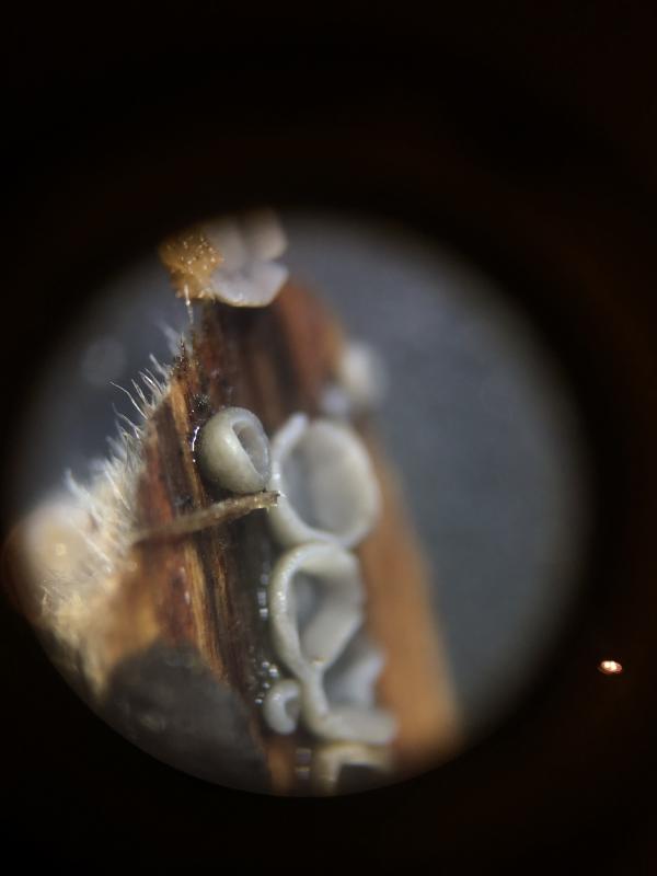
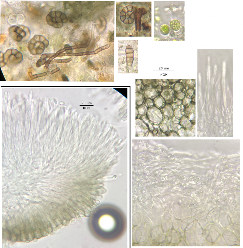
Preparation: Kept on wood in the fridge for several days. Slides prepared from three ascomata in the same cluster, mounted in tap water unless otherwise mentioned. My first attempt at staining/reactions, ~1% IKI (5% diluted ~1:5 with tap water) allowing a drop to pass under the cover slip of a water mount, and 3% KOH mounted directly.
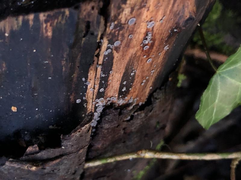
Habitat: Collected around one week ago (December), on hygric (almost saturated) unidentified deciduous wood, decorticated parts, in a pile of logs, mixed deciduous woodland, southern England. With teleomorphs of Calycina cf. citrina, Lachnum impudicum, Orbilia eucalypti, and Trichoderma strictipile on same wood pile. Some trebouxoid algae and helicoid conidia (seem different to M. nodosa microsclerotia) found around base of an ascoma.
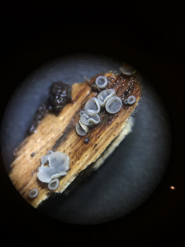
Ascomata: Apothecial, discoid, initially cupulate to urceolate, then +/- patelliform with thin rim, eventually often plane, mostly not very lobate in maturity, pale grey to whitish margin, darker grey interior, brownish exterior lighter towards margin and smooth, overmature more pale and margin less defined, <= ~1mm diameter, sessile, gregarious and some crowded.
Reactions:
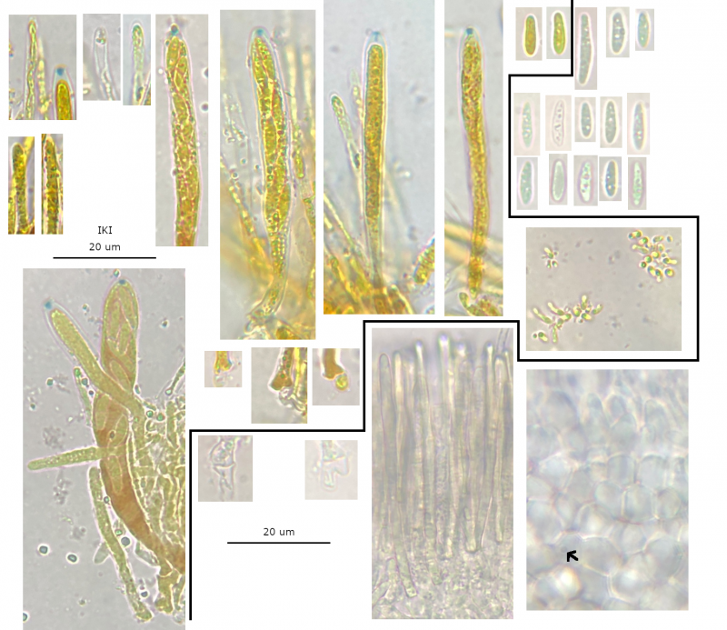
• IKI: apical rings bb 2?, cytoplasm of some asci going red-brown (depending on vitality?), cytoplasm of spores and paraphyses yellow.
• KOH: asci and paraphyses -, ectal excipulum greenish/olivaceous.
Subhymenium: Hyaline, densely woven hyphae.
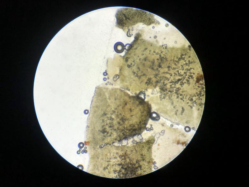
Ectal excipulum: Textura globose to angularis, except at margin globose to more isodiametric and hyphoid, some cells with a central guttule visible, marginal apical cells mildly clavate and protruding, brown up to margin, darker (almost black) at base.
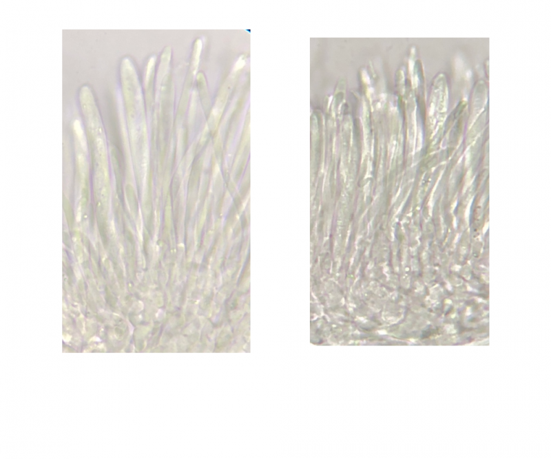
Marginal hairs: Cylindrical-clavate, plain, septate, (closer to hymenium some with longer irregular/asymmetric lanceolate apices?).
Asci: Cylindrical to mildly clavate, 8-spored, croziers +.
Paraphyses: Cylindrical-filiform, gradually slightly enlarged towards apex, large yellowish VBs, terminal cell longer ~2-3x, some branching identified, a few with crystals/exudate outside in IKI after squashing but may be unnatural.
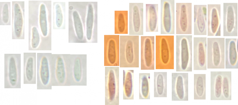
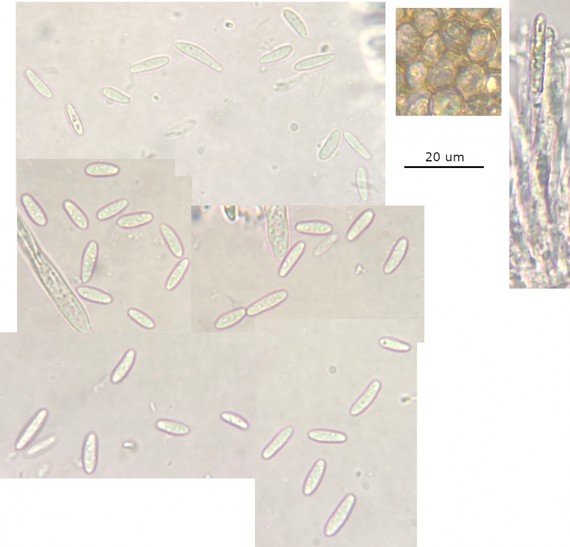
Spores: Narrowly-ellipsoid to allantoid (average q-value), inequilateral and occasionally slightly curved in side view, aseptate, multi-guttulate, often lots of small guttules and several slightly larger ones, guttules not restricted to poles, OCI (0.5?) 1 – 2 (3?), larger guttules shadowy?, greater range of lengths (one extreme spore ~15 µm long not included), measured
(7.5) 8.3 - 10.7 (12.1) × (2.2) 2.5 - 3.05 (3.1) µm,
Q = (2.8) 2.9 - 4.1 (4.2); N = 25;
Me = 9.4 × 2.8 µm, Qe = 3.4.
Subiculum: Maybe anchoring hyphae seen in macrophotos but not identified in slides.
Anamorph: No linked structures identified.


[quote]
I think this could be in the Mollisia olivaceocinerea group but I'm not sure how to separate the teleomorphs of the three species M. oblonga, M. nodosa, and M. mallochii.
[/quote]
There is no answer to that yet.
I don't think your find belongs to Mollisia olivaceocinerea (mallochii, nodosa, oblonga).
The spore shape is wrong.
The spores of Mollisia olivaceocinerea look in side view like a bread.
[quote]
Also, the OCI seems quite variable or hard to interpret with no experience,....
[/quote]
The OCI-evaluation is a big problem when the spores are tiny.
The best way to examine Mollisia is in a fresh vital stage, ideally when the asci shoot their spores.
In age it is possible, that the destruction of spore-vacuoles looks like oil.
Maybe you can examine the spores in Congo Red SDS? Drops of oil should remain there.
[quote]
• KOH: asci and paraphyses -,
[/quote]
No photo?
I have a collection that would fit microscopically, but macroscopically it looks different.
Has no official name or sequence, and is not in the key:
https://asco-sonneberg.de/pages/gallery/mollisia-clavispora-cf-230205-tr-iw195-01xsmjj42416.php?group_id=42416&position=87
Greetings
Ingo
It seems like it could be hard for me to resolve this on morphology. I certainly struggled to find a species in the keys that was a good match for both the spore size and other characters.
KOH reaction:
The last collage and the attached are all mounted in 3% KOH, but I can't be certain about a yellow reaction coming out in the mounting medium if it is only for a few seconds.
I still have some material so I can try the KOH again and (separately) the Congo red for the spores.
I was concerned about the vitality for the finer details of the asci and spores, it's a pity and I will consider this in future (and collecting ejected spores). Perhaps the best approach to progress here will be trying to collect some more vital material to examine (or just resign myself to genus).
It may well be that your species does not yet have a name.
This is really not rare.
To see the yellow reaction, I recommend 100x magnification.
https://asco-sonneberg.de/pages/gallery/mollisia-albogrisea-230507-diff-6nt-iw210-stma23048-dsm116342-mcol-04jj42177.php?group_id=42170&position=14
Greetings
Ingo
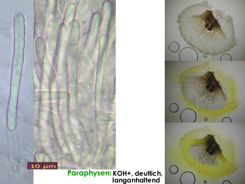
I tried the KOH again and there was no noticeable yellowing of the medium, the first photo is 150x and this appearance didn't change (I watched it at 100x).
I have also included some more photos of spores but focusing on the contents is certainly a challenge (I think getting a spore print will make this much easier). It seems there are 1 - 3 (4) smallish guttules, often towards the poles, and so far I would judge the OCI to be something like 0.5 - 1.
The CR (right side of photos) was also helpful but I need to practise using it as I didnt manage to get an even stain or clean it very well.