
12-11-2019 10:32
 Miguel Ángel Ribes
Hi againExactly at the same place than my previous
25-12-2019 17:54
Valencia Lopez Francisco JavierHola a todos/asEstas supuestas pezizas estaban en
28-07-2011 18:31
 Alex Akulov
Dear FriendsToday I made the pdf file of Velenovsk
12-07-2015 00:05
 Nedim Jukic
This one from the same locality as the previous on


30-05-2026 21:12
Philippe PELLICIERSur branche de mélèze (Larix) près de la neige,
31-05-2026 10:35
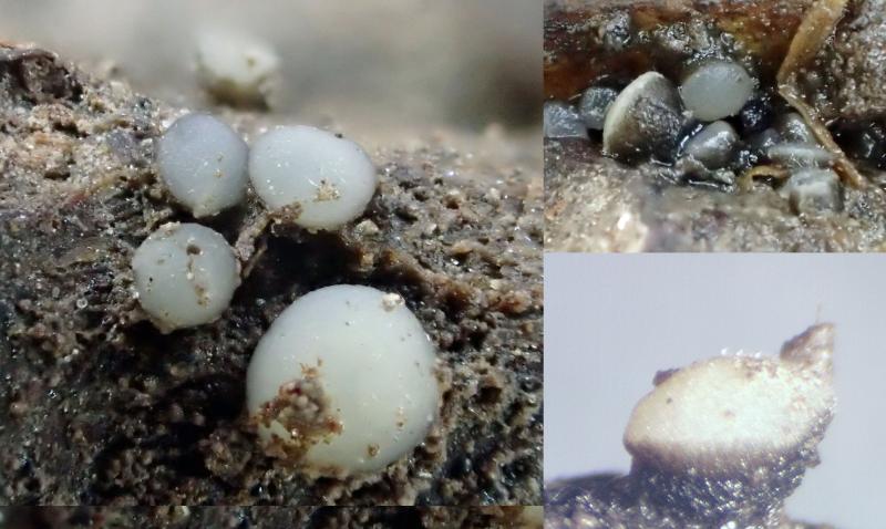
Hulda Caroline HolteHello,I collected this species growing on a rather
25-05-2026 16:35
 Bernard CLESSE
Bonjour à toutes et tous,J'ai trouvé récemment,
29-05-2026 15:35
daniel FERREBonjour à tous,Je voudrais votre aide pour cette
Vibrissea flavovirens?
Stefan Jakobsson,
08-07-2022 02:34
Can it be confirmed that this is V. flavovirens?
Hans-Otto Baral,
08-07-2022 08:39

Re : Vibrissea flavovirens?
I think the colour is not so important. Since I do not have an overniew on available measurements, I cannot easily say if such small measurements ever occurred, but I do not know a further species with these characters.
Stefan Jakobsson,
08-07-2022 11:49
Re : Vibrissea flavovirens?
Thank you!
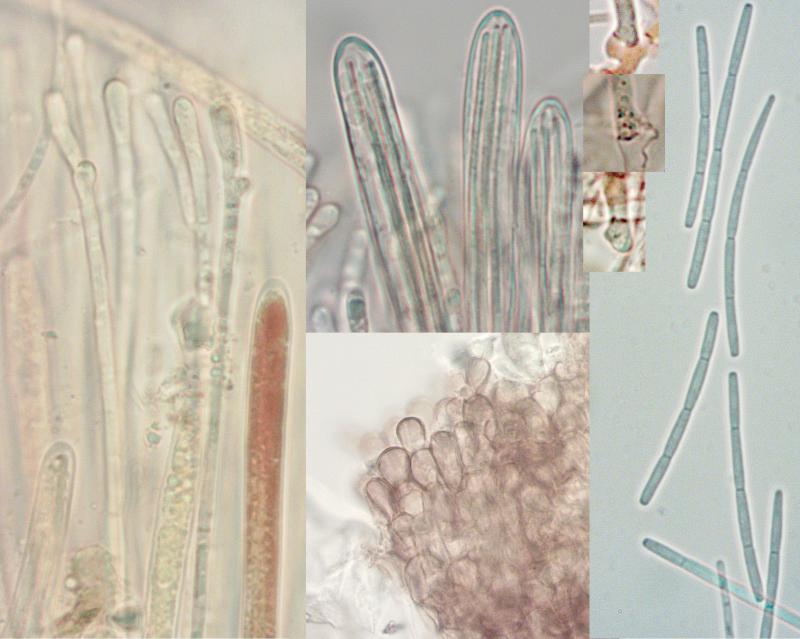
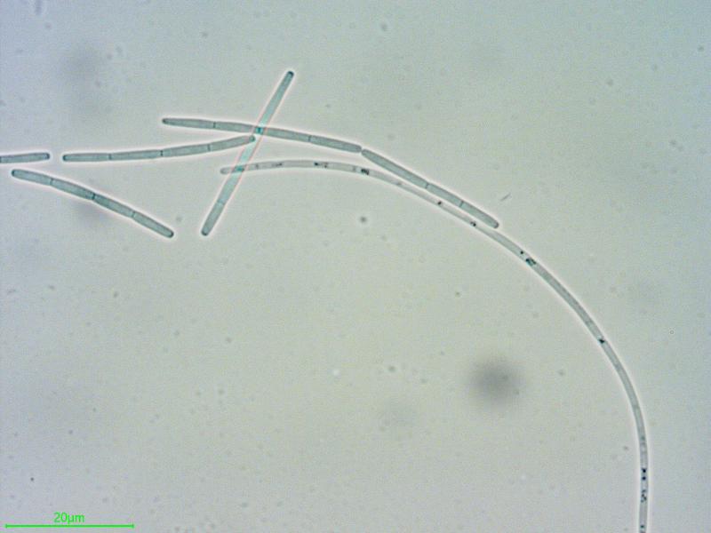
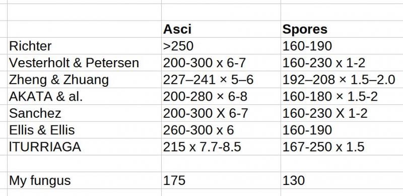
I looked up the asci and spore size in a few sources.
Hans-Otto Baral,
09-07-2022 09:42
Re : Vibrissea flavovirens?
Thanks for this survey. In my key I wrote for flavovirens:
asci *260-343 x 6.5-8.8 µm
spores *125-195 x 1.2-1.8 µm
spore fragments *(27–)30-42(–51) µm
Ascus measurements in your table are probably mostly in dead state, but the differences to living asci are apparently not very high.
In the type of V. minima Velen. on Salix, which I restudied and considered a synonym, I found asci +154 x 4.8-5.3 and spore fragments 27-48 µm. So your spore fragments are a bit shorter than usual.
In a collection from Sheffield (HB 9520) I measured spores *125-134 µm long (like yours), breaking into 4 part spores of *27-38 x 1.3-1.6 µm, 4-celled (ascus length not measured).
In the case there is a continuum of measurements among collections, I suspect that living asci much shorter than 260 µm also occur.
Identities in the literature are perhaps not certain. E.g. Zheng & Zhuang 2017 do not mention the number of spore cells and fragments, but the spore photo suggests flavovirens indeed.