
17-04-2026 19:16
 Enrique Rubio
Hi to everybodyI would appreciate any assistance r
14-04-2026 05:32
Ethan CrensonHi all, A few weeks back a friend pointed out som
17-04-2026 15:14
 Bruno Coué
Bonjour.Récoltes du 16/04/2026, sur feuilles mort
12-04-2026 15:52
Gernot FriebesHi,I'm looking for help with this anamorph collect
14-04-2026 21:52
Gernot FriebesHi,found on dead leaves of Carex elata. Conidia: 4
16-04-2026 22:09
Buckwheat PeteHello, I'd like to ask about this older specimen:
15-04-2026 19:33
Fátima Durán ManzanequeHi!! I need help, I found this Ascomycete but I d
14-04-2026 20:31
Gernot FriebesHi,can this be Psilachnum lateritioalbum on Phragm
12-04-2026 17:56
 Hardware Tony
Found on dead stems in February earlier this year
12-04-2026 12:22
 William Slosse
In a dune grassland in Oostduinkerke (Belgium), on
Erumpent on Wattle Part 2
Zuidland Peter,
25-06-2022 07:08
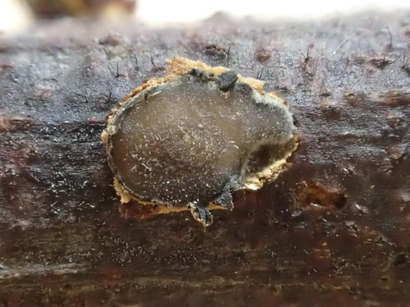
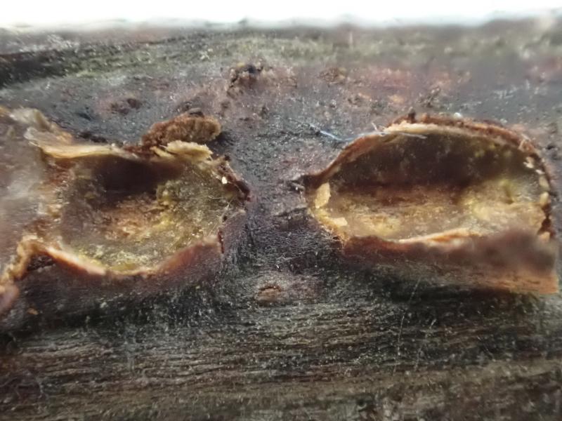
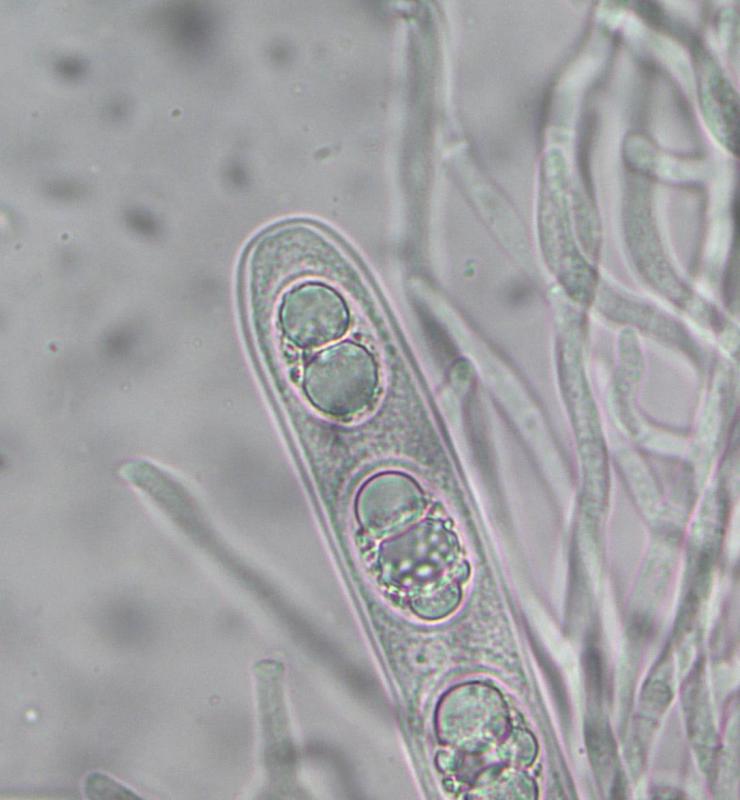
I view of possible confusion by poor images I representwhat I think is an erumpent I have found on Wattle. I have used Spooner's Heliotales of australia for years and I find nothing to help me in there, there is no budding like this on Heliotales according to him.
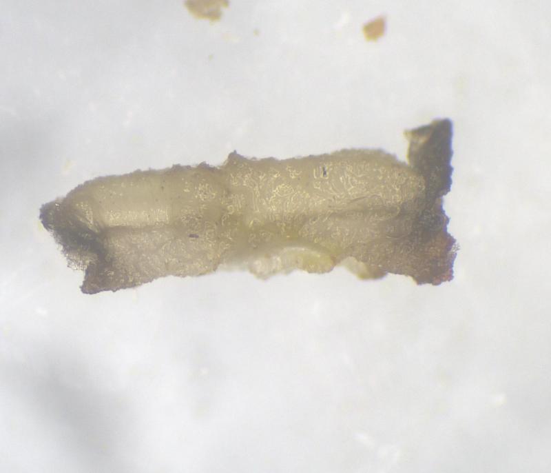
This sample forms under the bark, breaks through as it gets bigger and spreads across the wood surface. One image shows old craters with remnant excipulm around the edges under the bark.
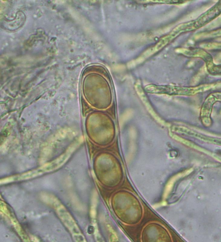
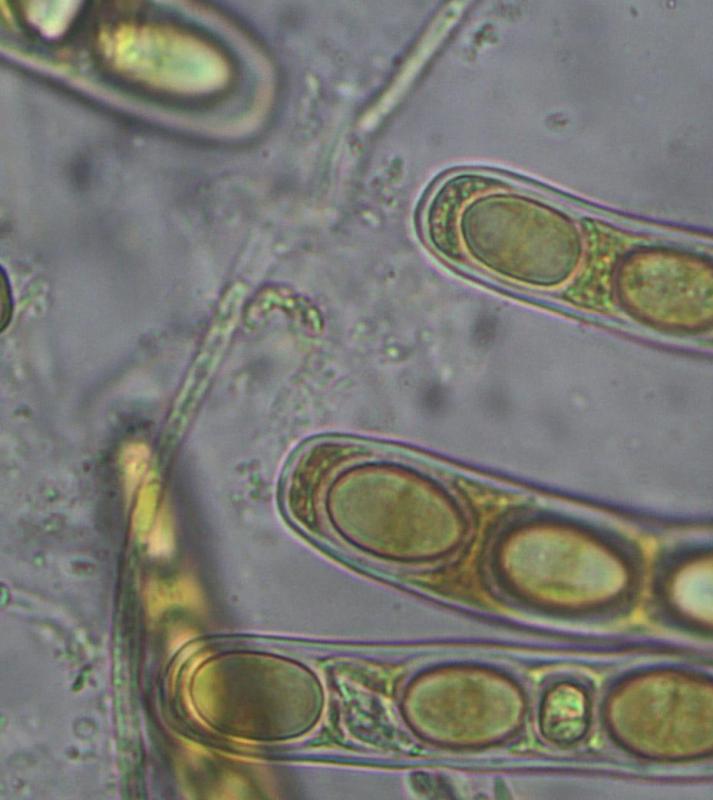
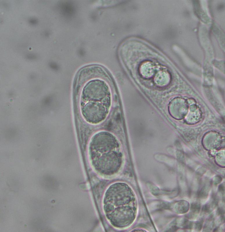
Fresh images from today, all microscopy in water; sample is being kept moist and cool for further work if needed.
SE Victoria Australia
Cheers
Pete
Hans-Otto Baral,
25-06-2022 09:23

Re : Erumpent on Wattle Part 2
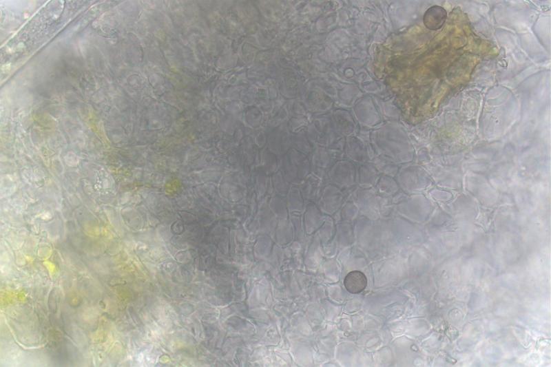
Helpful would be a closeup of the marginal lobes in section (last pic), if there are periphysoids. It could be an ostropalean fungus. Of course, free spores would be good and their measurements. Did you test IKI? MLZ is unsuitable, unless you pretreat with KOH.
Zuidland Peter,
25-06-2022 09:51
Re : Erumpent on Wattle Part 2
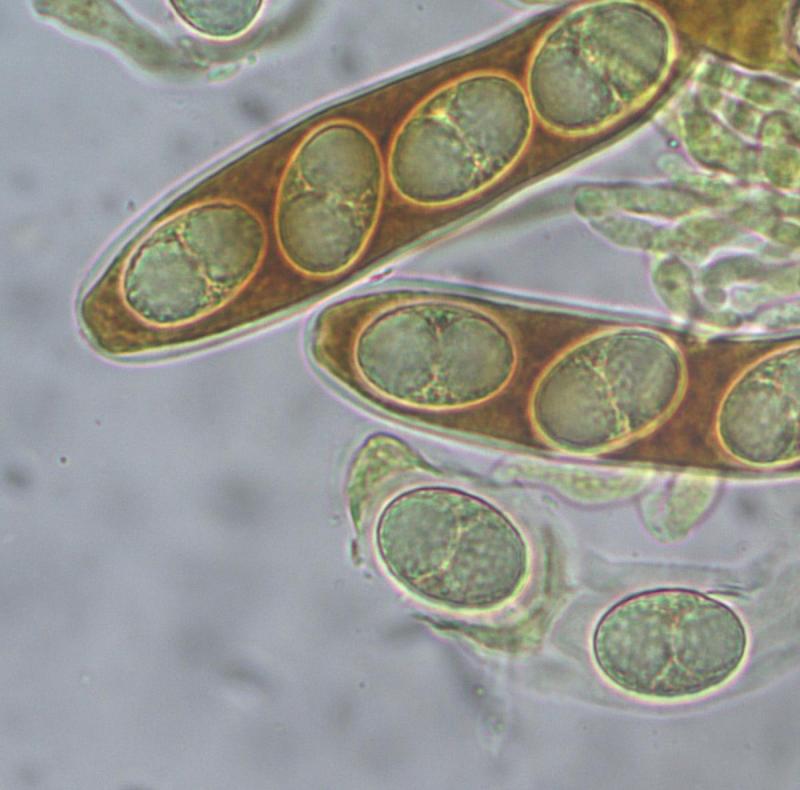
Thanks heaps Hans Otto,
I did check for IKI with Lugol's and it is the first micro image in my previous post on this( see below again); I have two more samples that I can try to check what you have asked for.
Take care
Pete
I did check for IKI with Lugol's and it is the first micro image in my previous post on this( see below again); I have two more samples that I can try to check what you have asked for.
Take care
Pete
Zuidland Peter,
26-06-2022 04:21
Re : Erumpent on Wattle Part 2
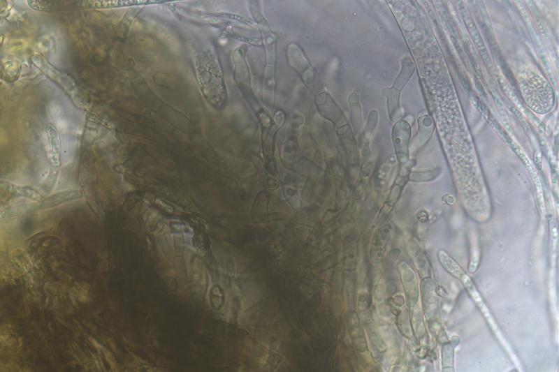
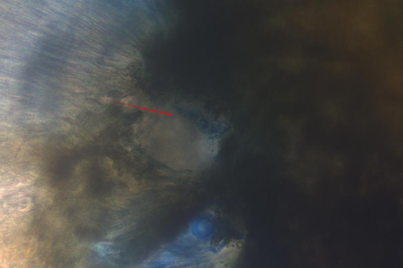
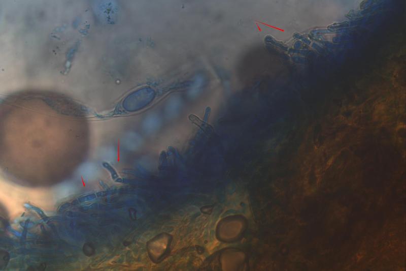
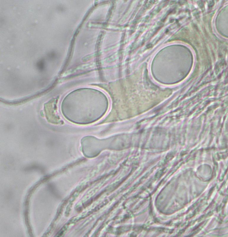
I think these are the periphysoids with arrows.
Pete
Pete
Zuidland Peter,
26-06-2022 04:24
Re : Erumpent on Wattle Part 2
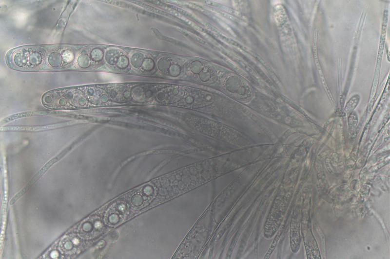
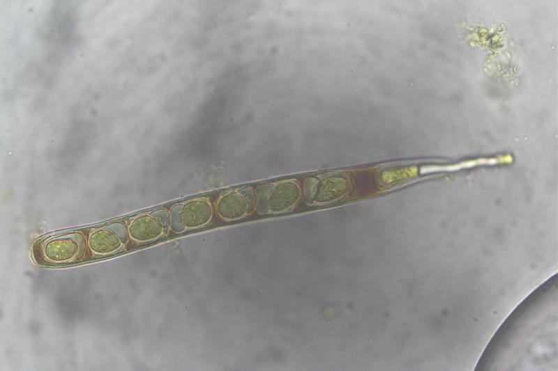
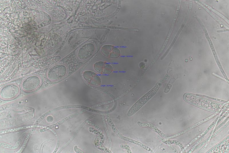
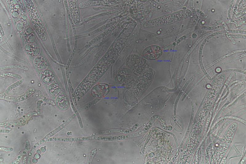
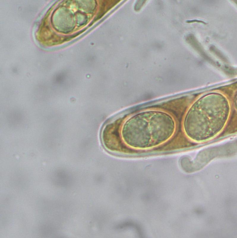
Free spores in H2O measured: 16.4 - 20.3 x 10.2 - 13.5um
many thanks
Pete
many thanks
Pete
Hans-Otto Baral,
26-06-2022 18:00
Re : Erumpent on Wattle Part 2
O.k., I am now a bit confused, do you think your wattle 1 and 2 are the same species?
I saw the IKI-ascus photo in 1 and wondered if it is a hemiamyloid apical ring or only plasma. It could be a ring, and then you should either show it in larger magnification or try KOH-pretreatment to see if the ring then stains blue in IKI.
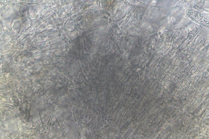
Periphysoids: I am not sure. They often form a compact layer of short parallel hyphae embedded in some gel.
Zuidland Peter,
27-06-2022 07:42
Re : Erumpent on Wattle Part 2
I am sorry that my poor skills as a non trained mycologist are causing you some grief or confusion, I don't mean to.
The specimen is the same in both series, I have no other asco near it nor with these spores.
Here is sample with just Lugol's again, from today and zoomed.
Pete
The specimen is the same in both series, I have no other asco near it nor with these spores.
Here is sample with just Lugol's again, from today and zoomed.
Pete
Zuidland Peter,
27-06-2022 07:45
Re : Erumpent on Wattle Part 2
Here is the sample heat treated with 4%KOH, cooled, remove most KOH apply Lugol's.
This leaves a clear looking asci with no stain.
This leaves a clear looking asci with no stain.
Zuidland Peter,
27-06-2022 07:47
Re : Erumpent on Wattle Part 2
Heat treat with KOH, cool, add Lugol's, remove Lugol's then add Lugols again.
This left the asci stained.
This left the asci stained.
Zuidland Peter,
27-06-2022 07:49
Re : Erumpent on Wattle Part 2
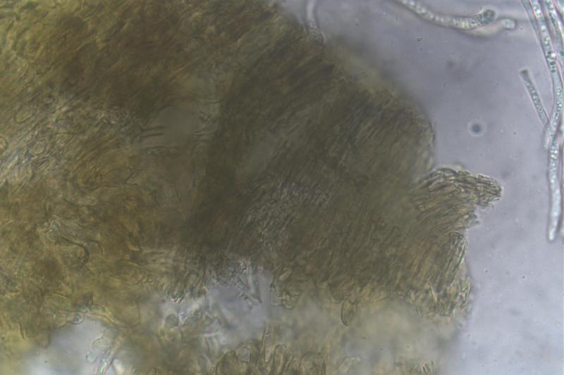
For the excercise I did it with MLZ too.
Heat with KOH, remove then add MLZ
One image looks to show a channel near the tip
Heat with KOH, remove then add MLZ
One image looks to show a channel near the tip
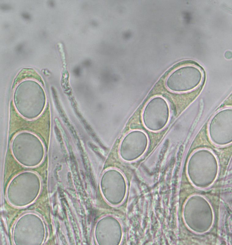
Hans-Otto Baral,
27-06-2022 08:21
Re : Erumpent on Wattle Part 2
Splendid and much better resolution! Now it is clear, the asci are inamyloid. The "channel" is an ocular chamber, maybe it is more typical of immature asci without spores or with beginning spore formation. In such asci it my be that the lateral ascus wall is thickened.