22-04-2026 20:54
 Enrique Rubio
Hi to everybody.This Pyrenopeziza grew in moist le
24-04-2026 03:16
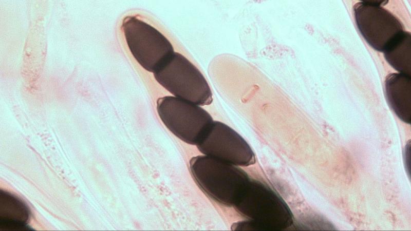
 David Chapados
Found while looking at something else from wood in
22-04-2026 01:06
 Richard VALERI
Bonjour à tous.Je vous présente cette Nectria s.
22-04-2026 20:17
 Marian Jagers
Is anyone familiar with the Hyphomycetes genus Pse

 When we are investigating characters of species under a light through microscope we do observe that in a 2D picture.
So we have to think in 3D but that is not always possible because our mindset cannot cope with the optical illusion we are looking at.
Accidentally I did find out that we can create a 3D picture by changing the focal distance from the lens to the object using a Plane Objective 100x/1.25 (photo 1 & 2). Probably by stacking photo's you will create the same effect.
The ring is elastic and the distance when the apical system is not fully developed is as follows: Diameter of the outer circular ring is 0,9 um; total diameter is 4,6 um and inner diameter is 2.8 um. Photo-3 is a ring clearly visible with a spore ready to enter.
Photo 4 the ring is connected to an ampty inner wall, photo 5 is the same situation but inside a still present outer wall.
Photo 6 shows the apical ring in the end phase with spore clicked inside and the outer wall still present.
Photo 7 & 8 show spores inside the ring and outer wall gone.
The ring itself is more oval than it is circular. (photo 9)
Kind regards,
Joop