

22-04-2026 20:54
 Enrique Rubio
Hi to everybody.This Pyrenopeziza grew in moist le
24-04-2026 03:16
 David Chapados
Found while looking at something else from wood in
22-04-2026 01:06
 Richard VALERI
Bonjour à tous.Je vous présente cette Nectria s.
22-04-2026 20:17
 Marian Jagers
Is anyone familiar with the Hyphomycetes genus Pse
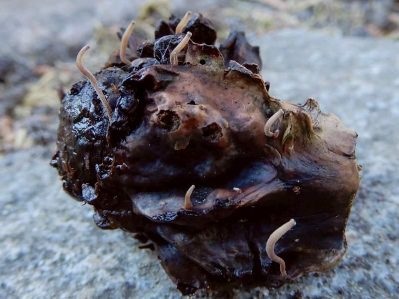
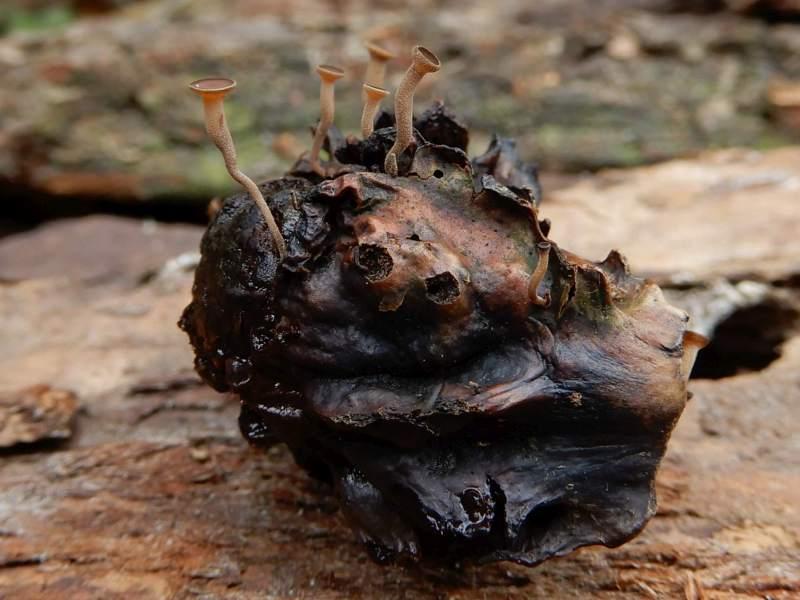
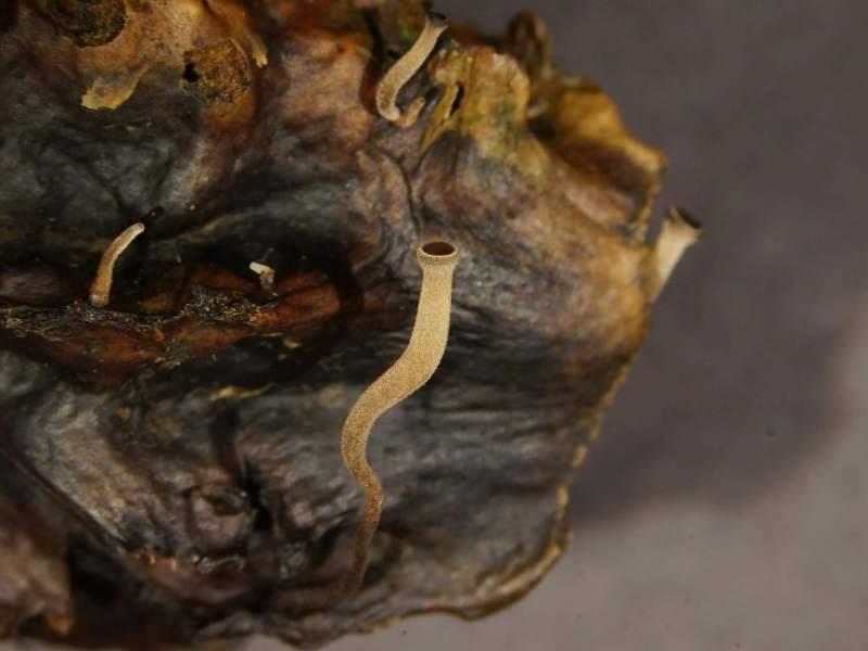
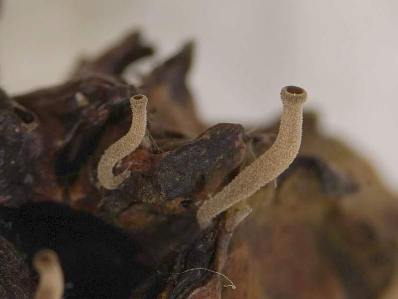
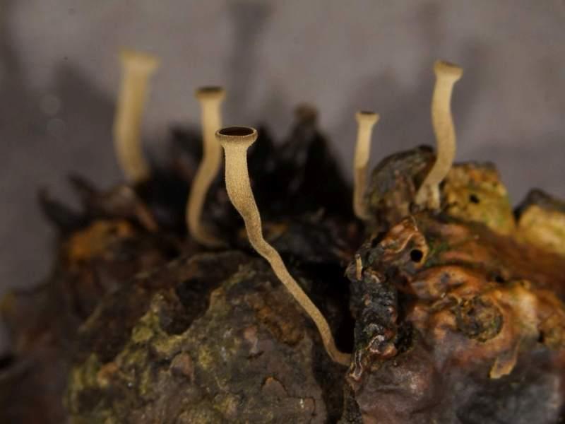
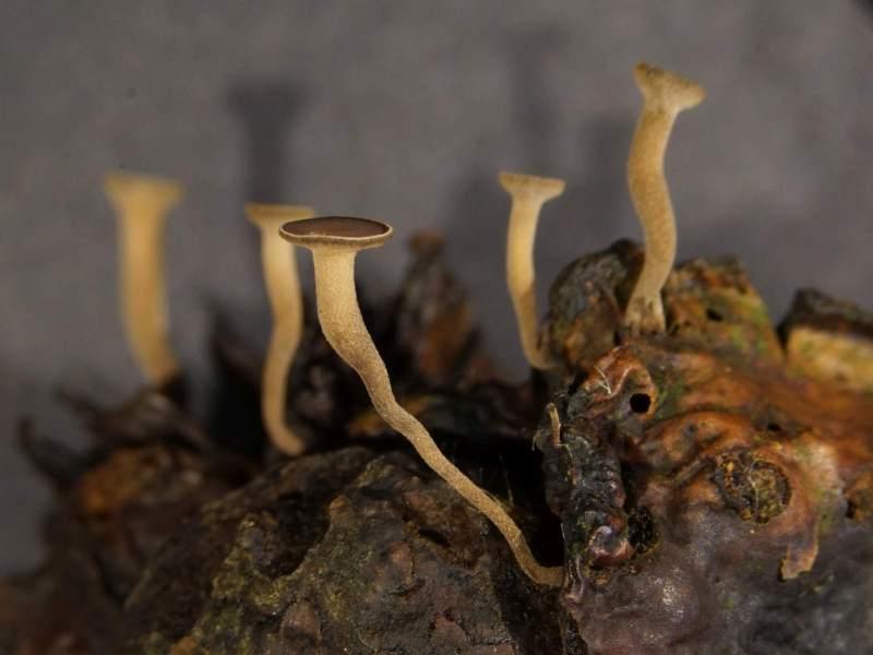
 Cultured on a Knopper gall (Knopper gall found in a forest nearby Utrecht, The Netherlands).
I started a moist chamber the second week of February. After about 3-4 weeks, very small, hairy stalks emerged from the substrate. They slowely grew into long stalked apothecia.
With the exception of Mycena rhenana/M. Cecediophila, which I actually hoped to culture, I cannot find information about fungi growing on Knopper gall.
I think this is Moellerodiscus lentus, but the spores are bigger and the substrate is unknown. So I am still in doubt of this name is correct. Anyone want to help me?
Greetings Marian
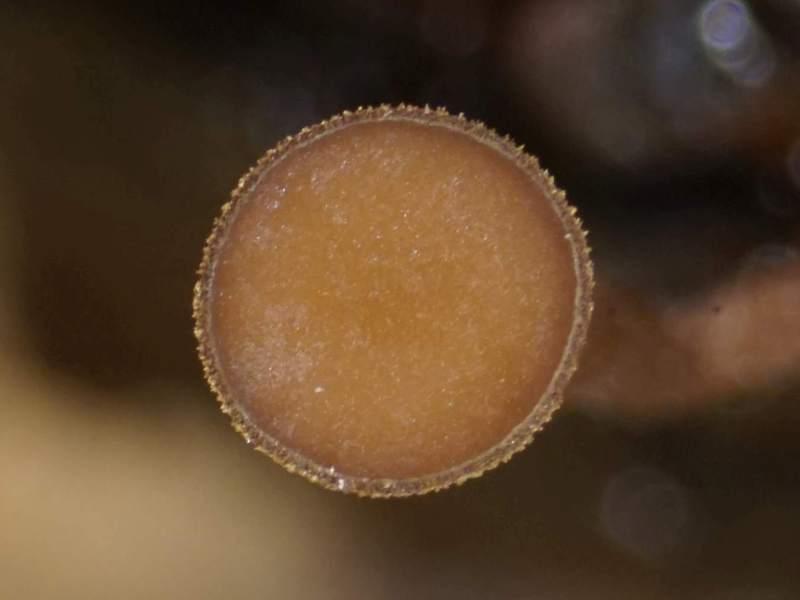
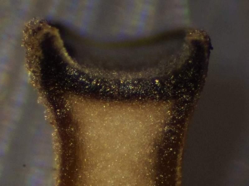
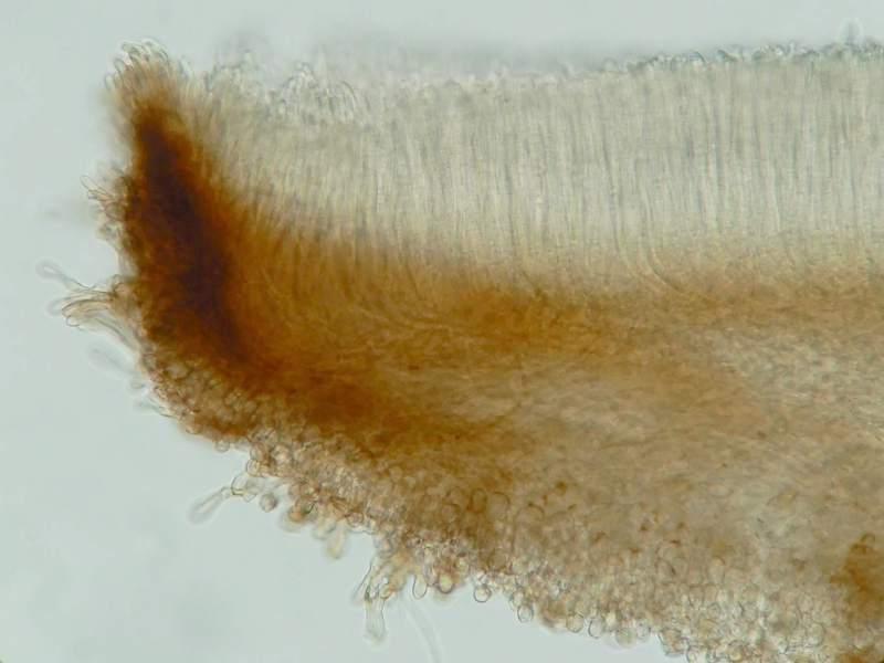
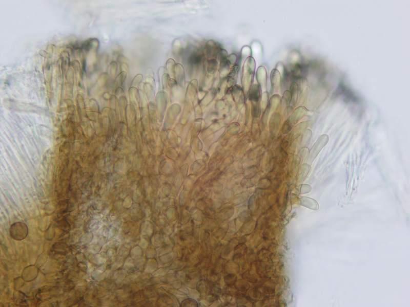
Apothecia 0.5-3,2 mm in diam, 0.5- 14 mm high, long stipitate, arising scattered, completely hairy, beige (darker after rehydrating or after superficial damaging).
Receptacle young barely wider than the stipe, later wider and cup-shaped, flattening by age, beige to brown, margin beige.
Hymenium yellowish brown, brown to reddish brown.
Stipe 0,5 to 13 mm long, in the middle ± 0.5-1,2 mm wide, young more or less cylindrical, older broader above and tapering toward the base, more often curved, beige, sometimes brown blackish at the base.
Microscopic characters:
Apothecium without ionomidotic reaction.
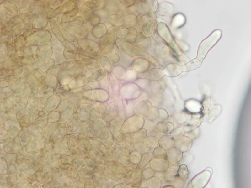
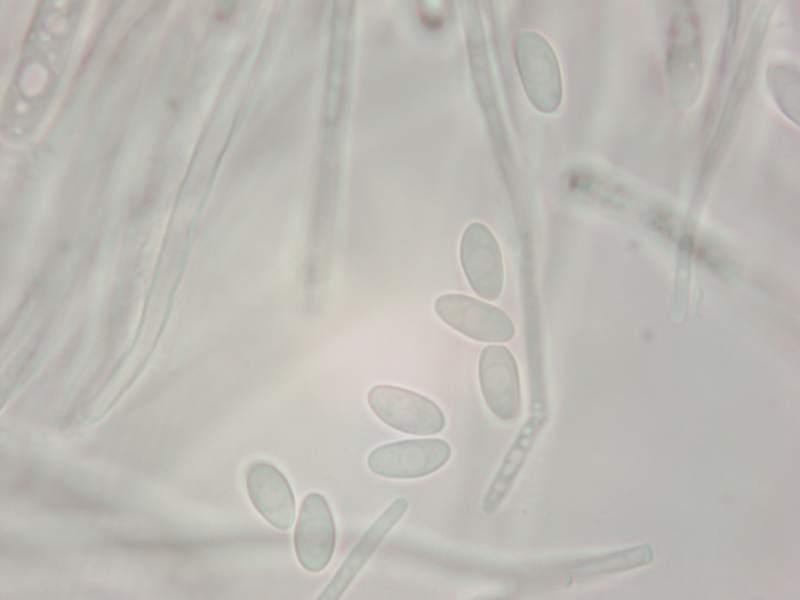
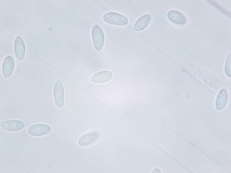
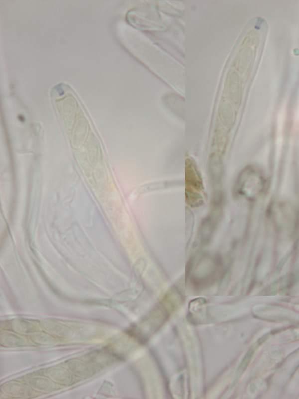
Spores: smooth, hyaline, aseptate, 8-10(-11)x(3,5-)3,7x4,5 µm.
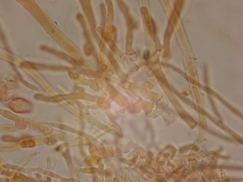
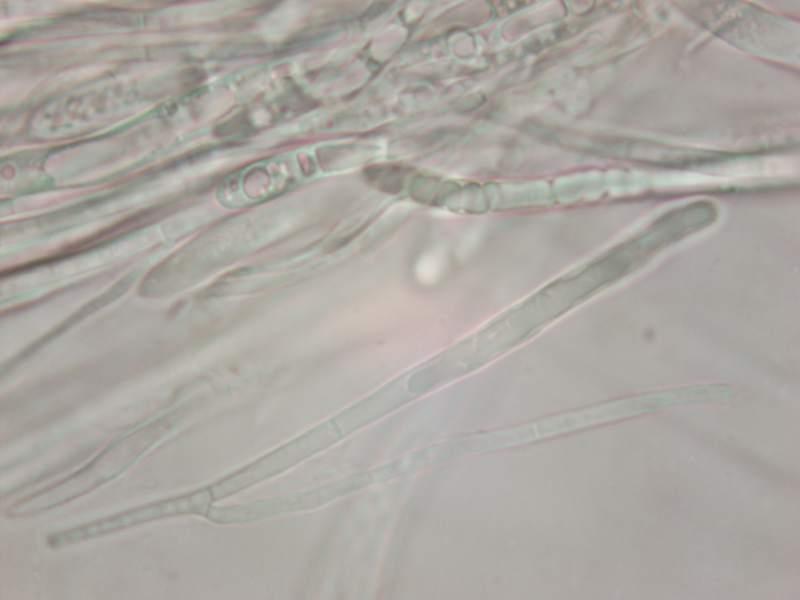
Asci: 8-spored, uniseriate or les often biseriate, 95-108x 7-8 µm, weakly J+ in Lugol, strongly J+ after treated with KOH, croziers present.
Paraphyses: equal to or slightly exceeding the asci, filiform, septate, branching, apex 2 -3 µm wide.
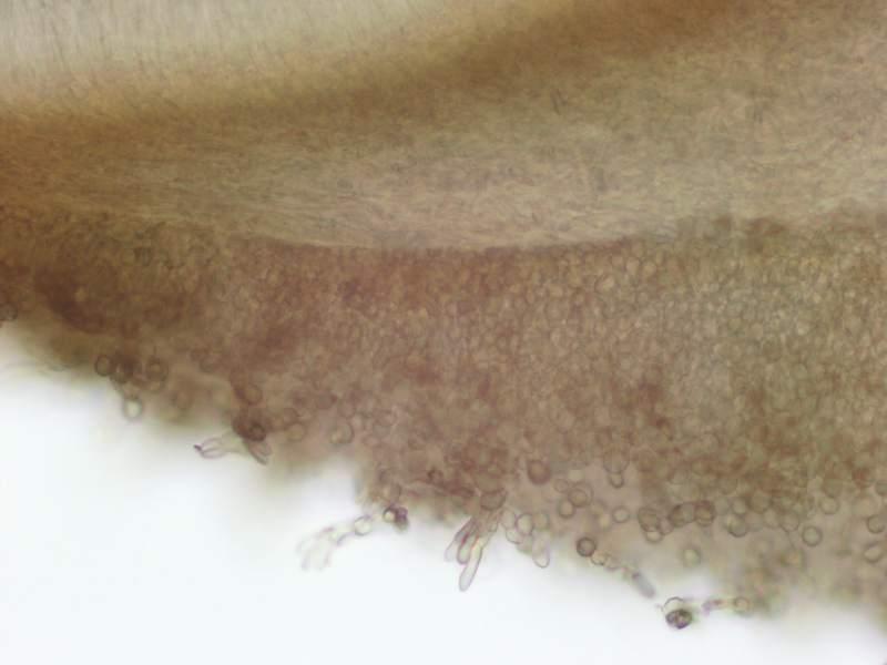
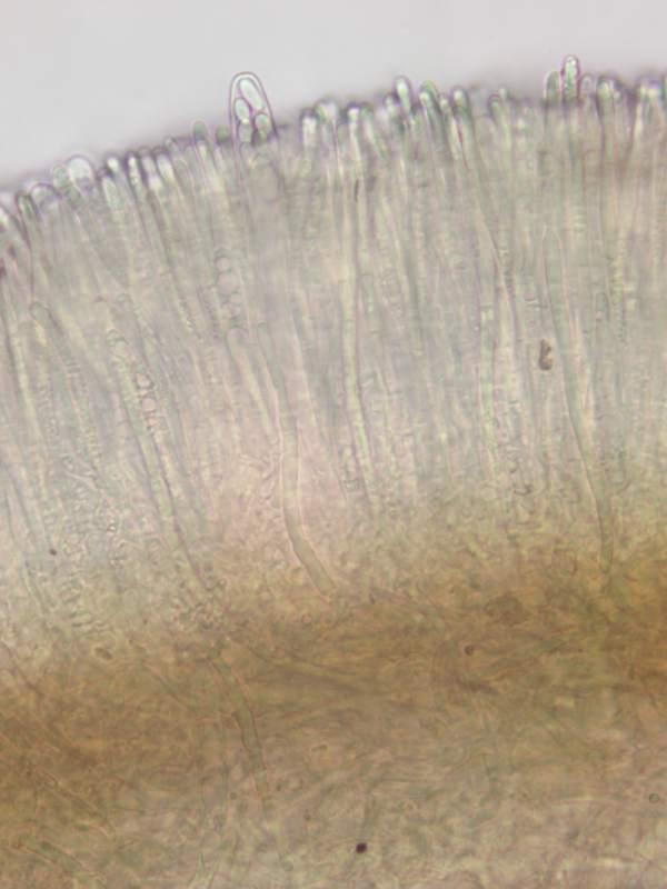
Subhymenium: consisting of vertically oriented, loosely interwoven hyphae, with light brown intracellulair pigment.
Medullary excipulum: consisting of interwoven hyphae, hyaline or with light brown intracellulair pigment.
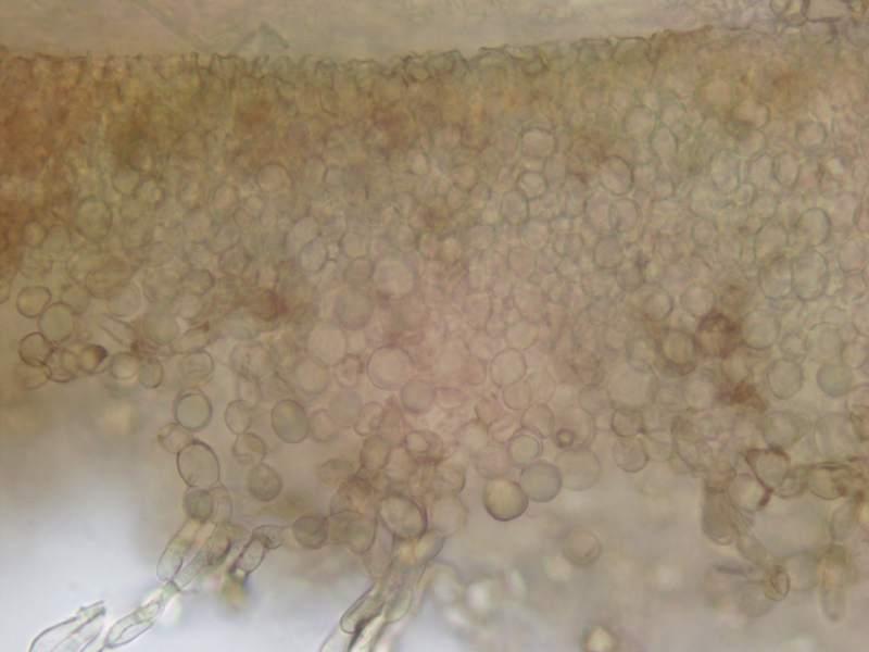
Excipulum: mostly textura globulosa, consisting of thickwalled roundish to oval, sometimes angular cells, outer cells 6-13x6-12 µm, scattered chain-like and oriented perpendicularly to the surface.
Margin apothecium composed of clavate terminal cells, hyaline or with light brown intracellulair pigment, 25x5 µm.
Stipe outer cells textura globulosa (simular to the receptacle).
Literature with a key to the genus I used: Yan-Jie ZHAO. 2013. Taxonomic Study of Lambertella (Rutstroemiaceae, Helotiales) and Allied Substratal Stroma Forming Fungi from Japan. Dissertation to the Graduate School of Life and Environmental Sciences, the University of Tsukuba.
Literature with a key to the species and a description: Dumont, K.P. 1976. Sclerotiniaceae XI. On Moellerodiscus (= Ciboriopsis). Mycologia 68(2): 233-267.


Nice description! But I feel it is not related to Moellerodiscus lentus (spores are not so wide), but to the species that grows on Eucalyptus. Of course, it is not the same species because it lacks of those beatiful yellowish VBs. Please compare to Enrique´s microphotos at Zotto´s Moellerodiscus folder "eucalypti H+ chlorin. Vbs".
Cheers,
Raúl