
27-04-2026 09:59
Pauline. PennaBonjour Can anyone advise me on these pycnidia fo

22-04-2026 20:54
 Enrique Rubio
Hi to everybody.This Pyrenopeziza grew in moist le
24-04-2026 03:16
 David Chapados
Found while looking at something else from wood in
22-04-2026 01:06
 Richard VALERI
Bonjour à tous.Je vous présente cette Nectria s.
22-04-2026 20:17
 Marian Jagers
Is anyone familiar with the Hyphomycetes genus Pse


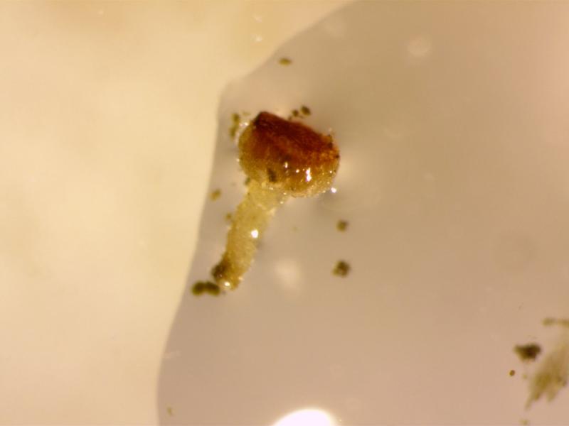
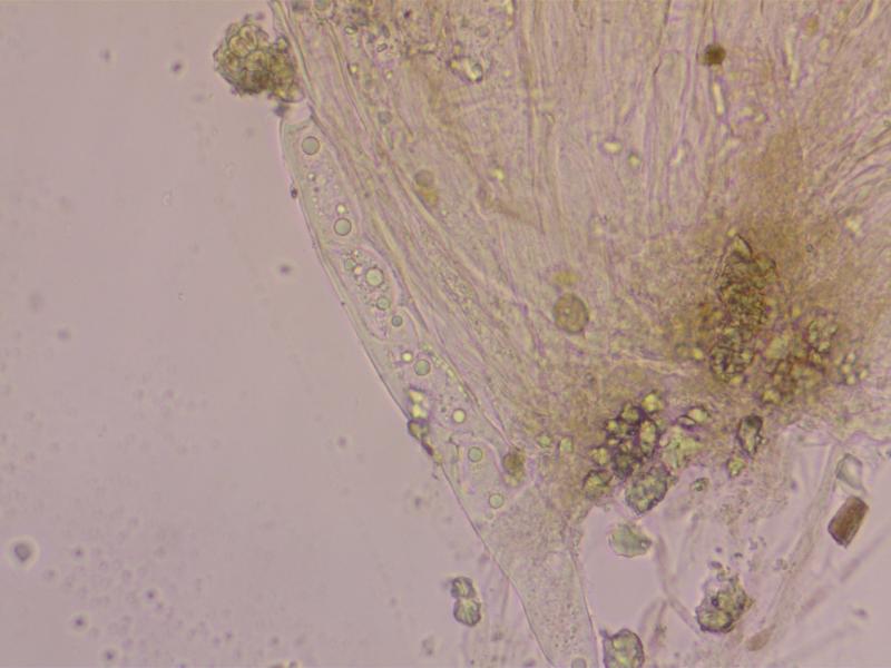
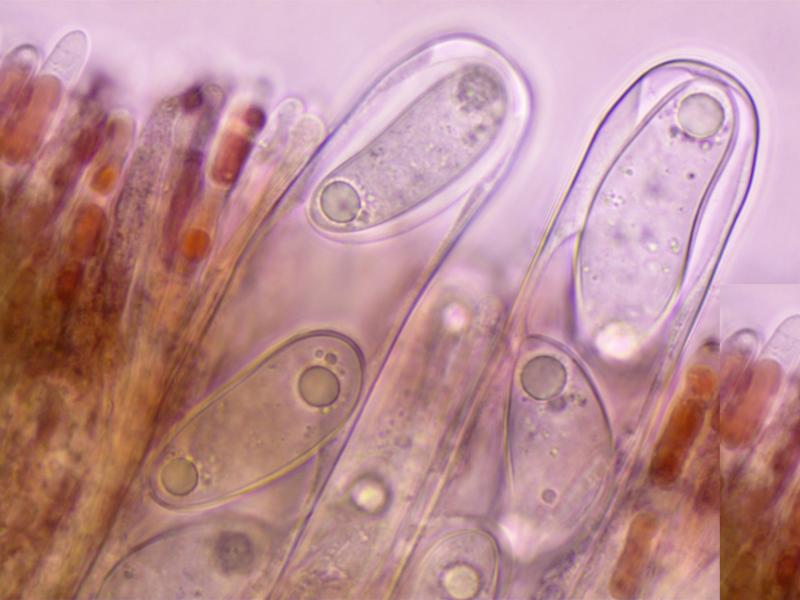
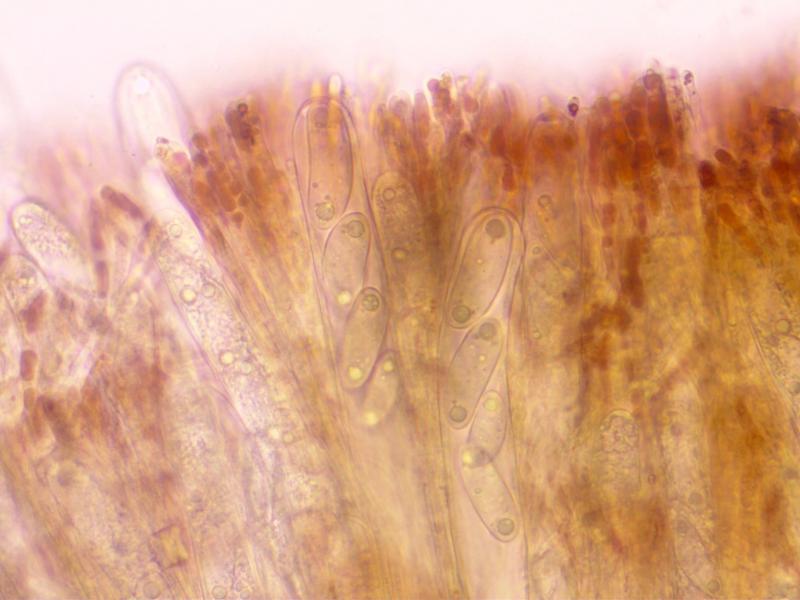
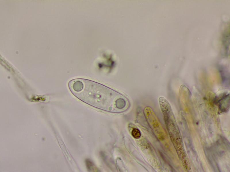
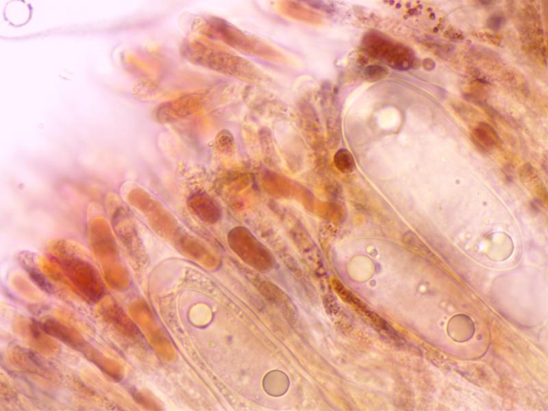
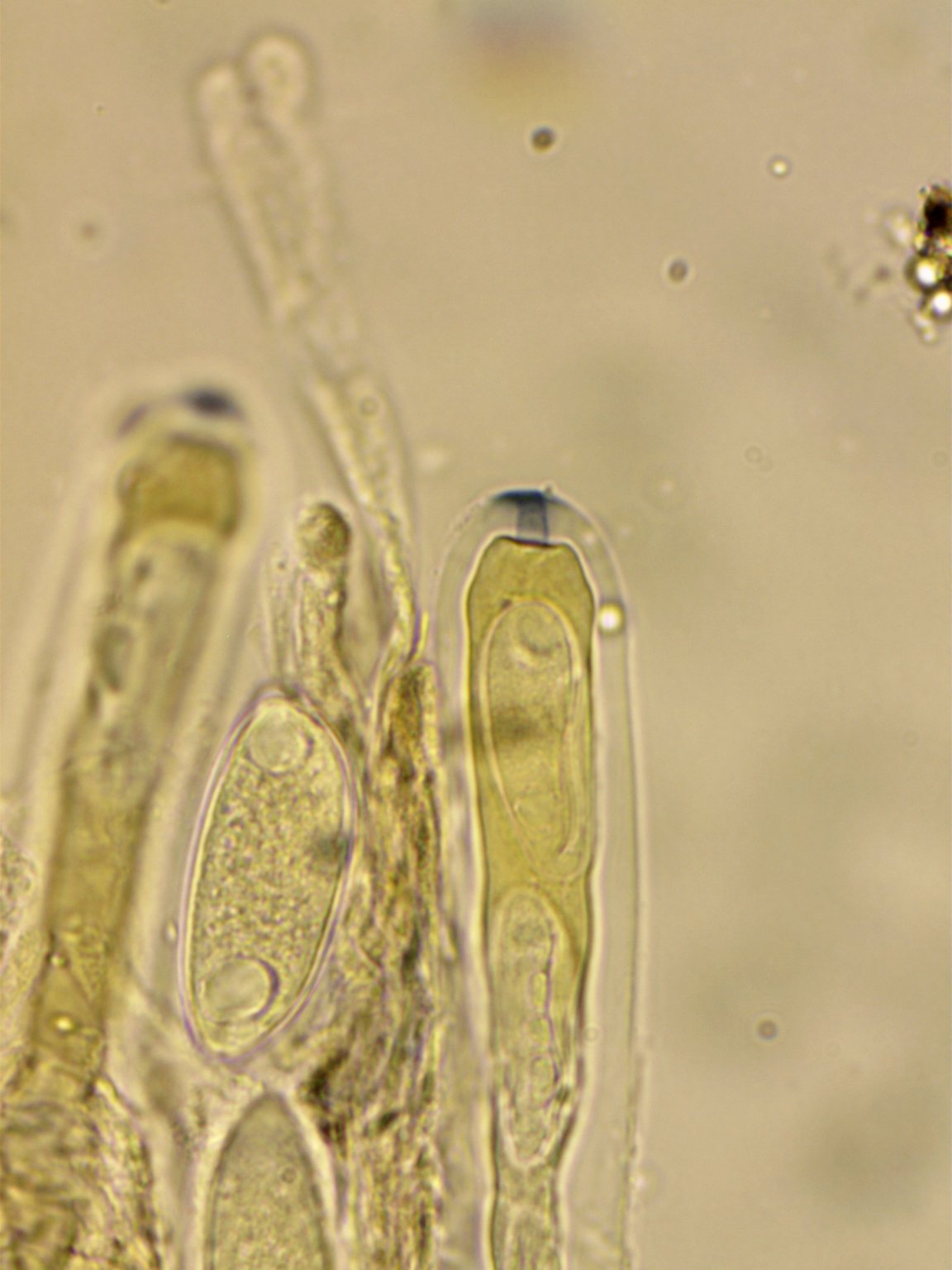
Hi All,
I have found a strange and distinctive Ascomycete growing on damp soil close to a brook. Only a single apothecium thus far.
Apothecium: c1x0.5mm. Hymenium convex, bright red, not clearly demarced from the excipulum.
Asci: Cylindric, 250-325x18-25. 4-5 spored, uniseriate, with differetial maturation of the ascospores. Ascus pore blueing in Melzer' after treatment with KOH.
Ascospores:44-50x15-19. Ellipsoid-cylindric, some curved and some with strangely attenuated apices. With polar guttules. With a gelatinous coat.
Paraphyses with red pigment, swollen to 6-7.
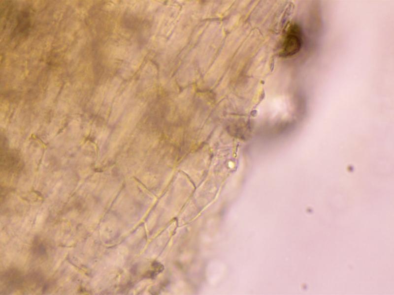
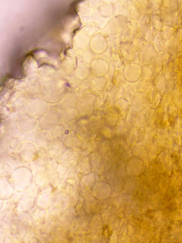
Excipulum: Textura Globosa.
I would be very interested in any thoughts on this. Thanks in advance.
Charles.
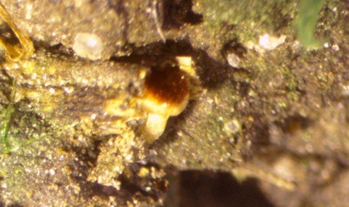
 rsz-red-stipitate-disco-asci-7-rhiwlas-pentraeth-7420-0001.jpg
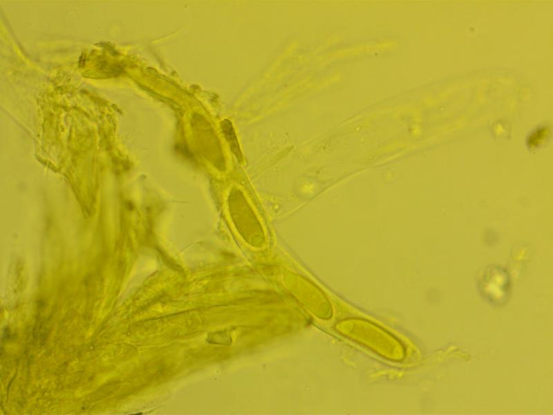
Hi Zotto,
Yes, the t. prismatica is from the stipe. Have not tested with Lugol and very little material remains (on a couple of slides) Will definitely look out for more material-not far from where I live in Pentraeth, Anglesey.
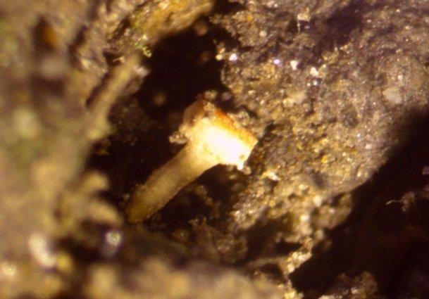
Hi Zotto,
Its the ascus width that I had got wrong-it should be 250-325x18-25, so really quite large. Being Sclerotiniaceous I imagine the fungus would have been associated with plant material perhaps with a sclerotium but, unfortunately it quickly became detached from the substrate. There was some Mnium hornum fairly close but I don't think the fungus was associated with it. The site was in broad leaved woodland with ash etc.