14-05-2026 05:36
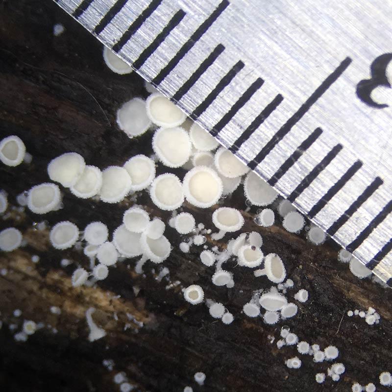
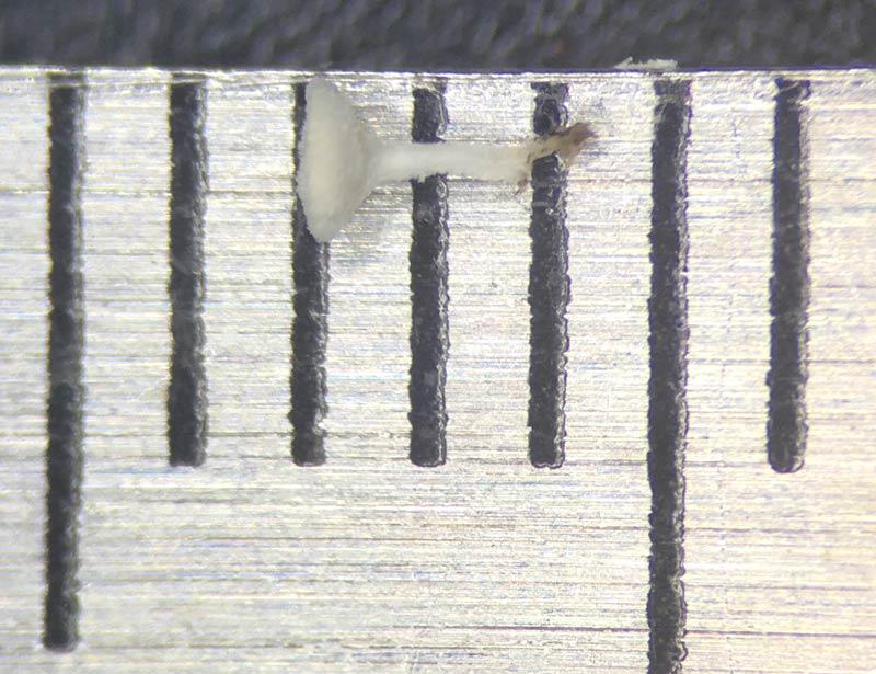
Ethan CrensonHi all, I haven't paid much attention to Lachnu
11-05-2026 12:32
 Bernard CLESSE
Pourriez-vous m'aider à identifier cette héloti
13-05-2026 15:26
 François Freléchoux
Bonjour,Voici une récolte faite il y a quelques j
12-05-2026 15:41
 Nicolas VAN VOOREN
Dear Ascolovers, especially interested in Pezizale
13-05-2026 12:05
 Thierry Blondelle
Bonjour à tous,J'aimerais avoir confirmation de c
10-05-2026 23:17
 Andreas Gminder
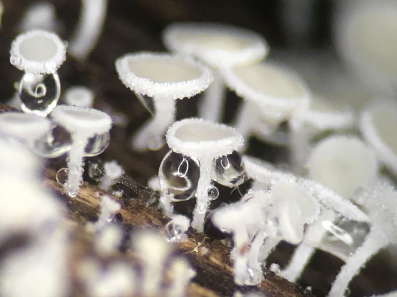
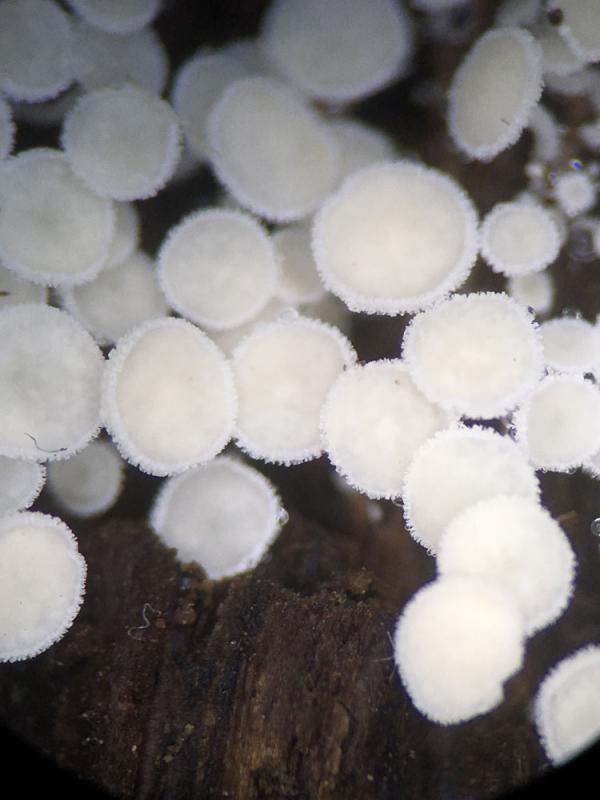
Hello,today we found in a moist steep decidous for
28-04-2026 20:07
 Lothar Krieglsteiner
... on twig in the air at standing Ceratonia siliq
27-04-2026 20:52
Lothar Krieglsteiner
Found on hanging tiwg of Olea europaea in dried-ou
11-05-2026 20:22
Lothar Krieglsteiner
on attached twig of standing Ficus caricaquite uns
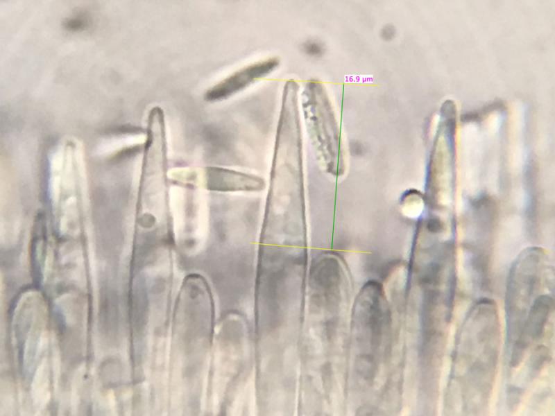
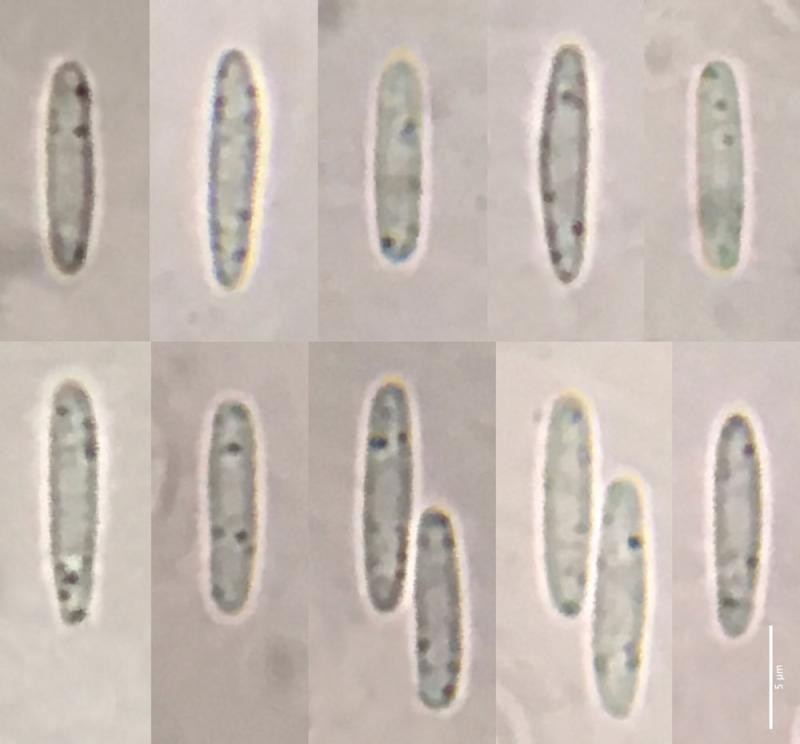
Spores
fusiform-cylindrical, straight, low oil content—just a few small guttules
7.7-11.9 x 1.7-2.5µm
Me: 10.3 x 2.2µm
Q: 3.7-5.4
Qe: 4.6
N=52
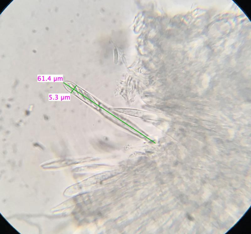
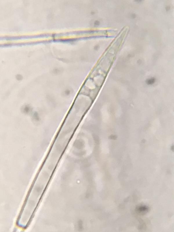
Asci
53-63 x 4.5-5.5µm
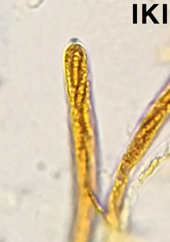
IKI+
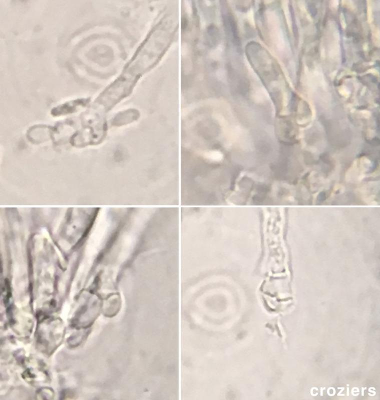
Croziers+
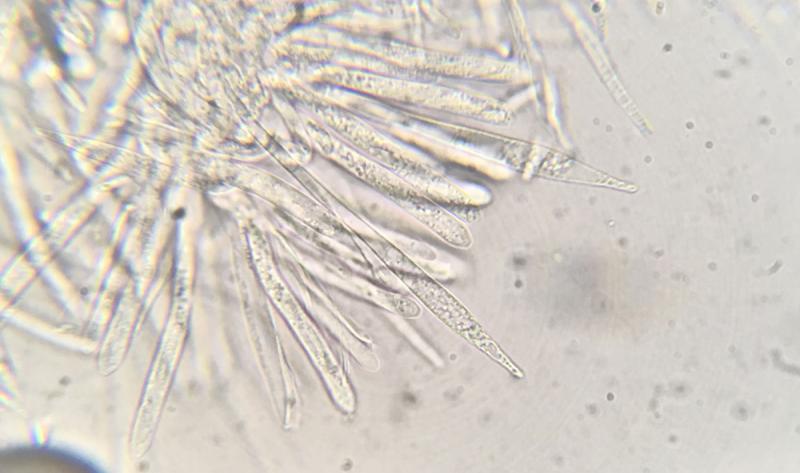
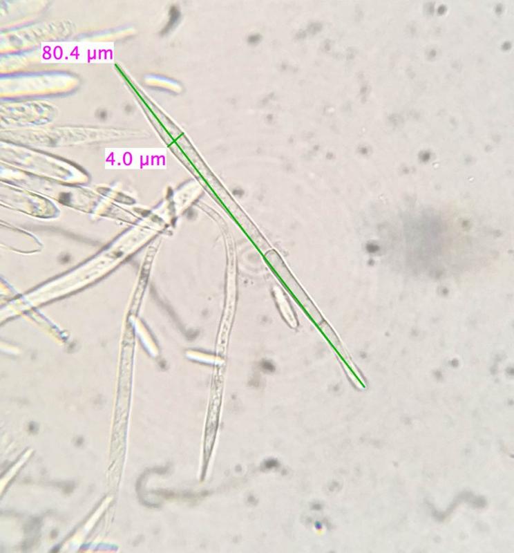
Paraphyses
68-111 x 3.5-4.9µm
lanceolate
extending up to 12.3-18.8µm above hymenium
as far as I can tell they are without VBs (or if there are VBs they are not very refractive). Please correct me if I am wrong.
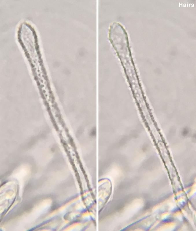
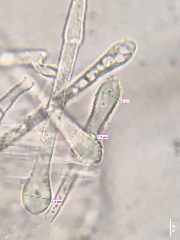
Occasionally, but not always capitate, echinulate ornamentation all the way to the ends
width at widest point 4.7-6.9µm
No oxylate crystals so far as I can tell.