
11-05-2026 12:32
 Bernard CLESSE
Pourriez-vous m'aider à identifier cette héloti
13-05-2026 15:26
 François Freléchoux
Bonjour,Voici une récolte faite il y a quelques j
12-05-2026 15:41
 Nicolas VAN VOOREN
Dear Ascolovers, especially interested in Pezizale
13-05-2026 12:05
 Thierry Blondelle
Bonjour à tous,J'aimerais avoir confirmation de c
10-05-2026 23:17
 Andreas Gminder
Hello,today we found in a moist steep decidous for
28-04-2026 20:07
 Lothar Krieglsteiner
... on twig in the air at standing Ceratonia siliq
27-04-2026 20:52
Lothar Krieglsteiner
Found on hanging tiwg of Olea europaea in dried-ou
11-05-2026 20:22
Lothar Krieglsteiner
on attached twig of standing Ficus caricaquite uns
29-04-2026 10:44
Lothar Krieglsteiner
growing at moist, drying-out soil at the side of a
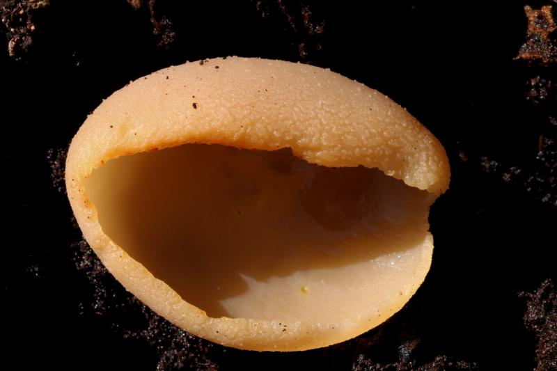
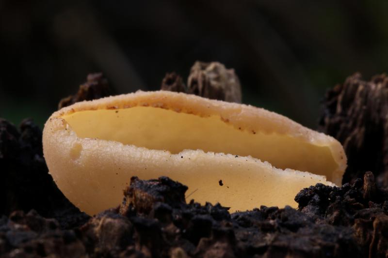
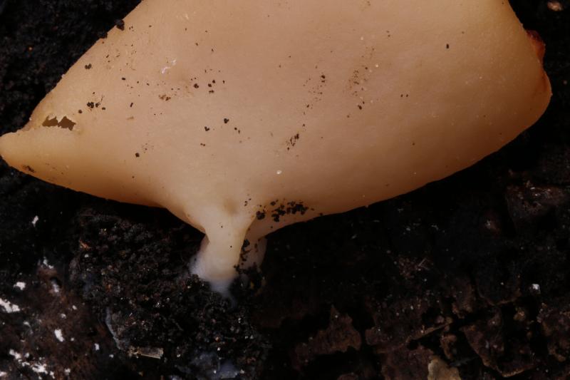
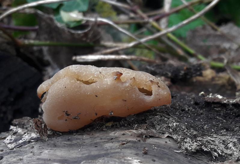
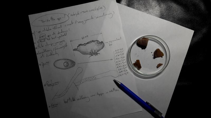
 this species grows on decaying stumps of Abele in my wild garden.
At first I thought Tarzetta sp but the species here grows on wood.
Short stipe to almost sessile.
Fruiting bodies >40mm
Odour: strongly mushroom.
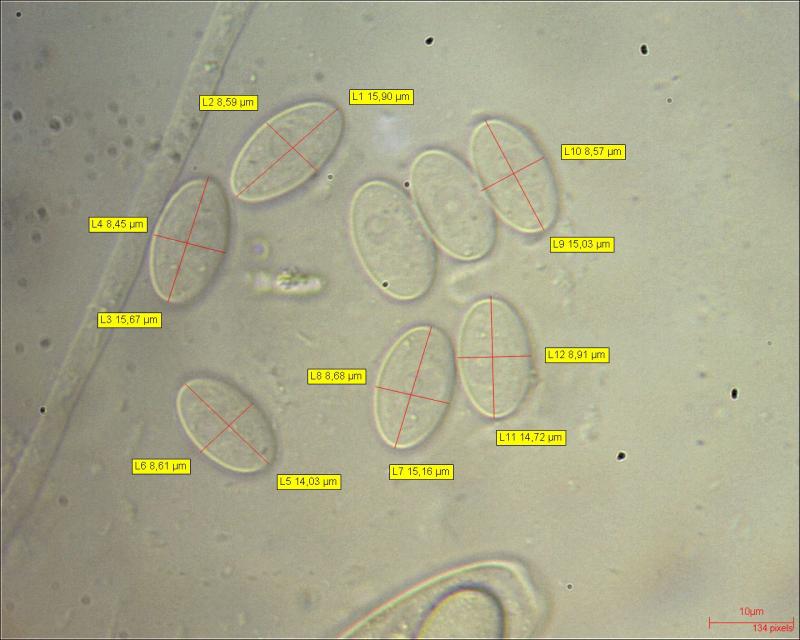
Spores: 14.71x8.32mu with one oildrop
Ascitops colouring bleu in Meltzer
Any idea?
Sincerely,
William
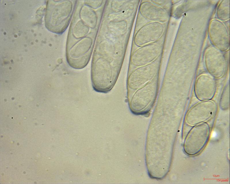
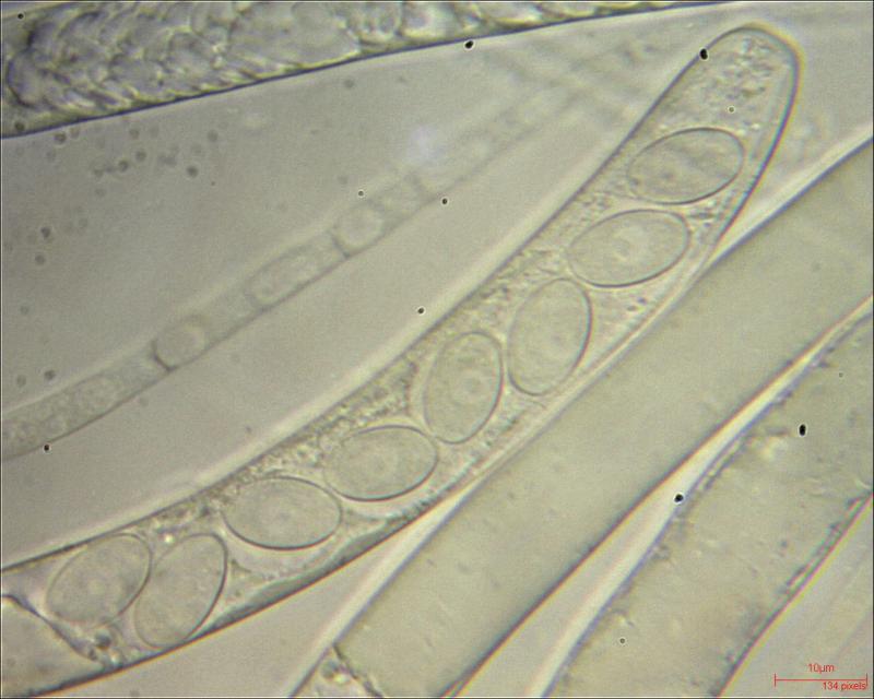


the asci from Tarzetta are not amyloid. In my opinion this is a species from the Peziza varia-group ("micropus") ?
Greetings Peter.
I have looked on it with the provisional key of B. Declercq and it looks indeed as a P.varia (syn. micropus).
Greetings,
William

the spores are actually eguttulate (it's a nucleus) and they also have a sheath (on the 7.photo). I had something from P. varia group with smooth spores and gel sheaths as well (bigger spores, though) and got some interesting comments here: http://ascofrance.fr/search_forum/43195
As for determination, I'm sorry I can't say more.
Viktorie

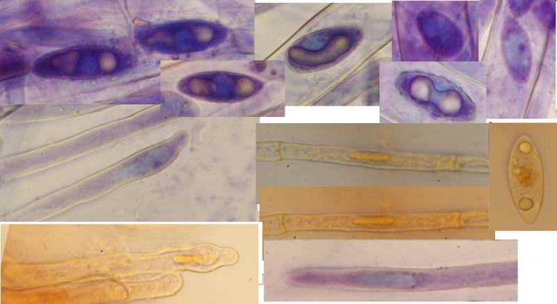
I'll attempt to answer, although with my rather short experience with microscopy I feel about the last person to ask.
Lipid guttules are more visible than nuclei, more clearly demarcated (at least in living spores), while nuclei look more like an "empty space". I didn't try yet any stains for LBs. As for stains for spore nuclei: In living material they are well enough visible in water. Sometimes I try acetocarmine - nuclei become dull carmine red, against orange colored rest of spore contents. Mr. Dougoud described the proper method as follows:
"L'observation des noyaux n'est pas très facile, il faut laisser agir le carmin acétique durant quelques minutes, puis dissocier le prélèvement par percussion sur le couvre objet, ajouter encore une goutte de carmin au bord de la lamelle, attendre encore un peu, retirer l'excédant de liquide et observer."
Some genera (e.g. Arpinia) are not carminophilous. I have almost no experience with staining nuclei in paraphyses, cannot distinguish them from other structures.
Giemsa solution can be used as well, T. Schumacher used it in his monographs of Scutellinia and Myriosclerotinia (with L.Kohn), staining method is described in the latter (Can. J. Bot. 63: 1610-1640). Never tried myself, here it's more problematic to obtain the solution when one doesn't work in a lab.
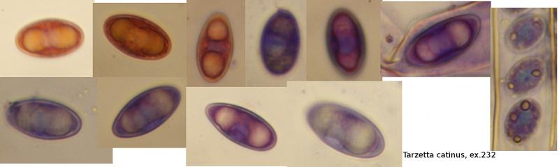
I tried CRB too, here's the result with Jafnea semitosta, but it stained only submature spores. Nucleus is blue, lipid bodies dirty yellowish, rest of cytoplasm violet. The other photo is of Tarzetta catinus - when a little of KOH is added to the CRB, spore cytoplasm discolors to dirty red, but the nucleus stays bluish for a while. Many operculates have thickwalled spores so that only submature spores can be stained. I suppose the stains might be more useful for inoperculates, where the variation of number of nuclei is perhaps bigger - it helped me to distinguish Dumontinia tuberosa (4 nuclei).
Hope it helped a bit.
Viktorie
(I took the liberty to download the scans from cybertruffle and convert it into double-sided pdf: http://tmp1.vize.name/myko/Vital_taxonomy.pdf )