

22-04-2026 20:54
 Enrique Rubio
Hi to everybody.This Pyrenopeziza grew in moist le
24-04-2026 03:16
 David Chapados
Found while looking at something else from wood in
22-04-2026 01:06
 Richard VALERI
Bonjour à tous.Je vous présente cette Nectria s.
22-04-2026 20:17
 Marian Jagers
Is anyone familiar with the Hyphomycetes genus Pse

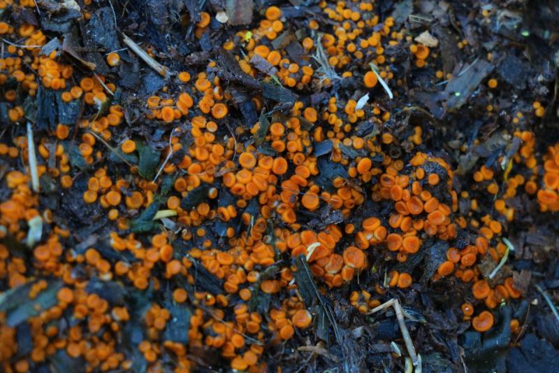
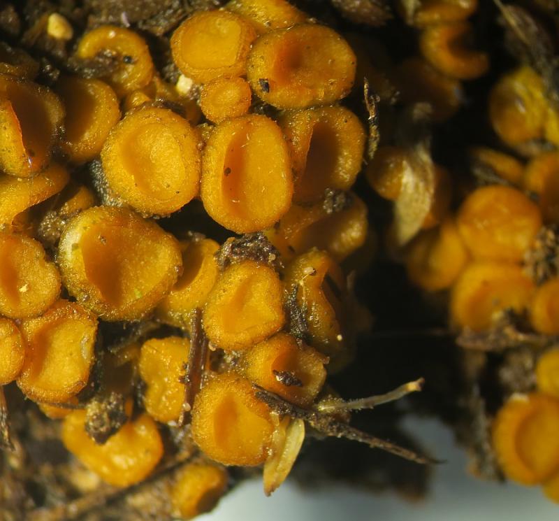
Cheilymenia sp. on wooden chips possibly mixed with excreta
Stephen Martin Mifsud,
18-02-2017 20:37
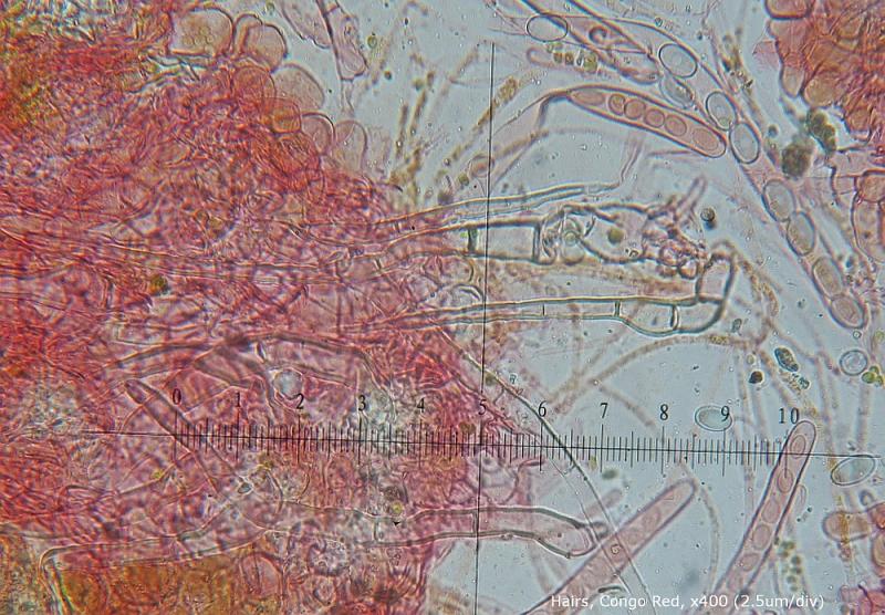
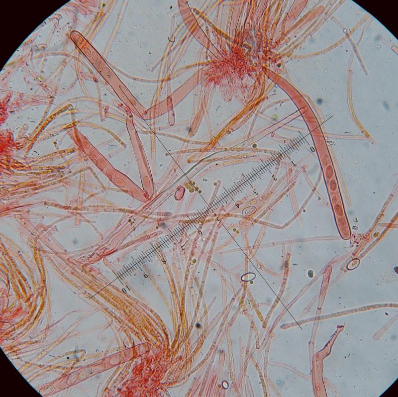
 Hi, I found clusters of amber=yellow discoid ascomata which are closed as a small bowl shaped strycture when young. Young specimens in particular had a coating of hyaline hairs, 1- (seldom 2-) septate with a bulbous, rooting base cell (or up to 3). The septum is usally close to the base.
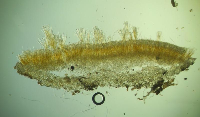
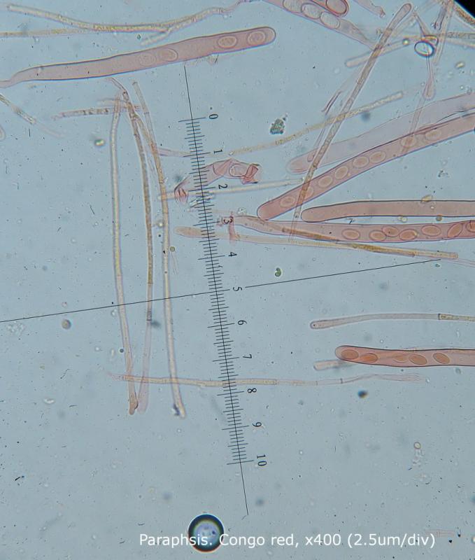
Asci J-ve, 190 - 230 x 12.5 - 13.9 µm
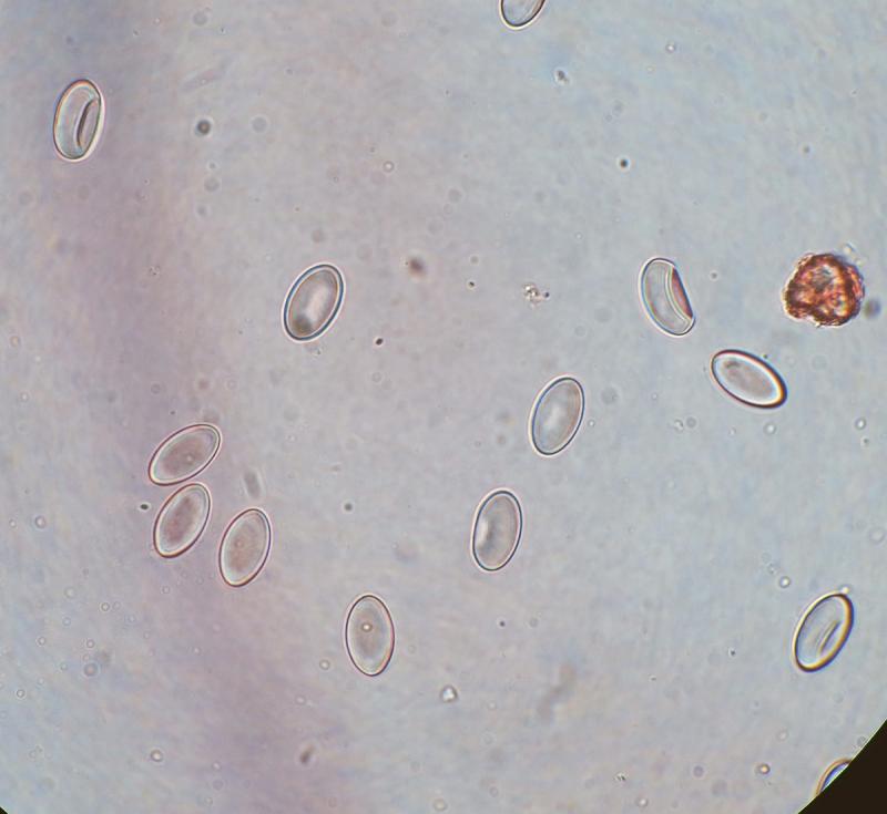
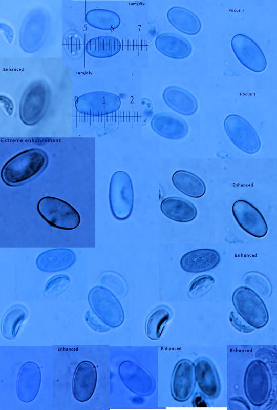
Ascospores, Broadly elliptic with a lateral crater-like depression at one side(artifact?), smooth or finely rugulose showing as cloudy surface at higher magnifications, 12.9 - 18 x 7.21 - 10.8 µm
(Average: 14.6 x 8.4 µm; Q: x1.42 - 2 µm (average x1.7), aseptate.
Excipulum one layer (I think, kindly confirm) of textura globulosa, 20-40um wide.
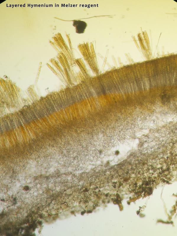
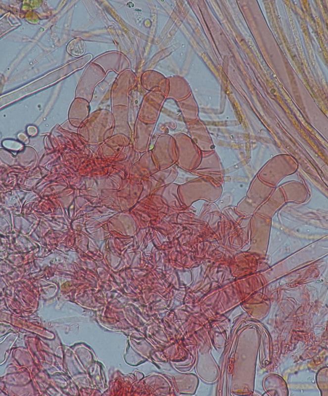
Interestingly there was a strange reaction with Melzer reagent. Shortly after applying the reagent, the hymenium became layered, where the uppermost part attained a yellow colour, followed by an unstained (or even faint blue) layer occupying about the upper third of the hymenium (or asci) and a reddish brown, more or less dextrinoid lower half of the hymenium and subhymenium. Exipulum remained unstained.
This cup fungus reminded my of Cheilymenia esp. its bulboid hairs, but I wish your advice. Specimens in fridge if you request further tests or observations.
Nicolas VAN VOOREN,
18-02-2017 20:42

Re : Cheilymenia sp. on wooden chips possibly mixed with excreta
No picture?
Viktorie Halasu,
18-02-2017 21:26

Re : Cheilymenia sp. on wooden chips possibly mixed with excreta
Hello, do you have a photo of spores' surface in LACB (without heating)?
Stephen Martin Mifsud,
19-02-2017 10:41
Re : Cheilymenia sp. on wooden chips possibly mixed with excreta
Nicolas, I wrote the text and then placed the images few mins later because sometime something wrong goes with the upload and the forum resets ( = loosing up the text).
Spore in LACB examined today under x1000 (oil immersion) magnification
Sizes confirmed (14-16um long) and there seems to be a very very fine striation or rugolosity which I tried to highlight but enhancing some images with post-processing.
The lateral crater-like depression is also confirmed. These spores are produced by inverting some living cups upside down on the glassslide for 12 hrs so these are ejected spores, fully mature. Stain left standing for 10mins then examined. No heat applied to the stain.
Stephen Martin Mifsud,
08-06-2020 11:01
Re : Cheilymenia sp. on wooden chips possibly mixed with excreta
Any fresh ideas on this Ascomycete (Cheilymenia sp.)