
28-04-2026 20:07
 Lothar Krieglsteiner
... on twig in the air at standing Ceratonia siliq
14-04-2026 05:32
Ethan CrensonHi all, A few weeks back a friend pointed out som
28-04-2026 20:33
Vitus SchäfftleinHello, I found Trochila ilicina on Ilex aquifoliu

30-04-2026 10:28
Rot BojanHello, by appearance I would say that I am dealing
27-04-2026 18:48
Tony MoverleyCollected 23rd April 2026, Norfolk, EnglandSwarms
27-04-2026 20:52
Lothar Krieglsteiner
Found on hanging tiwg of Olea europaea in dried-ou
28-04-2026 22:51
 Bernard CLESSE
Bonsoir à toutes et tous,Pourriez-vous m'aider à
29-04-2026 08:01
Lothar Krieglsteiner
... on twig attached to small tree of Citrus auran
29-04-2026 10:44
Lothar Krieglsteiner
growing at moist, drying-out soil at the side of a
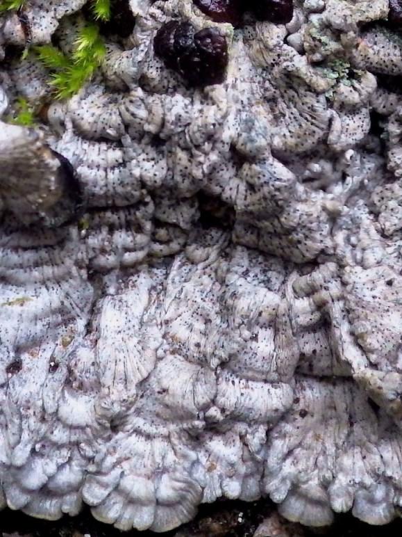
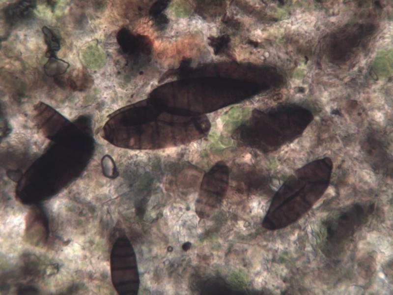
This fungus was found growing on the thallus of a Degelia (D. cyanoloma I think). It is visible as black dots immersed on the thallus of the host. Due to their small size was difficult to make a section of it and to observe it conveniently. It is completely strange for me, having some big and septated spores (no asci observed), dimensions up to 50 x 20 um. There are other structures present on the hymenium like some septate setae. I have no clue about what it can be, besides the fact that the spores look vaguely similar to those of the topic "Lichenicolous fungus on Bacidia - 2015-09-19", suggested to be of a Navicella sp.. Despite the long list of lichenicolous fungi presented in "Lichenicolous net" only few species are referred having Degelia spp. as host; one of each genera: Roselliniella, Stigmidium, Toninia, none seeming to be this one. Maybe someone know what it is. Any opinion will be appreciated.
Thanks in advance,
zaca
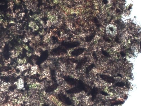
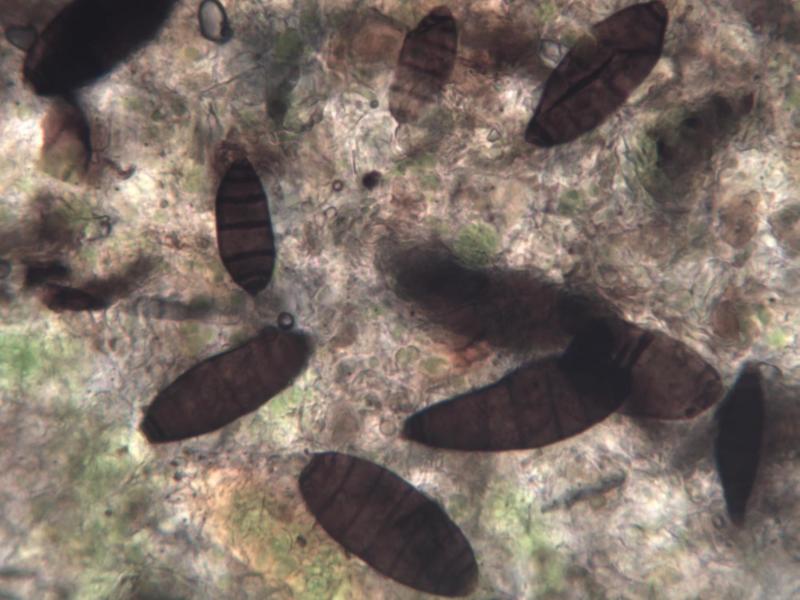

Curious. Are you confident that the ascospores originated in the small black immersed perithecia (if that's what they are)? I wonder if they could have originated elsewhere, though there are certainly a lot of them. The small perithecia remind me of Stigmidium degelii based on the macrophoto, but of course the spores do not match. Sometimes it is hard to find the spores in S. degelii just because of the small search area, and I have seen specimens that only seemed to produce pycnidia.
Good luck,
Kendra
Thanks for your comment.
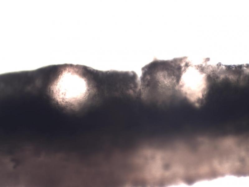
Unfortunately I'm not sure of anything connected with this fungus. The material is too small for adequate observation without proper means. I started by crushing some of the black dots and observed under the micro; then I realise the presence of these big spores and took photos of them. After I tryed to do a section, but the result was bad, as you can see; I then tryed to see the spores from that slide, but I didn't find any, because, as you said, the area for searching was too small and maybe it was hidden by the material from the thallus of the host. Therefore, I cannot assert that the spores observed are originated from the perithecia, but from where they came from, if I observed them as explained above?
Best regards,
zaca
Hi Zaca,
Sometimes I find spores of unclear origin (perhaps a nearby fungus) sitting on the surface of host tissue when I look at lichenicolous fungi. I wondered if that could be the case here, especially if the ascospores are surprisingly large compared to the size of the perithecia.
However, you may be right and the spores may have come from the same perithecia you observed. There are quite a lot of them in your photograph afterall. Unfortunately, in that case I have no idea what this fungus could be! Sorry - I hope that you can find a name for it.
Kendra
for your comment. I will make a new attempt and, if something new, I will report it here.
Best regards,
zaca