23-05-2026 11:44
 Charles Grapinet
Hello, I am having trouble identifying this copro
25-05-2026 16:44
François BartholomeeusenHi forum members,During an excursion organised by
25-05-2026 16:35
 Bernard CLESSE
Bonjour à toutes et tous,J'ai trouvé récemment,
22-05-2026 13:29
Gernot FriebesHi,I am curious to hear your opinion on this mater
23-05-2026 18:57
Sylvie Le GoffBonjour à tousRécolté sur une branchette de Sal
22-05-2026 14:44
 Lothar Krieglsteiner
in unripe condition citrine yellow, then soon fadi
22-05-2026 21:35
Steve ClementsBonjour, I expected this find on old wood on our
22-05-2026 18:12
Lothar Krieglsteiner
... in moist chamber from Portugal.As the fungus s
22-05-2026 20:08
Ethan CrensonHello all, Yesterday in NYC I was visiting an e
 Hi to all
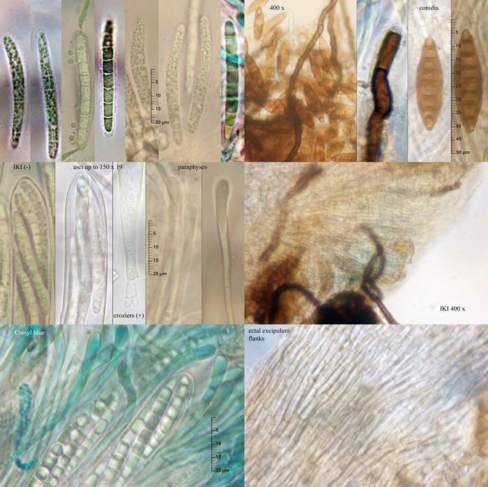
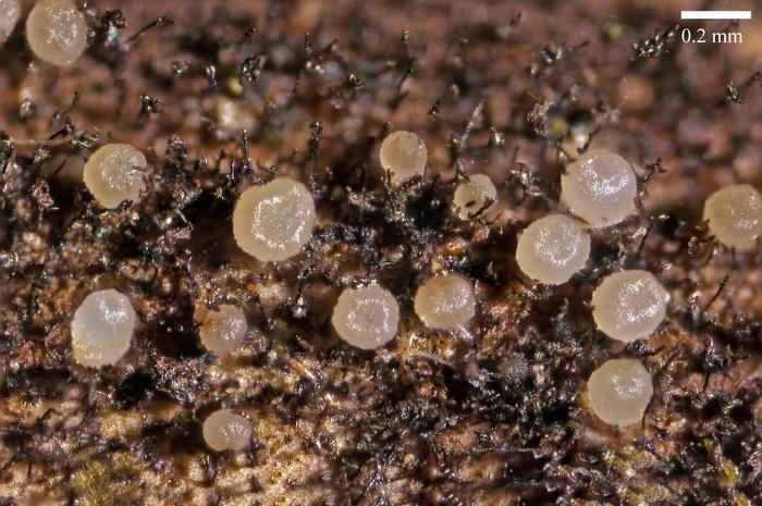
Few days ago I have found these small fruitbodys growing on old wet stems of Rubus grex fruticosus at the sea level. Here is my description of this fungus.
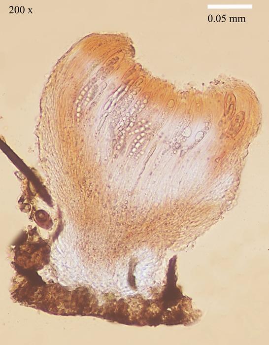
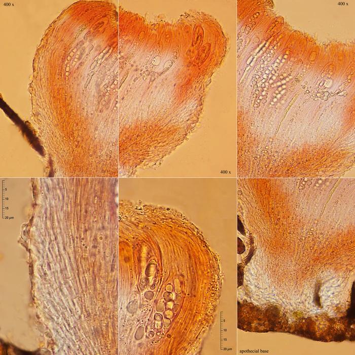
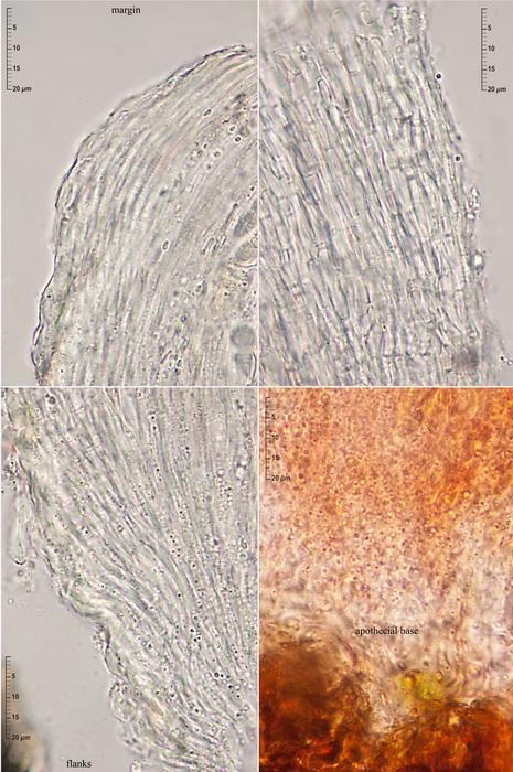
Apothecia superficial, gregarious, scattered among the blackish brown setae of the conidiophores of a dematiaceous mould (Pseudospiropes?), narrowly turbinate, more or less gelified, whitish to amber color, up to 0.30 x 0.25 mm, substipitate on a short and stout pseudostipe 0.12 high and 0.15 broad. Hymenium flattened, smooth or only slightly pruinose. Margin involute, glabrous, that exceeds the hymenium level. Excipulum glabrous and whitish.
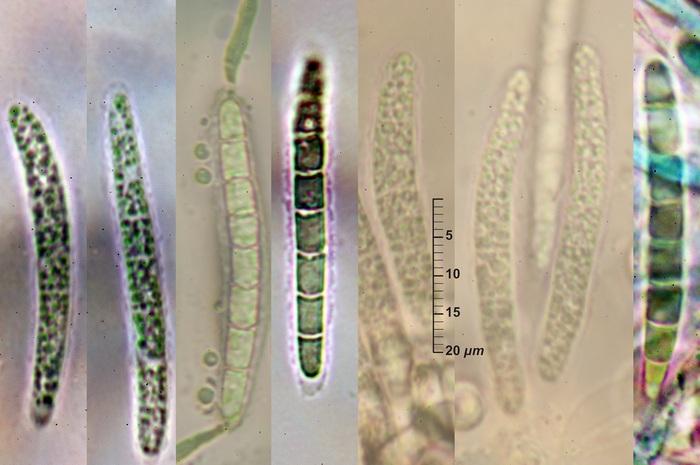
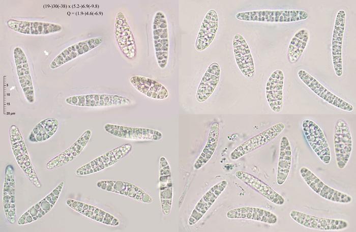
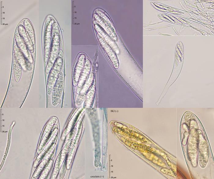
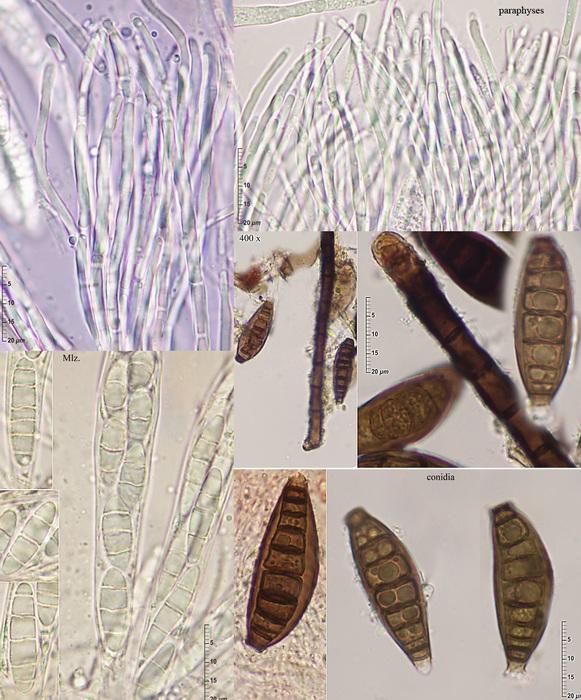
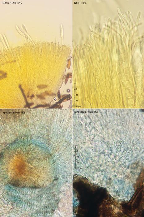
Asci narrowly clavate, inoperculate, unitunicate, 137-182 x 19-20 µm, 8-spored, IKI negative, arising from croziers. Ascospores biseriate, hyaline, smooth, with many small LBs, very polymorphic, ellipsoidal, cylindrical, subfusoid, obovoid, clavate, (19-)30(-38) x (5.2-)6.9(-9.8) µm; Q = (1.9-)4.6(-6.9), with 3(-5-6) cross septa only well visible in Melzer's reactive or congo red. A more or less smooth and complete gel sheath surrounds the fresh ejected ascospores. Paraphyses filiform, septate, not enlarged at their tips or only up to 2-2.5 µm, with cylindrical Vbs. Ectal excipulum textura oblita with cylindrical, elongate, septate, hyaline cells 2-2.5 µm, with a thin gel layer between them, wider at the apothecial base. Medullary excipulum is indistinguishable
The basal region of the ascomata stained blue in IKI. All the apothecial tissues extrude in fresh condition a bright yellow fluid in KOH 10%.
After reading Iturriaga & Korf's paper I don't know a species that fits well with this fungus.
Could you help me?
Thanks again

I have also my problems with Iturriaga's paper, I never came clear with it. But I have also problems to recognize more than one species in this genus. Maybe ther exist tropical species, but in our region I think it is always S. basitricha. Perhaps until someone finds genetical differences.
Did you see anything that is unusual in your fungus compared to S. basitricha on wood?
I remember an amyloidity of the spores in Iturriaga's paper, am I right? Perhaps to obtain only with overmature dead spores, similar as in D. connivens.
Hi Zotto
Yes. I see some differences with the 'common' basitricha on wood with more septate cylindrical ascospores with a narrower verrucose gel sheath and broader paraphyses tips. I have not seen the amyloid reaction on my fresh spores but I pressume that Iturriaga & Korf's observations were made allways on dried material.
This picture was take from an old collection from Robinia pseudoacacia
Thanks again