
14-08-2016 23:15
 Alex Akulov
Dear friendsCan you help me to find the descriptio

05-06-2026 11:02
Thomas Læssøehttps://svampe.databasen.org/observations/10596691
04-06-2026 11:36
Gernot FriebesHi,found on Vaccinium myrtillus.Asci: IKI –, 8-s
05-06-2026 12:10
 François Freléchoux
Capitotricha sp. sur Lonicea caerulea Caractères
19-05-2026 10:27
Patrice TANCHAUDBonjour, récolte récente sur terre retournée i
04-06-2026 18:39
Gernot FriebesHi,I collected this species in two different locat
22-05-2026 13:29
Gernot FriebesHi,I am curious to hear your opinion on this mater
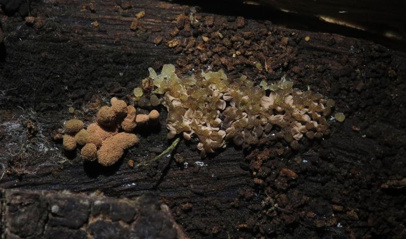
 This is my second post on this forum. I know it is dedicated to ascomycota but I've seen some posts on Myxomycota. I am requesting help on a Myxo. If this is not allowed on ascofrance, kindly let me know. I also plan to submit microfungi like Penicillium, Mucor, etc which I believe they are fine since they velong to Ascomycota.
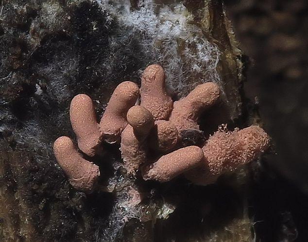
My specimen was in large numbers on a dead fallen branch, Shortly stipitate, pastel-pink, cylindrical, approx 2-4mm x 1mm, leaving a pale brown peridium when spores are liberated. Stipe very short, 0.5mm.
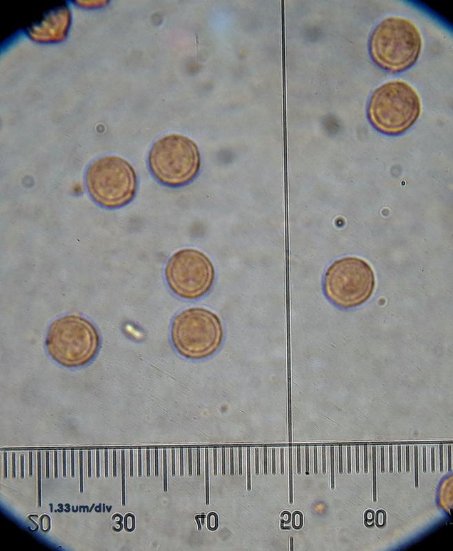
Spores circular, 7-8um
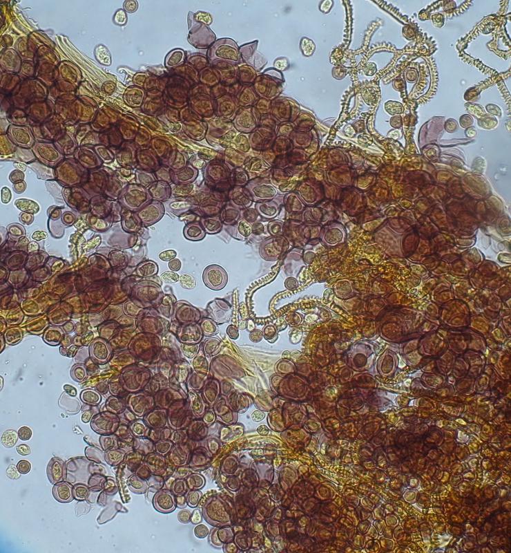
Cell of peridium? (or calyculus) shown in image #4 are 14-17um diameter with a yellow amorphous body. These cells rapture easily.
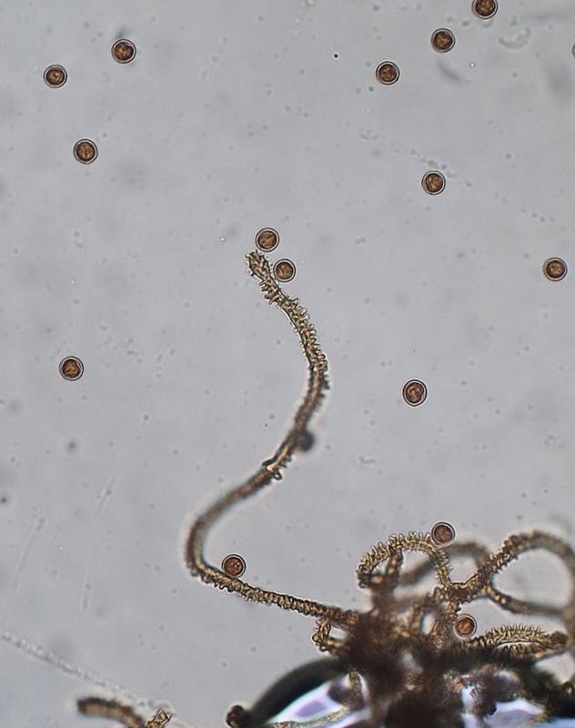
Capillitium with inflated ends, ovoid-capitate; with rims encircling half the diameter (4-5um), yellow under light microscope, quite elastic, but loosly attached to capitulum (in my opinion... to double check)
Personnellement je verrais plutôt dans le grnre Arcyria, par ex. A.virescens.
Cordialement.
Michel
http://www.google.fr/imgres?imgurl=http%3A%2F%2Fslimemold.uark.edu%2Fimages%2Fpictures%2Fmartin%2F96arcyriavirescens.jpg&imgrefurl=http%3A%2F%2Fslimemold.uark.edu%2Fmartin.htm&h=600&w=800&tbnid=i8UbZ-OyAwFxEM%3A&zoom=1&docid=qxk2eTqc4b9HQM&ei=0A4RVdWYN9XvaJWJgZAM&tbm=isch&iact=rc&uact=3&dur=1062&page=1&start=0&ndsp=27&ved=0CCAQrQMwAA

il manque trop de details pour être certain de l'espèce mais je pense qu'il faut aller dans la direction de Arcyria denudata, A. incarnata, A.major ou A. minuta si on tient compte de la dimension des spores.
Jacky

Hello together,
this Arcyria has a capillitium totally soluble from the calyculus (peridium) and rather pale red colour. This features are typical to A. incarnata. A. major has a strongly expanding capillitium which is brillant pink, A. denudata is dark red and has a capillitium not soluble from the peridium.
Yours, Lothar
P.S. Tubulifera has no capillitium at all (Liceales)
Thank you very much for your replies and education. I had seen tubilifera looking similar on google images and garasped on it. Now I found a key to Orders/families(/genera), and of course it is Trichiales with its true capillitium. I also found a key for Arcyria and it had keyed as A. incarnata. Good job and thanks again.
I have two question please (you can answer in French ;-) )
1) What are the structures in photo 4 please? Like small amorphous crystals in a thin-walled cell? think they were towards the base and so maybe (?) calyculus tissue?
2) How woud you distingusih between a persistent not soluble capillitium net and capillitium totally soluble from the calyculus ?
Dear Stephen,
1) What are the structures in photo 4 please? Like small amorphous crystals in a thin-walled cell? think they were towards the base and so maybe (?) calyculus tissue?
... first: Myxos do not have a tissue. The only thing that are cells in myxo fruitbodies are the spores. Myxo plasmodia, their vegetative stage, are one large cell with many nuclei. So, cell borders are missing also in the fruitbodies.
2) How woud you distingusih between a persistent not soluble capillitium net and capillitium totally soluble from the calyculus ?
The best is blowing. In A. incarnata you will be able to push the capillitium to anywhere, and only the cup remains - as is visible in your foto. In A. denudata you can blow and blow and blow and blow - and the sporocarp remains as it was, only losing some spores.
Yours, Lothar
P.S. Sorry, I am not able to write in French - ...