
17-04-2026 19:16
 Enrique Rubio
Hi to everybodyI would appreciate any assistance r
14-04-2026 05:32
Ethan CrensonHi all, A few weeks back a friend pointed out som
17-04-2026 15:14
 Bruno Coué
Bonjour.Récoltes du 16/04/2026, sur feuilles mort
12-04-2026 15:52
Gernot FriebesHi,I'm looking for help with this anamorph collect
14-04-2026 21:52
Gernot FriebesHi,found on dead leaves of Carex elata. Conidia: 4
16-04-2026 22:09
Buckwheat PeteHello, I'd like to ask about this older specimen:
15-04-2026 19:33
Fátima Durán ManzanequeHi!! I need help, I found this Ascomycete but I d
14-04-2026 20:31
Gernot FriebesHi,can this be Psilachnum lateritioalbum on Phragm
12-04-2026 17:56
 Hardware Tony
Found on dead stems in February earlier this year
12-04-2026 12:22
 William Slosse
In a dune grassland in Oostduinkerke (Belgium), on
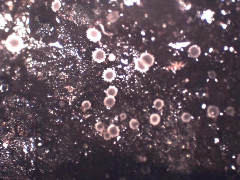
Hello Everyone,
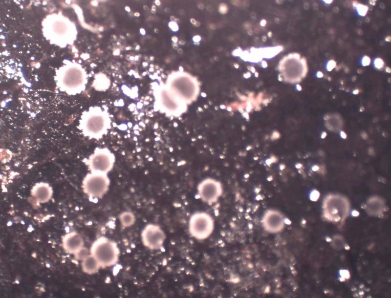
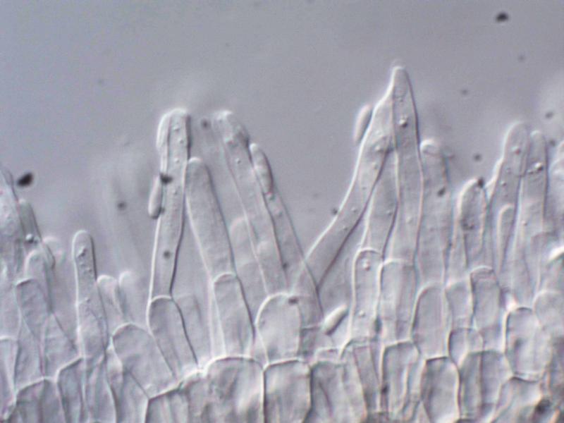
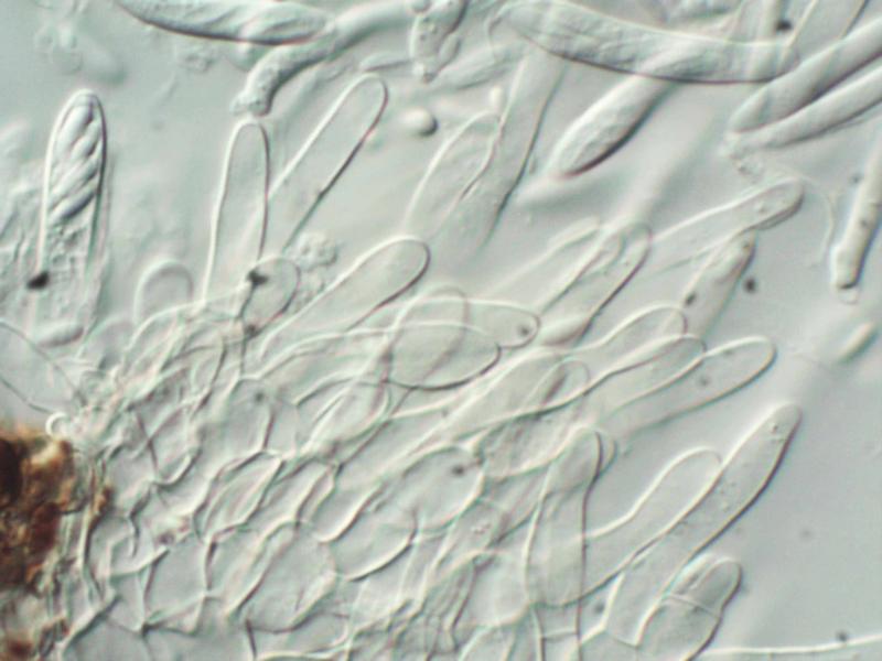
I would appreciate some help with a delightful minute species of probably Calycellina.Found in abundance on decaying leaves of Alnus glutinosa from a very wet location in North Argyll.Apothecia very small, ranging from 100µm to about 1 mm. brown . Sessile with a marked basal ring of dark cells. Excipular structure of thin-walled rounded cells, almost 'prismatica' Apothecium surrounded be a very variable fringe of delicate hairs, mostly very short, occasionally clearly visible as ion the photo. Asci with pore that is Iodine +ve, Spores narrowly elliptical about 8 x 3. Paraphyses slender. Hairs, paraphyses and spores mostly without inclusions. Pictures taken from fresh material in water.
Apologies fro the lack of scale bars: at the time the measurement system was not functioning.
Reagrds to all, Peter Wilberforce
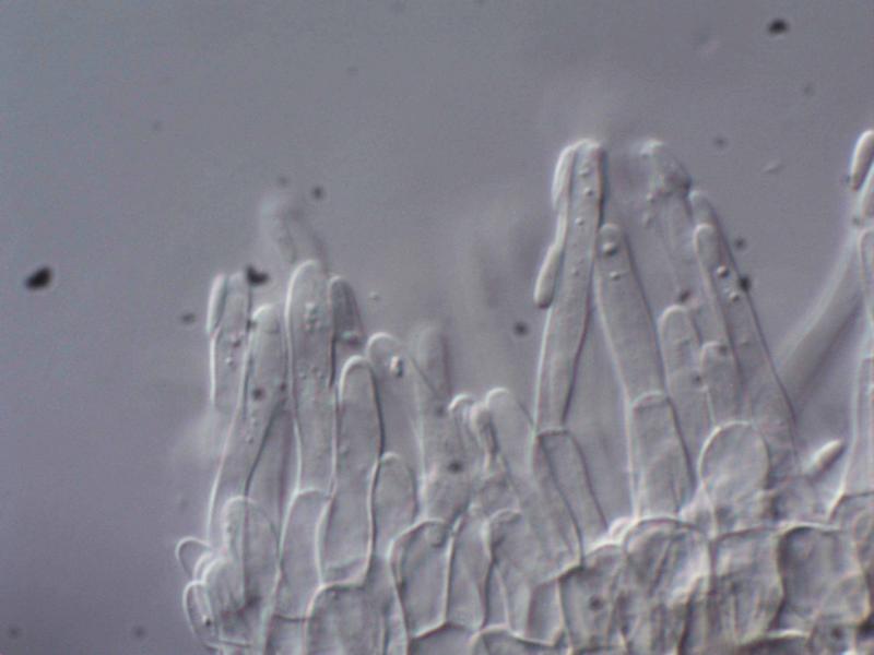
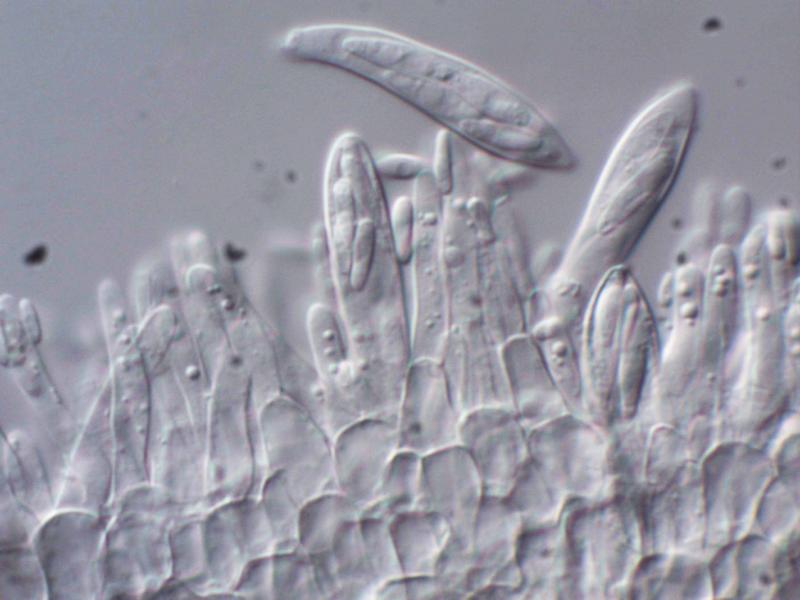
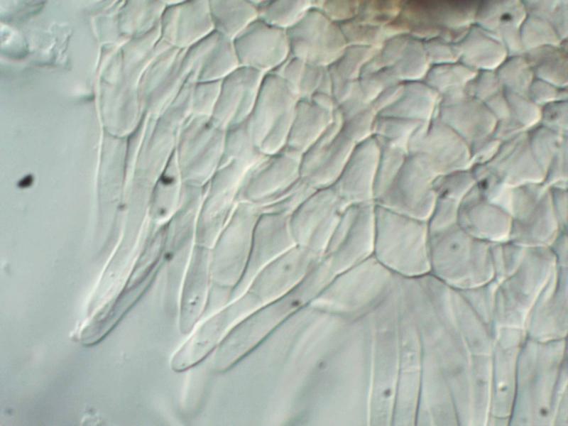

do you also have a photo of the more basal excipulum? The absence of elongate VBs in the paraphyses and hairs excludes a Calycellina. My idea on this substrate is Pyrenopeziza betulina (on Betula leaves) or the similar P. fuckelii (= Microscypha monticola) on Salix leaves.
But I might be wrong ....
Alnus is certain?
Zotto
Dear Zotto,
Many thanks for commenting on this unusual species. There is no doubt at all about the identitiy of the host leaves: Alnus glutinosa. The location is a very wet and midge-ridden area in a narrow glen alongside a stream.
I tried at the time to get sections, but found the apothecia very fragile. This was using a freezing stage. I have quite a bit of dried material, I'll try soaking a bit up and try sections again.
The hairs were generally very short and only visible under a stereo mic. The picture shows a most unusual long-haired sprecimen. In all cases the hairs seem to be 2-celled: a basal squat cell and an apical elongated finger-like cell. Always hyaline with few inclusions.
Thanks for trying to help with this interesting fungus.
In a few weeks time Ill try and collect more material.
Very kind regards,
Peter W