
29-05-2026 15:35
daniel FERREBonjour à tous,Je voudrais votre aide pour cette
28-05-2026 16:15
James MitchellHello,Does anyone have the original publication of
28-05-2026 11:06
Thomas Læssøehttps://svampe.databasen.org/observations/10596750
23-05-2026 11:44
 Charles Grapinet
Hello, I am having trouble identifying this copro
25-05-2026 16:44
François BartholomeeusenHi forum members,During an excursion organised by
26-05-2026 21:25
Dirk GerstnerHello everyone, I'm completely stumped by this li
26-05-2026 22:44
Ethan CrensonHi all, I think I have Incrucipulum capitatum her
22-05-2026 14:44
 Lothar Krieglsteiner
in unripe condition citrine yellow, then soon fadi
25-05-2026 16:35
 Bernard CLESSE
Bonjour à toutes et tous,J'ai trouvé récemment,
22-05-2026 13:29
Gernot FriebesHi,I am curious to hear your opinion on this mater
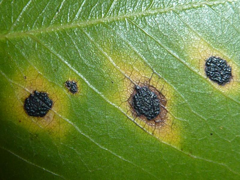
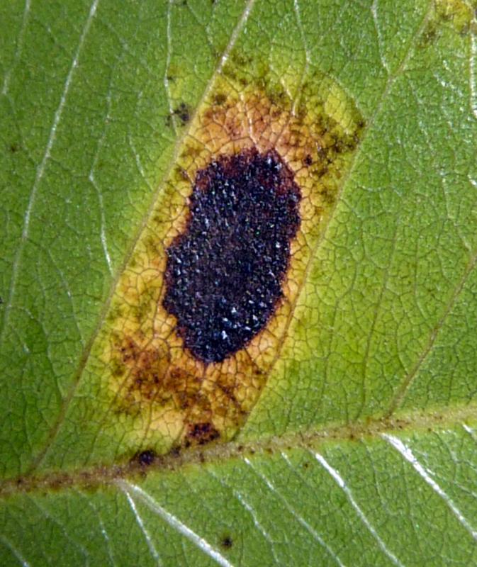
Here a ascomycete associated to leaf spots,in Cassia fistula; have applanate ascostroma, 1 mm. high,
Ascospores biseriate, hyaline, oblong. May be a Phyllachora sp.?
Phyllachora canafistulae F.L. Stevens & Dalbey, Bot. Gaz. 68: 55 (1919).
syn. Phyllachora azuanensis Petrak & Ciferri, Annales Mycologici 30: 235 (1932).
Anamorph: unknown.
Teleomorph: infected areas not clearly delimited. Blackened regions 0.5-3mm diam, scattered
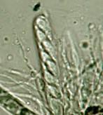
over the adaxial surface of leaves, irregular in outline, sometimes surrounded by a narrow ring of dead leaf tissue to 0.5mm wide, black, sometimes verrucose (probably a feature of the leaf epidermal architecture), slightly raising the substrate surface, 1- to 8-loculate, the individual locules domed, the ostioles usually inconspicuous, but eventually becoming slightly papillate. Ascomata hologenous, the ostioles epigenous. Upper wall of stroma 30-65µm thick, composed of cuticle, epidermis and some palisade cells completely occluded by dark brown melanized tissue, in a continuous layer above and between the ascomata. Lower wall of stroma to 45µm thick, of similar composition. Mesophyll tissue completely invaded by thin-walled dark brown fungal cells. Ascomatal wall 10-25µm thick, composed of strongly flattened dark brown fungal cells, with an inner layer of flattened hyaline cells near the apex from which the periphyses develop. Ascomata 160-200µm diam, globose to pyriform, slightly taller than wide, with the hymenium extending over the whole of the lower wall. Paraphyses copious, to 3.5µm wide,
gradually tapering, very thin-walled, sometimes branched near the base or with a few knob-like branches near the apices. Asci 88-130 x 11-23µm, cylindrical to saccate, short- to long-stalked, apex obtuse, very thin-walled, without visible apical structures, 8-spored. Ascospores arranged obliquely uniseriately, sometimes partially biseriately, 14-18(-20) x 6-8(-11)µm, mostly fusiform to narrowly ovoid, but often with a small proportion irregularly shaped (giving the impression that germination started within the ascus before the laying down of ascospore walls was complete), hyaline, aseptate, relatively thick-walled, with a gelatinous sheath 2-3µm thick, not clearly visible in all specimens.
Typification: Puerto Rico: Mayaguez, on Cassia fistula, 14 Jun. 1915, F.L. Stevens 7022 (ILL
- holotype, BPI!, K!, MAPR, NY! - isotypes of P. canafistulae). Dominican Republic: Cordillera Central: Azua: San Juan de la Maguana: near Arroyo Manacle, in living leaves of Barbiera pinnata, 25 Aug. 1929, E.L. Ekman 3665 (BPI! - holotype, S! - isotype of P. azuanensis).
Distribution: Bermuda, Brazil (?), Costa Rica, Cuba (fide Kreisel, 1971), Dominican Republic,
Grenada, Jamaica, Panama, Puerto Rico, Trinidad.
Host species: Cassia fistula L., C. grandis L. fil., C. javanica L. (fide Stevenson, 1975),
C. javanica subsp. nodosa (Roxb.) K. & S. Larsen (syn. C. nodosa Roxb.). ?Clitoria pinnata (Pers.) ined. (syn. Barbieria pinnata (Pers.) Baillon).