27-02-2026 17:51
 Michel Hairaud
Bonjour, Quelqu'un peut il me donner un conseil p

28-02-2026 14:43
A new refrence desired :Svanidze, T.V. (1984) Novy

29-11-2024 21:47
Yanick BOULANGERBonjourJ'avais un deuxième échantillon moins mat

27-02-2026 16:17
 Mathias Hass
Hi, Found this on Betula, rather fresh fallen twi

27-02-2026 12:56
Åge OterhalsFound on fallen cones of Pinus sylvestris in midle

27-02-2026 11:21
 Yannick Mourgues
Hi to all. Here is a specie that can may be relat
Melanconium sur feuille d'Hedera helix
Alain GARDIENNET,
17-02-2013 08:38
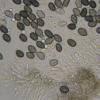
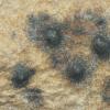
J'ai trouvé ces acervules sur feuille de Hedera helix.
J'ai regardé tous les coelomycetes classiques sur feuille, sans succès.
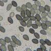
La seule hypothèse que je me permets d'avancer est Melanconium hederae, qui vient usuellement sur bois. Mes conidies mesurent environ : 6-9 x 4-5,5 µm.
Est-ce possible ?
Alain
Alain GARDIENNET,
17-02-2013 11:37
David Malloch,
17-02-2013 16:24

Re : Melanconium sur feuille d'Hedera helix
Hello Alain,
This fungus seems to be one of the species belonging in Sphaeropsis, Coniothyrium or Microsphaeropsis. The conidia appear to have a truncate base as though they were produced holoblastically or by annellides, indicating one of the first two genera. However the basal differentiation could be an elongation produced by squeezing out through a phialide. If it is phialidic it may be a species of Microsphaeropsis. Try mounting it in Congo Red to see the exact mode of conidiogenesis. Especially, look for annellations on older conidiogenous cells.
I tried to find it in the host index of Sutton's Coelomycetes, but there is nothing like that on Hedera.
Dave
Alain GARDIENNET,
17-02-2013 21:15
Re : Melanconium sur feuille d'Hedera helix
Hi David,
Thanks, I'm less alone...
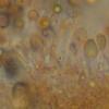
Here are two new pictures showing conidiogenese. The conidiophores are up to 15 x 2,5 µm, sometimes septate. Could you be able to see the base here ? I don't think they are phialidic.
I can check again if necessary.
Have you read notes in Grove 1937, p6 (Coniothyrium hederae) and p17 (Sphaeropsis helicis) ?
In both cases, Melanconium hederae is mentioned.
Alain
David Malloch,
18-02-2013 02:11

Re : Melanconium sur feuille d'Hedera helix
Hi Alain,
This is a complicated problem with no modern literature for guidance. Grove was very certain of the fungus he called Melanconium hederae and did not think it was the same as Coniothyrium hederae. In Saccardo (Syll. Fung. 3:307) the two species seem to be treated as synonyms. To me the pycnidia in your photograph look more like Coniothyrium or one its more recent segregate genera. Melanconium.species should have more of an aecervulus.
I agree with you that the conidiogenesis does not appear to be phialidic, but I don't see evidence of annellides either.
Dave
Alain GARDIENNET,
19-02-2013 09:21
Re : Melanconium sur feuille d'Hedera helix
Hi Dave,
In your proposals, Coniothyrium was the better, indeed.
But I have searched again in Melanconium. Some of Melanconium species show single acervules. And I have been able to compare, it's very closed.
Thus, somebody who has a data of Melanconium hederae on twigs says to me that it looks like ( no differences in shape, color, size ...)
Perhaps could it be a record of Melanconium hederae on leaves ?
Thanks again, it's important to have a discussion, because, as you said, the modern litterature is so much poor.
Alain
Luc Bailly,
21-12-2024 21:12
Re : Melanconium sur feuille d'Hedera helix
I recently found it on Hedera helix branch: https://www.inaturalist.org/observations/255797728
Just posting it for double checking.
Cheers!
